Radiology Rounds – 11/22/22

We are back with another #RadiologyRounds for you!

A middle-aged male presents with sub-acute fatigue, fevers and dyspnea on exertion. His admission labs are notable for severe neutropenia (ANC < 500)

This patient has evidence of a halo sign which represents local vessel invasion or hemorrhage

The patient undergoes evaluation. RVP, sputum culture and serum galactomannan and 1-3-B-d-glucan are negative. A BAL with transbronchial biopsies are obtained.

The patient was found to have pulmonary mucormycosis and was started on IV liposomal amphotericin-B.

Want to learn more about this case? Take a listen to our Fellow’s Case Files from Baylor College of Medicine

29. Long COVID Roundtable

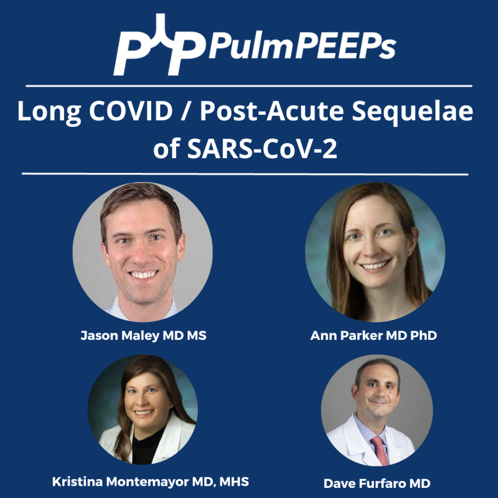
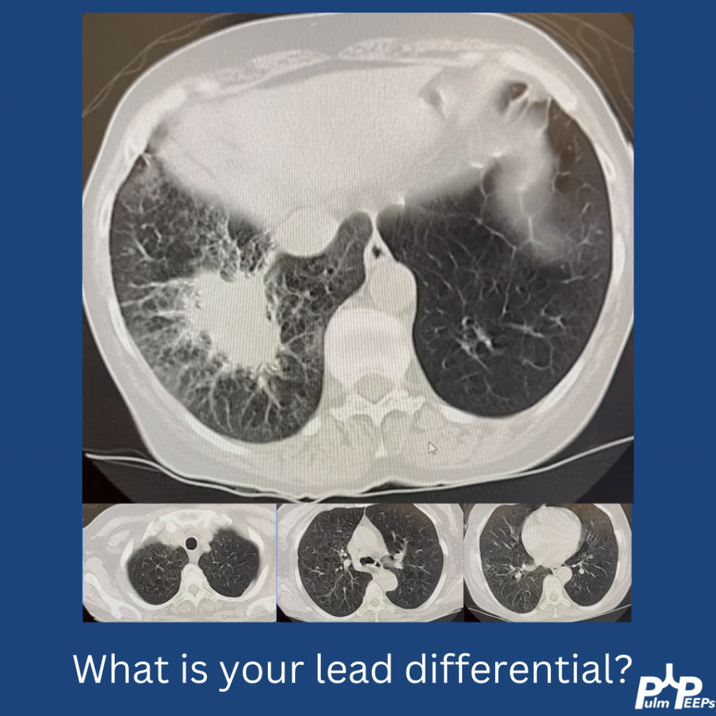
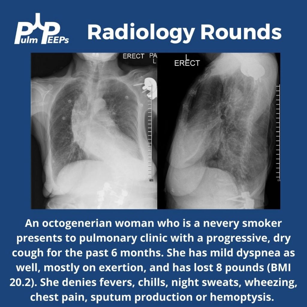
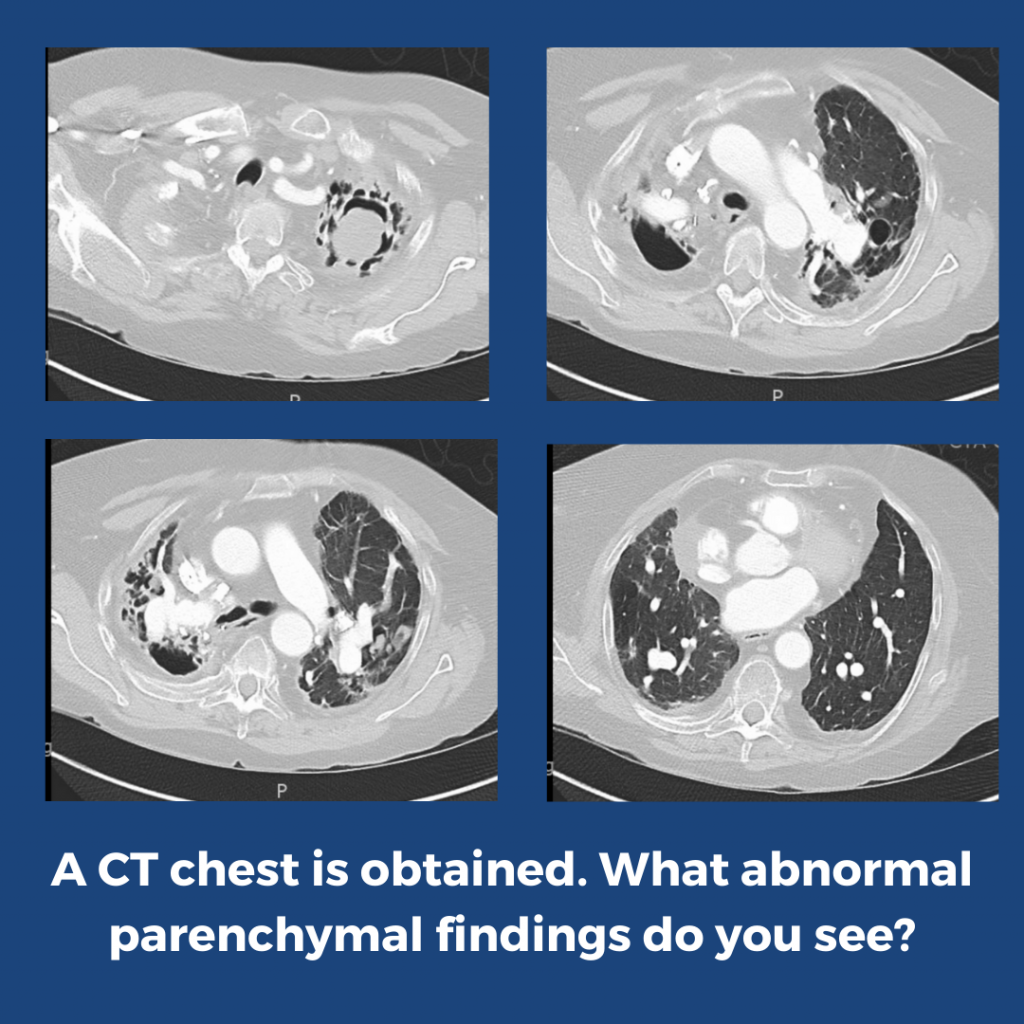
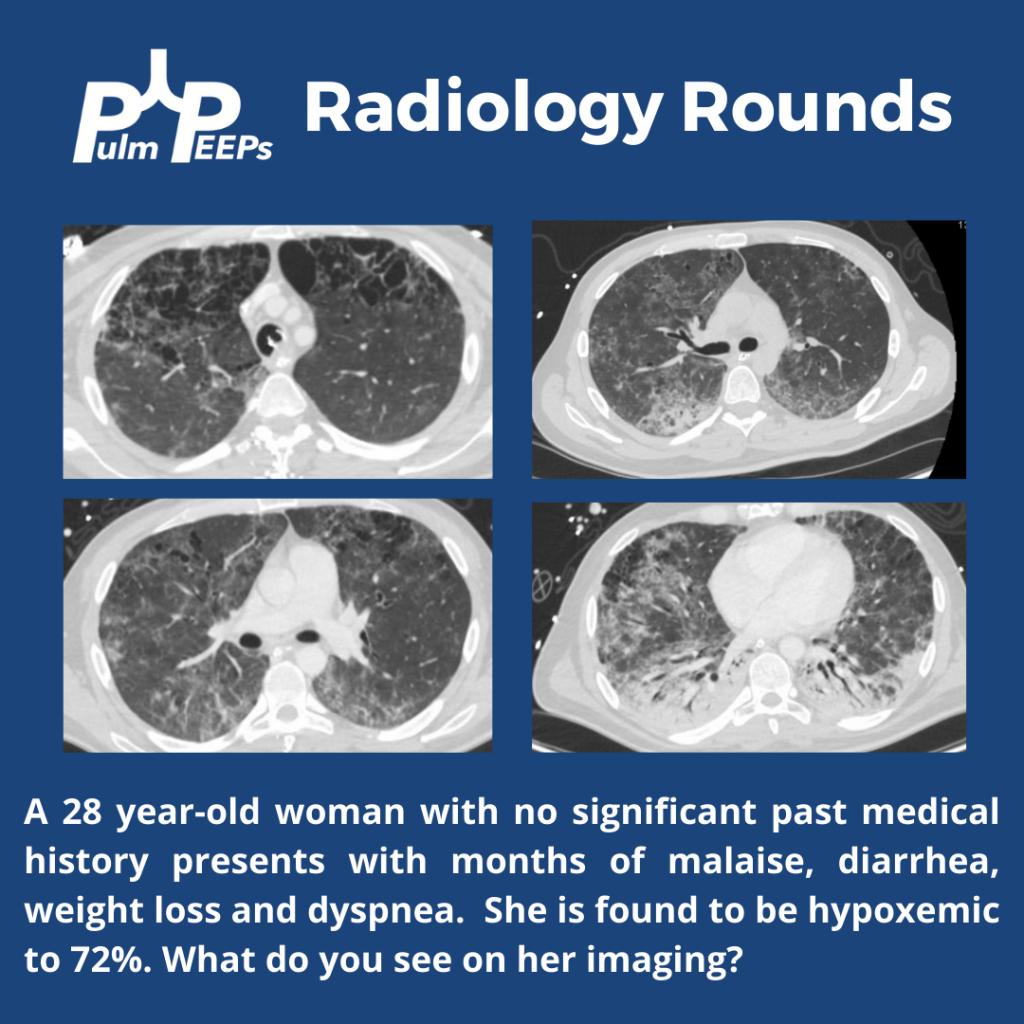
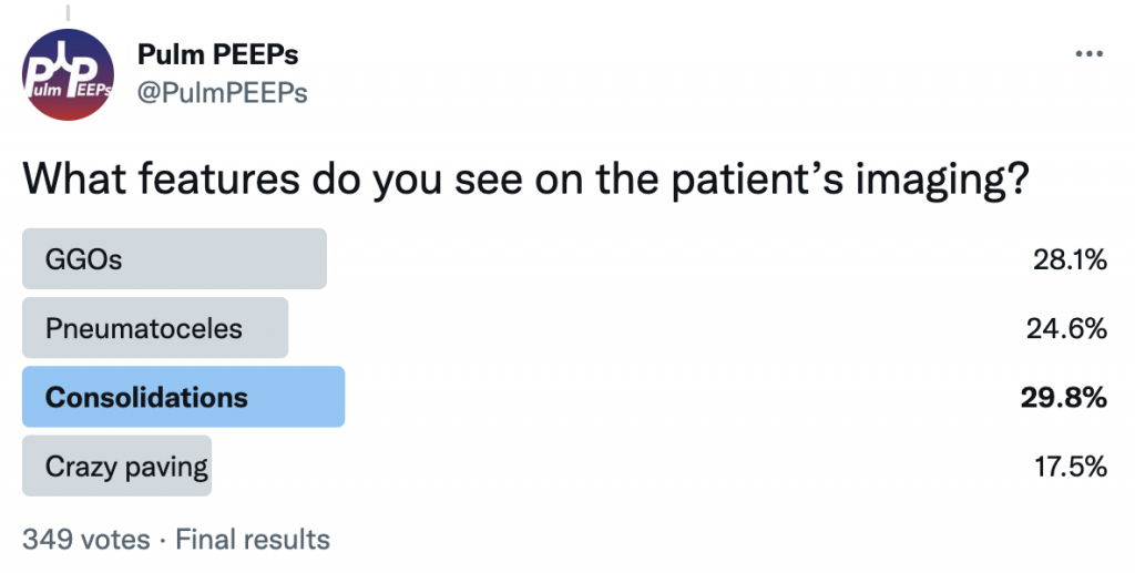
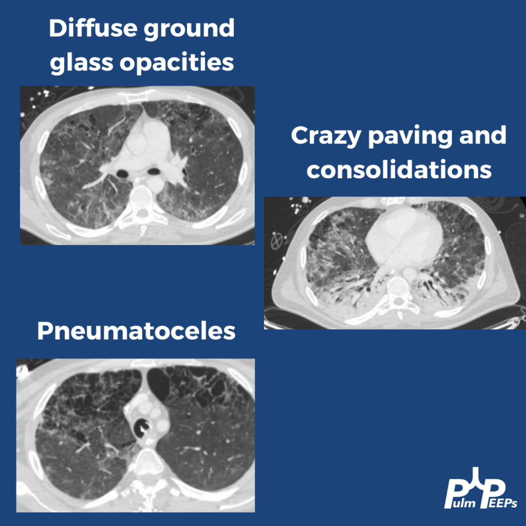
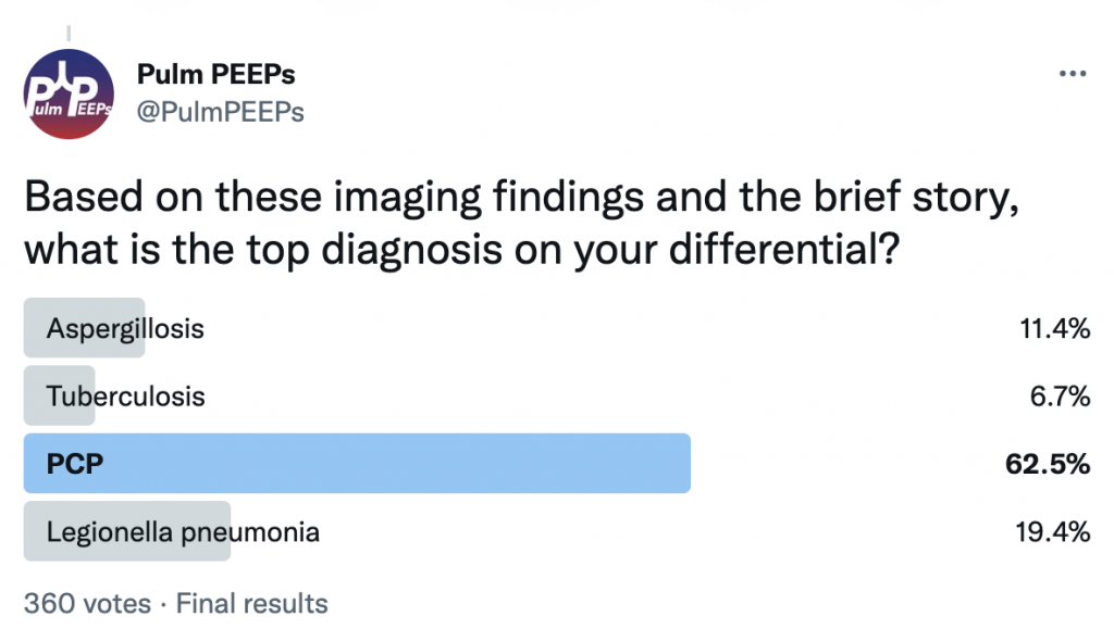
This week on Pulm PEEPs, Dave and Kristina are joined by Jason Maley and Ann Parker, two pulmonary and critical care physicians who are leaders in treating patients with Long COVID, or Post-Acute Sequelae of SARS-CoV-2. Both of them help run the Long COVID clinics at their respective institutions and are part of broader consortiums dedicated to patient care. They also both participate in research to improve outcomes for patients with Long COVID and Post-Intensive Care Syndrome. In this conversation, we cover the diagnosis of Long COVID, common symptoms, abnormal test findings, possible mechanisms of disease, the impacts of variants and vaccines, treatments, and the natural history of this condition. We hope this will be helpful for providers, patients, and family members.

Meet Our Guests

Jason Maley is an Assistant Professor of Medicine at Beth Israel Deaconess Medical Center and Harvard Medical School. He is the Director of the BIDMC Critical Illness and COVID-19 Survivorship Program, and the Co-Chair of the American Academy of Physical Medicine and Rehabilitation Postacute Sequeleae of SARS-CoV-2 infection (PASC) initiative. He is NIH funded to study post-COVID patients.

Ann Parker is an Assistant Professor of Medicine at Johns Hopkins and is the Co-Director of the Johns Hopkins Post-Acute COVID-19 team. She is NIH funded with her research focusing on survivors of respiratory failure and critical illness.

Key Learning Points

Long COVID or Post-Acute Sequelae of SARS-CoV-2 or Post-COVID condition

  • Long COVID was first described this way by patients so this is the common nomenclature that is used. It is also referred to as Post-Acute Sequelae of SARS-CoV-2 or Post-COVID condition
  • Defined by patients that have not returned to their baseline health 3 months after their acute episode of COVID-19
  • Major organizations in describing this disease and doing research are:
    • World Health Organization
    • Multiple patient-led organizations
    • CDC – INSPIRE
    • NIH – RECOVER

Post-COVID Clinic

  • Seeing patients across the spectrum of illness. Not all patients had to be critically ill or hospitalized
    • The standard patient has changed over time and now the vast majority had a mild initial illness, but afterward had unusual and persistent symptoms
  • Patients are generally referred by their PCP or self-referred
  • The criteria for being seen in clinic are very loose to make sure patients are not excluded
    • Many patients do not have a confirmed case of COVID since patients early in the pandemic often did not have a positive test available, and now many people are testing positive at home
  • Initial records review to make sure that can help patients
  • Standardized questionnaires
    • Screening for physical impairment, mental health impairment, and cognitive impairment
  • Rehabilitation and multi-disciplinary based approach
  • It is extremely important to be aware of the bias in patient populations in Post-COVID clinics
    • The population that can make it to clinic may not, and does not, represent all patients who have had COVID or have Long COVID. Patients may be limited in their ability to get to clinic based on their physical condition, financial resources, location, support, and language barriers.

Overlap of Long COVID and PICS

  • These conditions are very similar and certainly have a lot of overlap
  • For patients coming out of the ICU, screening should start with looking for known PICS symptoms.
    • These domains are mental health, physical impairment, and cognitive function
  • There may be some unique aspects, such as:
    • Severe persistent fatigue
    • Extreme changes in taste and smell

Common symptoms

  • Many symptoms are complex and multifactorial
  • Neuropsycholgoicl impairment – termed “brain fog”
    • Difficulty with concentration, and cognition
  • Anxiety
  • Depression
  • Persistent shortness of breath
    • Dyspnea can be reported even with just talking for long periods of time
    • “Deep breaths are just not satisfying”
  • Cough
  • Chest pain
  • Dysautonomia
    • Palpitations, dizziness, orthostasis
  • Fatigue
  • Post-exertional malaise
  • Cognitive blunting or “brain fog”
  • Changes in sleep
  • Headaches

Common findings on testing in patients with Long COVID

  • Shortness of breath
    • Some may have impaired diffusion (low DLCO) on PFTs
    • However, often patients have normal or near-normal PFTs
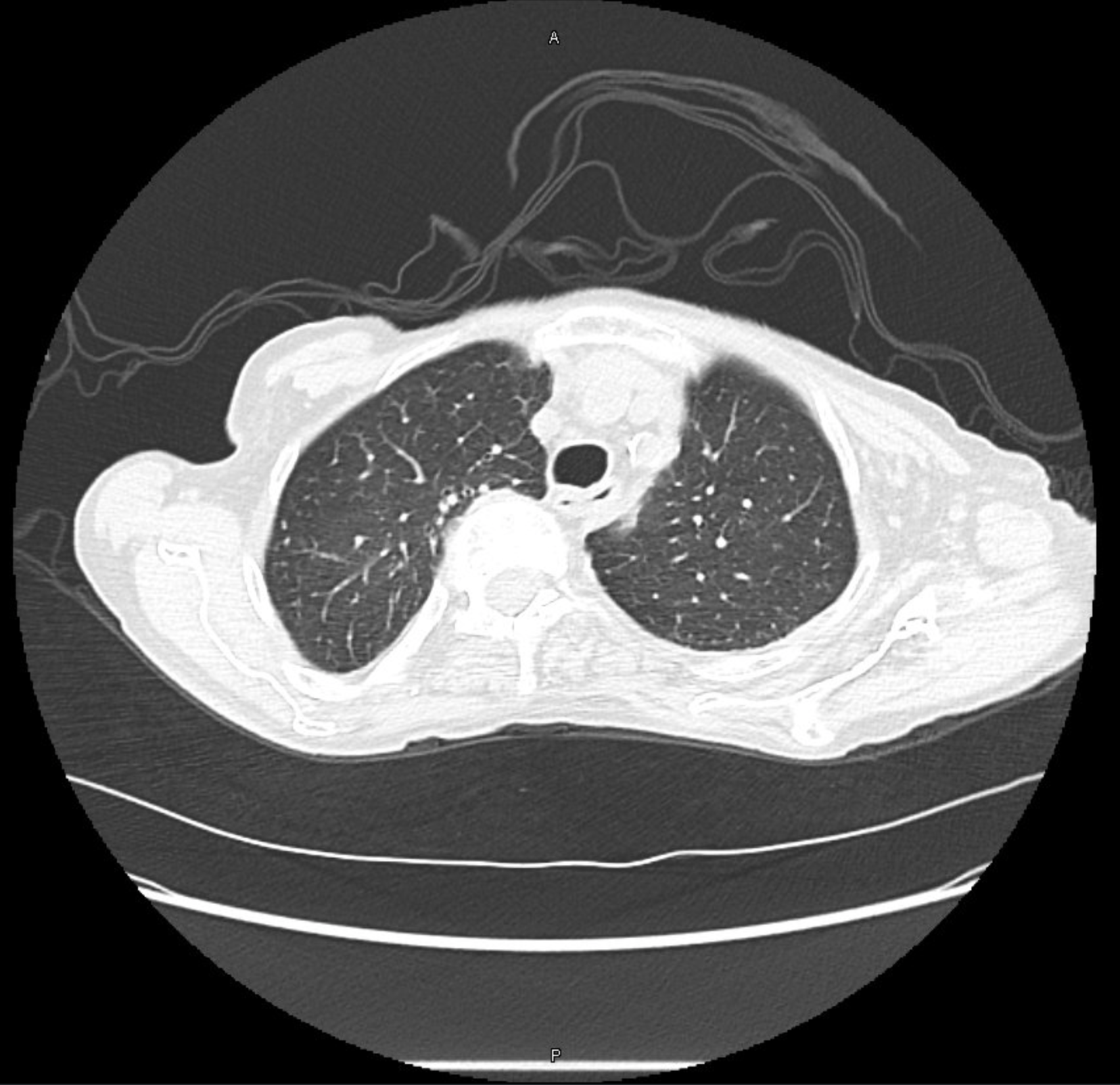
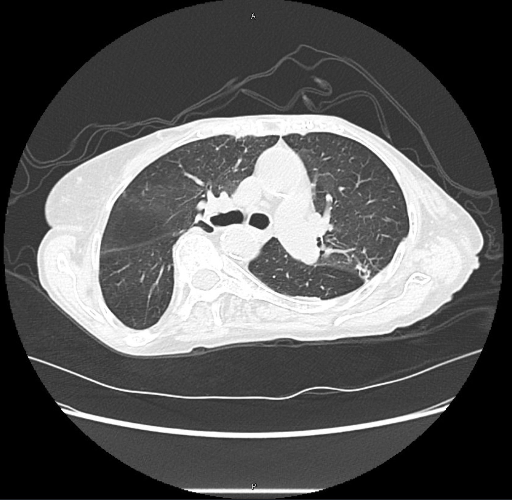
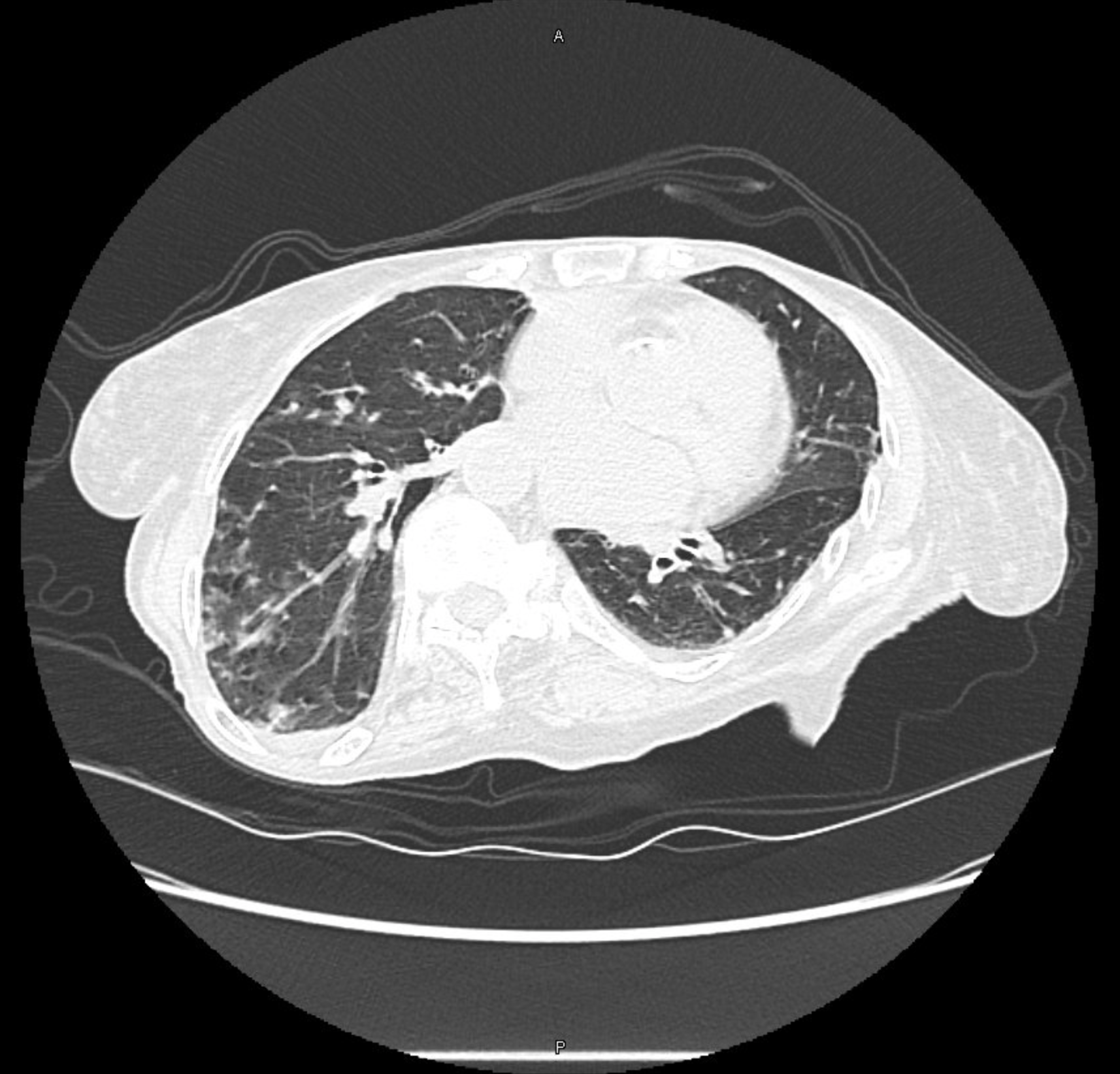
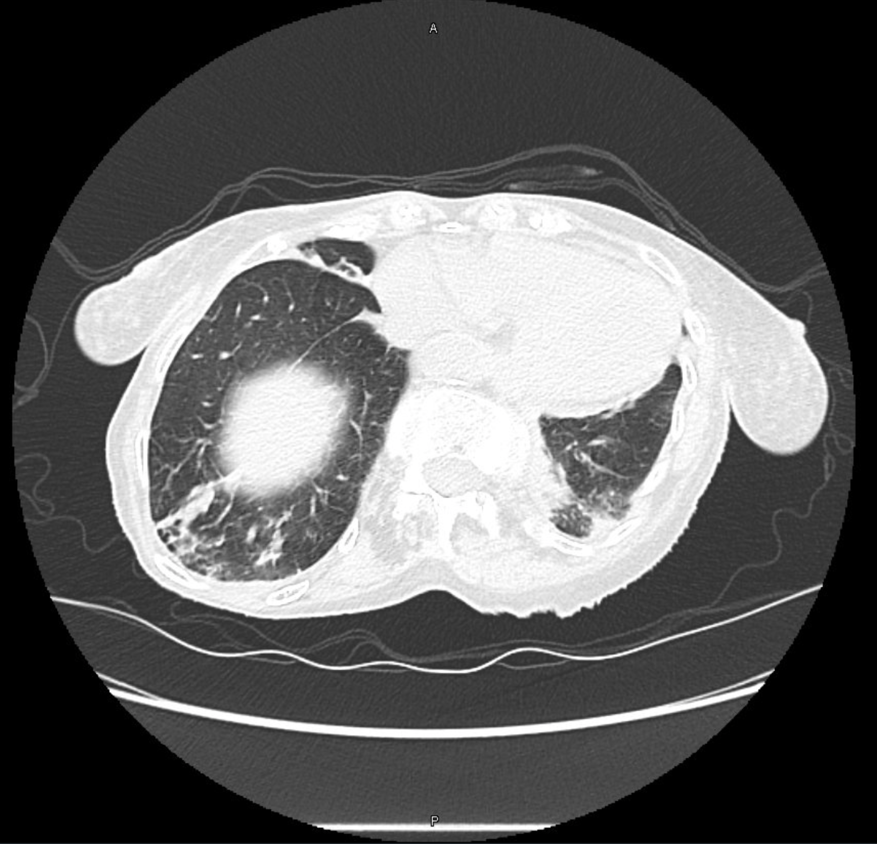
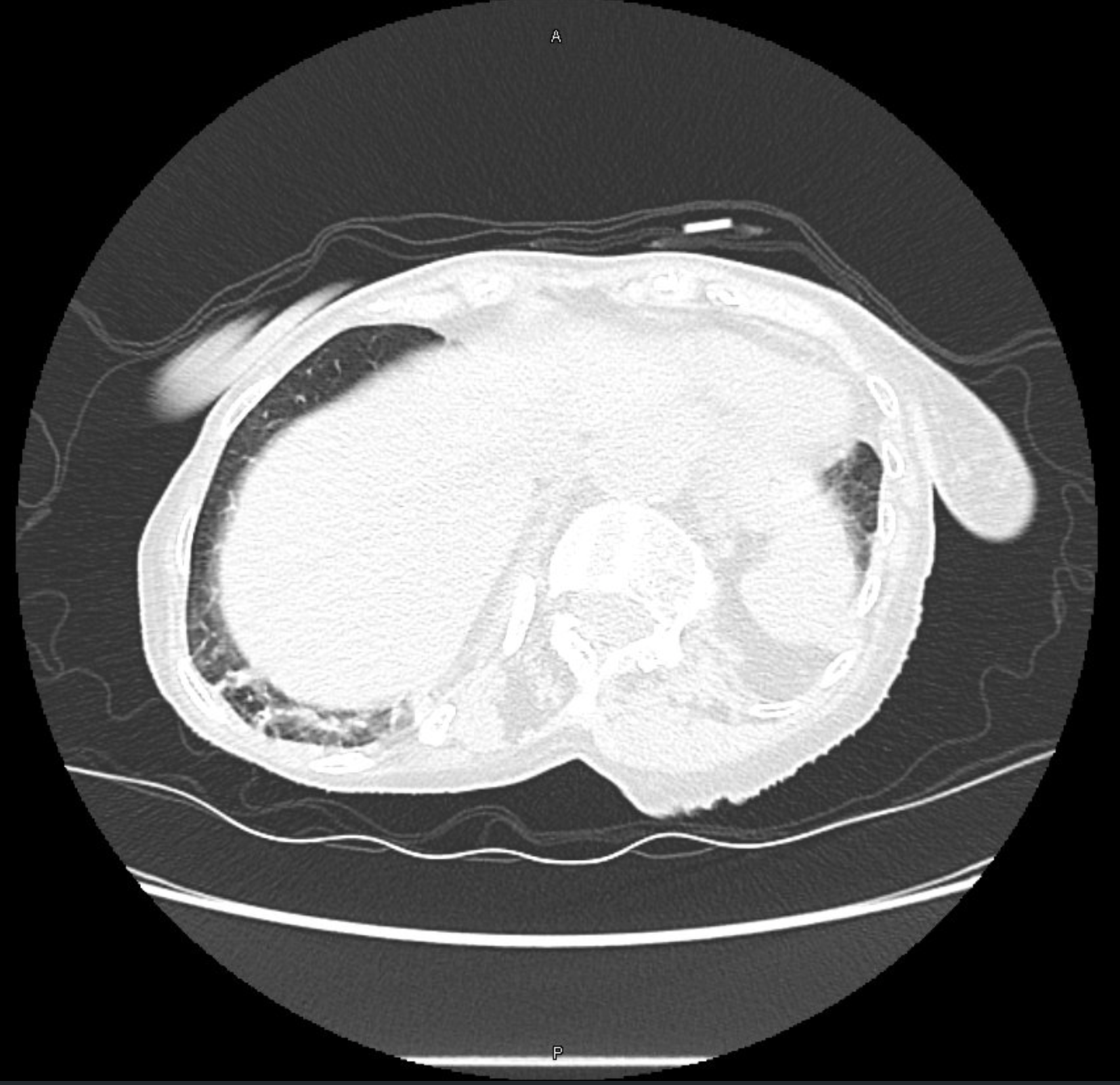
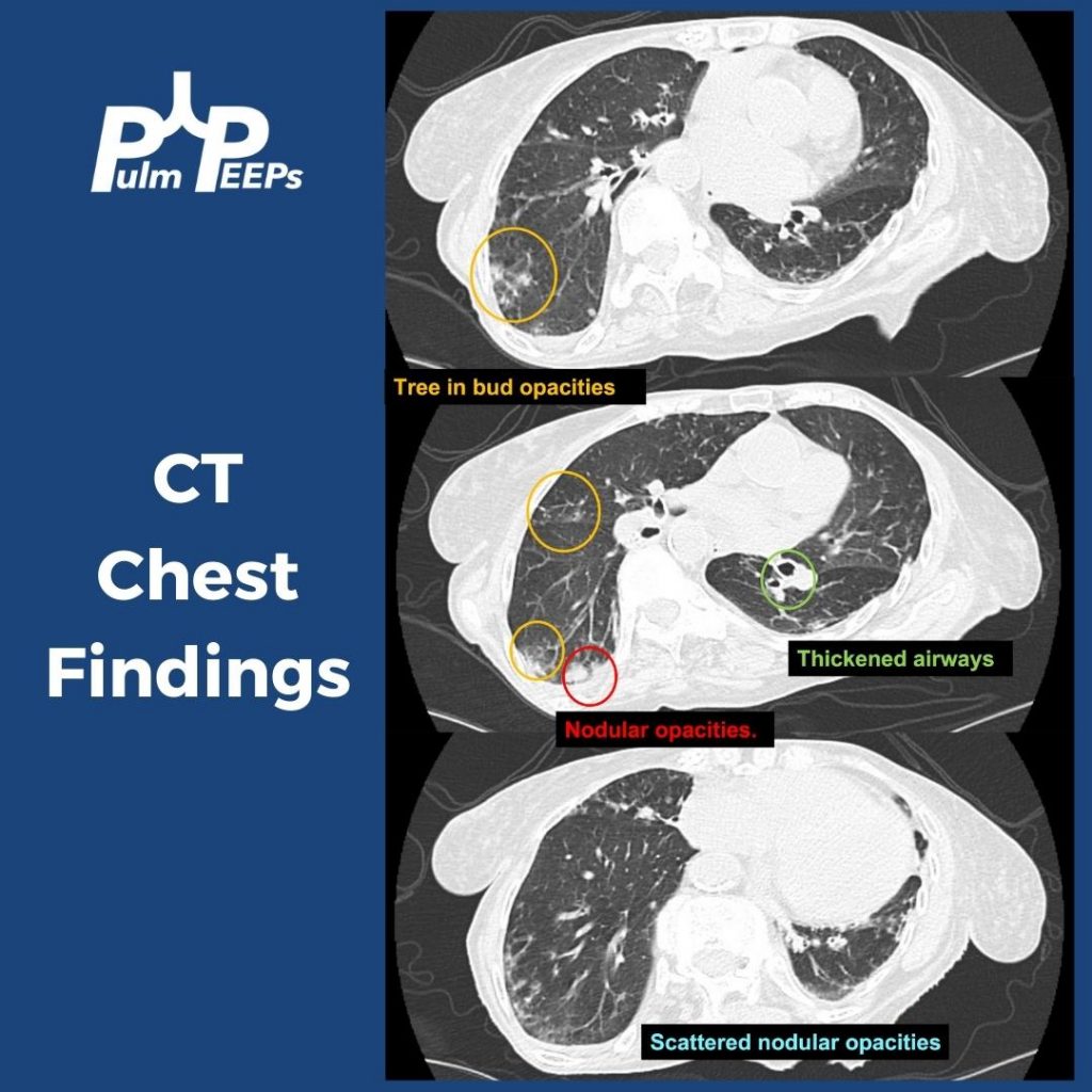
    • 10 – 20 % have air trapping on inspiratory/expiratory chest CTs that could indicate bronchiolitis
    • One study showed that CPETs showed impaired oxygen extraction
      • Preserved cardiac output to exercise and no evidence of deconditioning
      • This study indicated an issue at the peripheral level (ex: vascular, mitochondrial) with oxygen extraction.

Variants

  • It is very difficult to say if variants differ in rates of Long COVID given that often patients do not get sequencing to know the variant and because there is overlap in the timing of variants
  • Further testing will continue on this going forward

Vaccines

  • Reduced risk of Long COVID with vaccination
    • Boosting further decreases the risk compared to just the initial vaccination
  • There is a variable response to getting vaccinated if a patient has Long COVID
    • Most patients tolerate it well and some patients have relief of symptoms
    • There are some patients who can develop worsened Long COVID symptoms

References and further reading

  1. Chippa V, Aleem A, Anjum F. Post Acute Coronavirus (COVID-19) Syndrome. In: StatPearls. StatPearls Publishing; 2022. Accessed November 14, 2022. http://www.ncbi.nlm.nih.gov/books/NBK570608/
  2. Crook H, Raza S, Nowell J, Young M, Edison P. Long covid—mechanisms, risk factors, and management. BMJ. 2021;374:n1648. doi:10.1136/bmj.n1648
  3. Durstenfeld MS, Sun K, Tahir P, et al. Use of Cardiopulmonary Exercise Testing to Evaluate Long COVID-19 Symptoms in Adults: A Systematic Review and Meta-analysis. JAMA Network Open. 2022;5(10):e2236057. doi:10.1001/jamanetworkopen.2022.36057
  4. Nalbandian A, Sehgal K, Gupta A, et al. Post-acute COVID-19 syndrome. Nat Med. 2021;27(4):601-615. doi:10.1038/s41591-021-01283-z
  5. Soriano JB, Murthy S, Marshall JC, Relan P, Diaz JV. A clinical case definition of post-COVID-19 condition by a Delphi consensus. Lancet Infect Dis. 2022;22(4):e102-e107. doi:10.1016/S1473-3099(21)00703-9
  6. Sudre CH, Murray B, Varsavsky T, et al. Attributes and predictors of long COVID. Nat Med. 2021;27(4):626-631. doi:10.1038/s41591-021-01292-y

Radiology Rounds – 11/8/22

This week’s #RadiologyRounds is brought to you by our newest contributor, Nick Ghionni! We’ll dive into a classic Radiology Sign and talk about what it represents, and how it helps inform your differential.

In this case, the patient underwent a biopsy that showed no malignancy but grew NTM! Given that NTM can be superimposed on malignancy, repeated biopsies were done that corroborated. She is being treated with close monitoring.

Radiology Rounds – 10/25/22

This week’s #RadiologyRounds is coming from the pulmonary clinic. Follow us on Twitter to see the case and answer our polls live!

Given the patient’s weight loss and persistent symptoms despite trying some empiric therapies, a chest CT was obtained. PFTs were also ordered 🙂

The patient’s CT had tree-in-bud opacities, nodular consolidations, scattered micronodular opacities, and airway thickening.

The pt had an induced sputum but could not produce a sample. She underwent bronchoscopy + lavage and her AFB smear was positive with negative TB NAAT. The culture ended up growing M. chimaera! Does that explain her symptoms? Here are the diagnostic criteria for pulmonary NTM:

All other testing was negative and the patient was diagnosed with pulmonary NTM. After a long discussion about treatment (an interesting topic for another day!!) she was started on triple antibiotic therapy and after 9 months her cough had resolved and she was gaining weight.

24. Fellows’ Case Files: Baylor College of Medicine

Welcome to another episode in our Pulm PEEPs Fellows’ Case Files series! The purpose of this series is to highlight and amplify the incredible clinical work that is done by pulmonary and critical care fellows, share fascinating cases, and assemble a diverse network of pulmonary and critical care educators. Today we’re headed to Baylor College of Medicine to hear about a fascinating case. Tune in, let us know what you think on Twitter, and let us know if you have a great case to share!

Meet Our Guests

Benjamin Moss completed his internal medicine residency training at Baylor College of Medicine in Houston, Texas, and is currently a senior pulmonary and critical care fellow there.

Philip Alapat is an Assistant Professor of Medicine and the Program Director of the Pulmonary and Critical Care Fellowship at Baylor. He completed his residency and fellowship training all at Baylor.

Patient Presentation

A 60s-year-old man with seropositive RA on Rituximab presents with dyspnea and cough, and overall “not feeling well.”  For the past week, he has had malaise, body aches, and subjective fever. For the past 3 days, he has had acutely worsening dyspnea that is worse with exertion, but present at rest and a cough with scant sputum production. He had been on Methotrexate previously but within the last year developed pancytopenia and MTX was stopped and he was switched to adalimumab/Humira. His pancytopenias did not resolve, and he was ultimately diagnosed with Felty syndrome (a triad of RA, neutropenia, and splenomegaly) and switched to rituximab every 6 months with his last dose being 4 months ago. During the last week, he tried taking prednisone 10 mg a day but his symptoms did not improve.

X-ray on presentation (L) X-ray 8 months ago (R)

Key Learning Points

**Spoilers Ahead** If you want to think through the case on your own we advise listening to the episode first before looking at these points.

Image Source: Reference 1: https://doi.org/10.1007/s00134-019-05906-5

References and Further Reading

  1. Azoulay E, Russell L, Van de Louw A, et al. Diagnosis of severe respiratory infections in immunocompromised patients. Intensive Care Med. 2020;46(2):298-314. doi:10.1007/s00134-019-05906-5
  2. Cornely OA, Alastruey-Izquierdo A, Arenz D, et al. Global guideline for the diagnosis and management of mucormycosis: an initiative of the European Confederation of Medical Mycology in cooperation with the Mycoses Study Group Education and Research Consortium. Lancet Infect Dis. 2019;19(12):e405-e421. doi:10.1016/S1473-3099(19)30312-3
  3. Ibrahim AS, Spellberg B, Walsh TJ, Kontoyiannis DP. Pathogenesis of mucormycosis. Clin Infect Dis. 2012;54 Suppl 1:S16-22. doi:10.1093/cid/cir865

Radiology Rounds – 8/30/22

Time for another #RadiologyRounds! This case is authored by PulmPEEPs associate editor @TessLitchman. Great teaching ahead!

Trick question (sorry)! All of these features are present.

A bronchoscopy was performed and the patient was diagnosed with PCP. Additional testing confirmed a new diagnosis of HIV.

This patient was treated with high-dose Bactrim and IV steroids, in addition to being started on ART.

10. A Case of Fevers, Night Sweats, and Dyspnea

This week, we have another great case episode on Pulm PEEPs! We are joined by Emily Fridenmaker who helps us think through a fascinating case presenation.

Meet Our Guests

Emily Fridenmaker is a Pulmonary, Critical Care, and Sleep Medicine fellow at the University of Kentucky College of Medicine. She went to medical school at West Virginia school of medicine and did her internal medicine residency at Charleston Area Medical Center

Patient Presentation

A middle-aged woman presents with 2-3 weeks of mild but progressive shortness of breath with exertion associated with low-grade fevers, worsening night sweats, and fatigue. Further history reveals a progressive non-productive cough and weight loss. She has a past medical history of neurologic dysfunction over two years and a working diagnosis of chronic inflammatory demyelinating polyneuropathy and is pursuing treatment in Mexico due to cost limitations. She has been receiving prednisone 10 mg daily and azathioprine. Aside from travel history to Mexico, her social history is notable for prior employment in a candle factory, and for hiking with some cave exploration. She is a former 20 pack-year smoker and has rare alcohol use.

Key Learning Points

References and links for further reading

  1. McKinsey DS, McKinsey JP. Pulmonary histoplasmosis. Semin Respir Crit Care Med. 2011;32(6):735-744. doi:10.1055/s-0031-1295721
  2. Cuellar-Rodriguez J, Avery RK, Lard M, et al. Histoplasmosis in solid organ transplant recipients: 10 years of experience at a large transplant center in an endemic area. Clin Infect Dis. 2009;49(5):710-716. doi:10.1086/604712
  3. Wheat LJ, Slama TG, Norton JA, et al. Risk Factors for Disseminated or Fatal Histoplasmosis. Ann Intern Med. 1982;96(2):159-163. doi:10.7326/0003-4819-96-2-159
  4. Smith JA, Kauffman CA. Endemic fungal infections in patients receiving tumour necrosis factor-alpha inhibitor therapy. Drugs. 2009;69(11):1403-1415. doi:10.2165/00003495-200969110-00002
  5. Poplin V, Smith C, Milsap D, Zabel L, Bahr NC. Diagnosis of Pulmonary Infections Due to Endemic Fungi. Diagnostics. 2021;11(5):856. doi:10.3390/diagnostics11050856
  6. Wheat LJ, Freifeld AG, Kleiman MB, et al. Clinical Practice Guidelines for the Management of Patients with Histoplasmosis: 2007 Update by the Infectious Diseases Society of America. Clinical Infectious Diseases. 2007;45(7):807-825.
  7. Azar MM, Hage CA. Clinical Perspectives in the Diagnosis and Management of Histoplasmosis. Clinics in Chest Medicine. 2017;38(3):403-415. doi:10.1016/j.ccm.2017.04.004
  8. Threadcraft MA, Case R. Vape-Associated Pulmonary Injury (VAPI) Presenting With a “Miliary” Pattern on Imaging. Cureus. 13(2):e13385. doi:10.7759/cureus.13385
  9. Raoof S, Amchentsev A, Vlahos I, Goud A, Naidich DP. Pictorial essay: multinodular disease: a high-resolution CT scan diagnostic algorithm. Chest. 2006;129(3):805-815. doi:10.1378/chest.129.3.805
  10. Sharma BB. Miliary nodules on chest radiographs: A diagnostic dilemma. Lung India. 2015;32(5):518-520.
  11. Purek L, Laroumagne S, Dutau H, Maldonado F, Astoul P. Miliary mesothelioma: a new clinical and radiological presentation in mesothelioma patients with prolonged survival after trimodality therapy. J Thorac Oncol. 2011;6(10):1753-1756. doi:10.1097/JTO.0b013e31822e295a

Radiology Rounds – 2/22/22

This week we’re sharing a distinct radiology pattern on chest X-ray and CT scans that should raise its own differential. Make sure to listen to our case episode next week to hear more about this patient and the diagnostic workup.

References and links for further reading

  1. Raoof S, Amchentsev A, Vlahos I, Goud A, Naidich DP. Pictorial essay: multinodular disease: a high-resolution CT scan diagnostic algorithm. Chest. 2006;129(3):805-815. doi:10.1378/chest.129.3.805
  2. Sharma BB. Miliary nodules on chest radiographs: A diagnostic dilemma. Lung India. 2015;32(5):518-520.
  3. Threadcraft MA, Case R. Vape-Associated Pulmonary Injury (VAPI) Presenting With a “Miliary” Pattern on Imaging. Cureus. 13(2):e13385. doi:10.7759/cureus.13385
  4. Purek L, Laroumagne S, Dutau H, Maldonado F, Astoul P. Miliary mesothelioma: a new clinical and radiological presentation in mesothelioma patients with prolonged survival after trimodality therapy. J Thorac Oncol. 2011;6(10):1753-1756. doi:10.1097/JTO.0b013e31822e295a