67. Fellows’ Case Files: Northwestern University

Listen in today to another stop on our Fellows’ Case Files journey. We’re at Northwestern University for another great case presentation. Tune in, check out our associated infographic, and let us know what you think!

Meet Our Guests

Jamie Rowell is a first-year clinical fellow in the Northwestern PCCM program. She completed medical school at the Medical University of South Carolina and her internal medicine residency and Chief Residency at the University of Vermont Medical Center.

Cathy Gao is an Instructor of Medicine at Northwestern and completed her PCCM fellowship there last year. Her research focuses on using machine learning applied to ICU EHR data to characterize patient trajectories and identify potential interventions to improve outcomes.

Clara Schroedl is an Associate Professor of Medicine in Pulmonary and Critical Care and Medical Education. She is the program director of the Northwestern PCCM fellowship program, with an interest in medical education and simulation.

Case Presentation

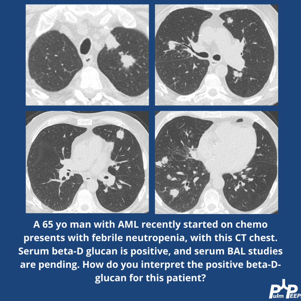
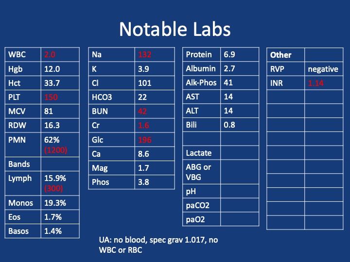
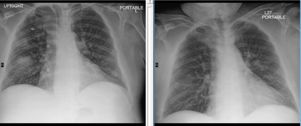
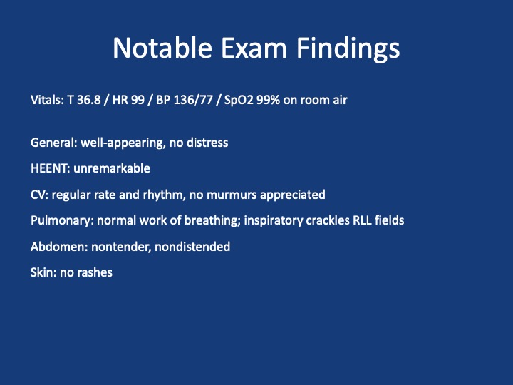
A 25-year-old previously healthy woman presents with recurrent episodes of right chest pain and cough. In October she was treated with antibiotics and felt somewhat better but in December, she presented again with chest pain, and again was treated with antibiotics. The pain improved but she still felt breathless. In February, again she had intense chest pain interfering with life, and was given NSAIDs and took high dose TID without clear benefit.

One month later, she coughed up some bloody mucus, so now she is presenting for evaluation. The chest pain is worse with deep breaths and improves in between these episodes. She only notes it on her right side. At this point, she does sometimes feel short of breath; she used to run 5 miles but is now struggling to run two miles. She denies any unusual exposures. She went to school in central rural Ohio for a while. She has no history of pulmonary infections, no exposure to mold or animals, and no history of vaping.

Key Learning Points

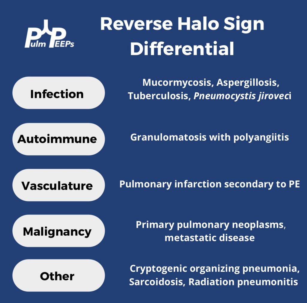
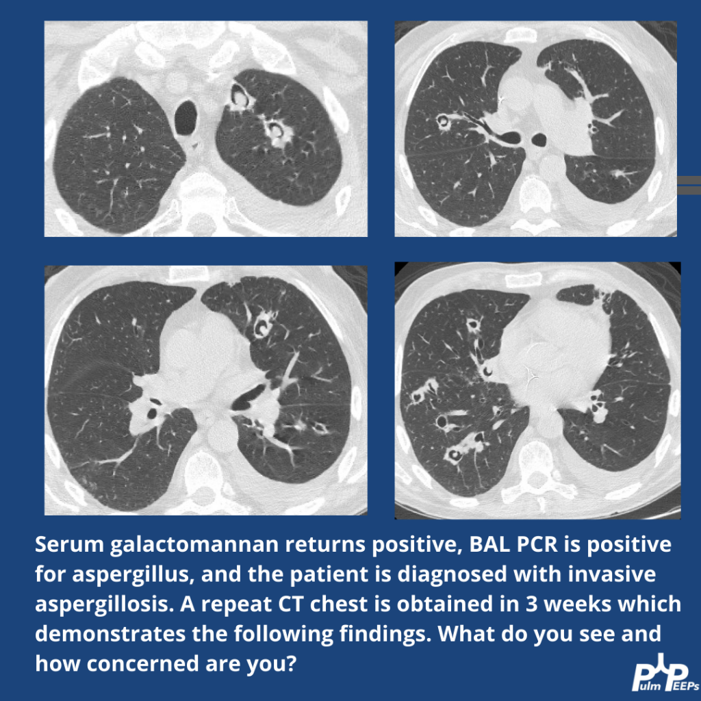
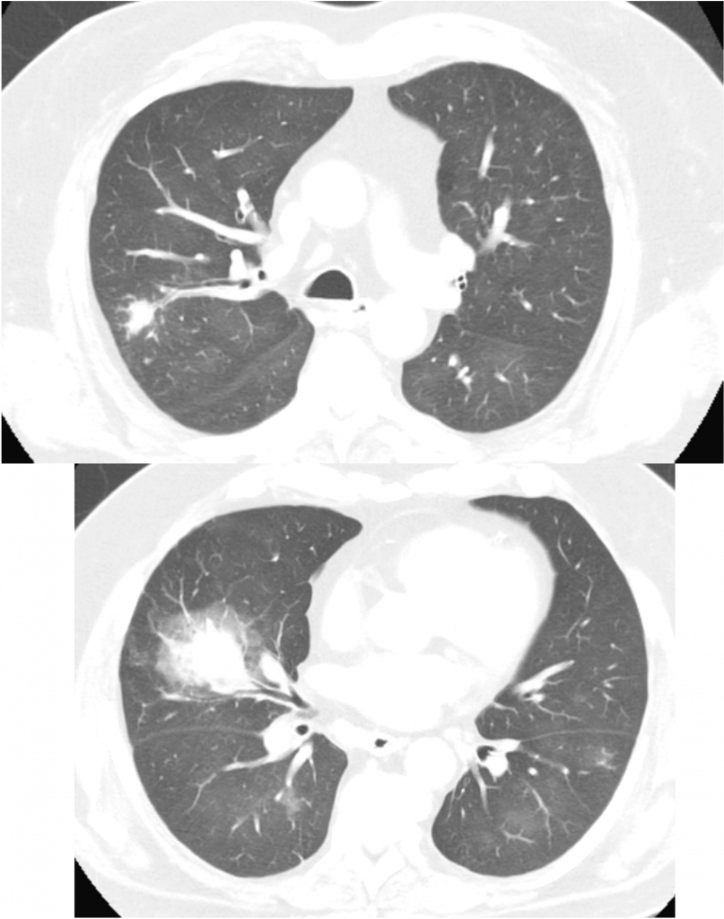
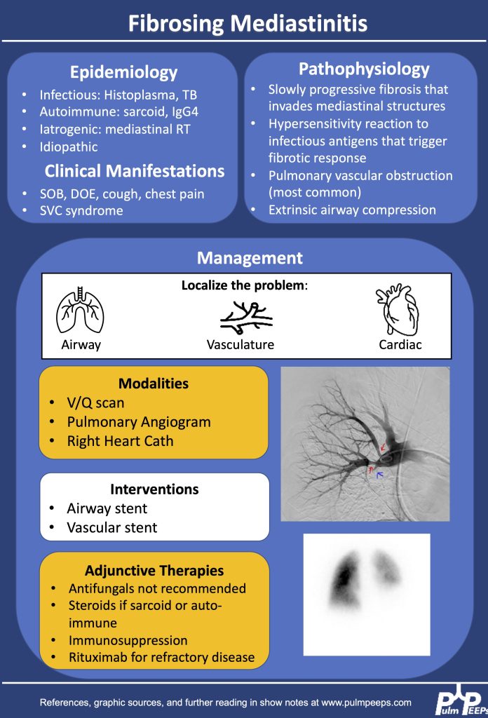
1.Making the diagnosis of Fibrosing Mediastinitis :

–Etiologies: histoplasmosis, sarcoidosis, tuberculosis, IgG4, Behcet, ANCA vasculitis

–Imaging modalities: CT chest, perfusion studies, pulmonary angiogram

–Imaging characteristics:  infiltrative, heterogeneous, fibrotic process that crosses fat planes and encroaches on nearby structures causing airway or vascular stenoses  

2. Management strategies:

–No curative therapies. Goal to relieve symptom burden

–Airway stents

–Vascular stents

–Rituximab

–Antifungals, steroids generally not considered effective

References and Further Reading

Kern et al. Bronchoscopic Management of Airway Compression due to Fibrosing Mediastinitis. Annals of the American Thoracic Society 2017. 14: 1235-1359 

Welby JP, Fender EA, Peikert T, Holmes DR Jr, Bjarnason H, Knavel-Koepsel EM. Evaluation of Outcomes Following Pulmonary Artery Stenting in Fibrosing Mediastinitis. Cardiovasc Intervent Radiol. 2021 Mar;44(3):384-391. doi: 10.1007/s00270-020-02714-z. Epub 2020 Nov 17. PMID: 33205295.

Westerly, BD Targeting B Lymphocytes in Progressive Fibrosing Mediastinitis. Am J Respir Crit Care Med. 2014 Nov 1; 190(9): 1069–1071.

https://rarediseases.org/rare-diseases/fibrosing-mediastinitis/#complete-report

https://pubmed.ncbi.nlm.nih.gov/21422386/

https://academic.oup.com/cid/article/30/4/688/421789

https://pubmed.ncbi.nlm.nih.gov/22033450/

https://www.sciencedirect.com/science/article/pii/S2352906715300087

https://www.atsjournals.org/doi/pdf/10.1513/AnnalsATS.201610-782RL

Radiology Rounds – 1/16/24

We are back with another #RadiologyRounds for 2024 featuring high-yield imaging findings and teaching points for you to review

In this immunosuppressed patient, you find a reverse halo sign, right pleural effusion and left lower lobe consolidation.

Serum fungal markers are negative but given the reverse halo sign, you empirically start Amphotericin B given concern for pulmonary mucormycosis.

Given the profound neutropenia, the patient was predisposed and mucor was identified on lung tissue biopsy and IV Amphotericin-B was continued.

Radiology Rounds – 11/21/23

We’re back with another #RadiologyRounds by Pulm PEEPs Associate Editor Tess Litchman. An immunosuppressed 65-year-old man presents with neutropenic fever. He is started on empiric broad-spectrum antibiotics with vancomycin and zosyn. Serum beta-D-glucan is positive.

Further, workup reveals a positive serum galactomannan and a BAL PCR that is positive for Aspergillus. The patient is diagnosed with invasive aspergillosis and started on voriconazole. A repeat CT is obtained in 3 weeks. What do you think of the evolving findings?

24. Fellows’ Case Files: Baylor College of Medicine

Welcome to another episode in our Pulm PEEPs Fellows’ Case Files series! The purpose of this series is to highlight and amplify the incredible clinical work that is done by pulmonary and critical care fellows, share fascinating cases, and assemble a diverse network of pulmonary and critical care educators. Today we’re headed to Baylor College of Medicine to hear about a fascinating case. Tune in, let us know what you think on Twitter, and let us know if you have a great case to share!

Meet Our Guests

Benjamin Moss completed his internal medicine residency training at Baylor College of Medicine in Houston, Texas, and is currently a senior pulmonary and critical care fellow there.

Philip Alapat is an Assistant Professor of Medicine and the Program Director of the Pulmonary and Critical Care Fellowship at Baylor. He completed his residency and fellowship training all at Baylor.

Patient Presentation

A 60s-year-old man with seropositive RA on Rituximab presents with dyspnea and cough, and overall “not feeling well.”  For the past week, he has had malaise, body aches, and subjective fever. For the past 3 days, he has had acutely worsening dyspnea that is worse with exertion, but present at rest and a cough with scant sputum production. He had been on Methotrexate previously but within the last year developed pancytopenia and MTX was stopped and he was switched to adalimumab/Humira. His pancytopenias did not resolve, and he was ultimately diagnosed with Felty syndrome (a triad of RA, neutropenia, and splenomegaly) and switched to rituximab every 6 months with his last dose being 4 months ago. During the last week, he tried taking prednisone 10 mg a day but his symptoms did not improve.

X-ray on presentation (L) X-ray 8 months ago (R)

Key Learning Points

**Spoilers Ahead** If you want to think through the case on your own we advise listening to the episode first before looking at these points.

Image Source: Reference 1: https://doi.org/10.1007/s00134-019-05906-5

References and Further Reading

  1. Azoulay E, Russell L, Van de Louw A, et al. Diagnosis of severe respiratory infections in immunocompromised patients. Intensive Care Med. 2020;46(2):298-314. doi:10.1007/s00134-019-05906-5
  2. Cornely OA, Alastruey-Izquierdo A, Arenz D, et al. Global guideline for the diagnosis and management of mucormycosis: an initiative of the European Confederation of Medical Mycology in cooperation with the Mycoses Study Group Education and Research Consortium. Lancet Infect Dis. 2019;19(12):e405-e421. doi:10.1016/S1473-3099(19)30312-3
  3. Ibrahim AS, Spellberg B, Walsh TJ, Kontoyiannis DP. Pathogenesis of mucormycosis. Clin Infect Dis. 2012;54 Suppl 1:S16-22. doi:10.1093/cid/cir865