
We are back with our first #RadiologyRounds of the new academic year. We have a young, immunocompetent man presenting with fever, weight loss, and abdominal pain.
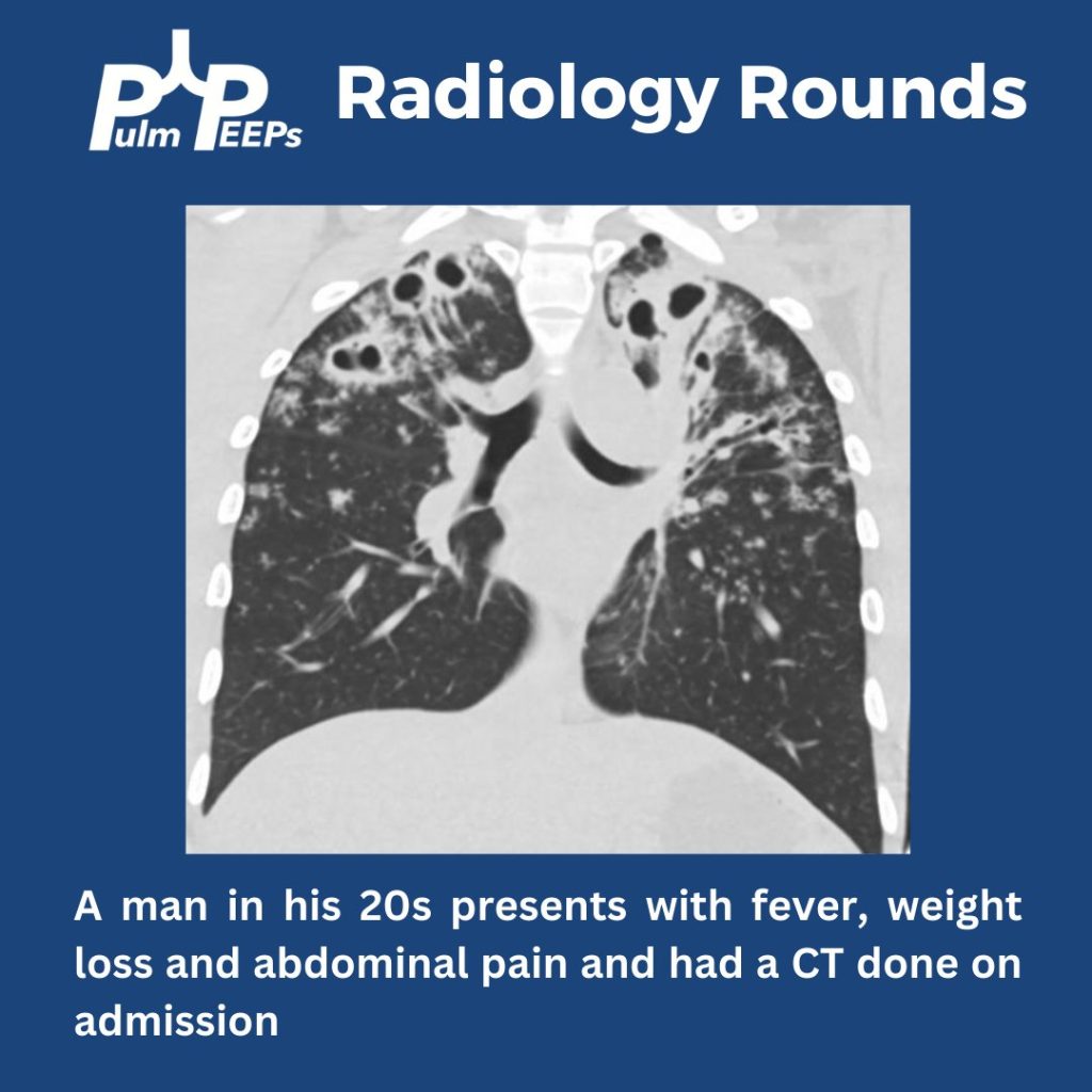
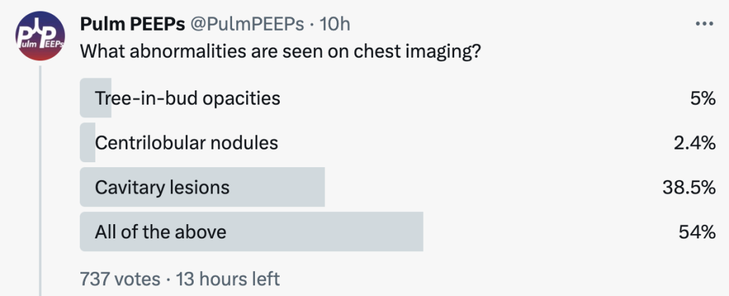
What abnormalities are seen on his chest imaging?
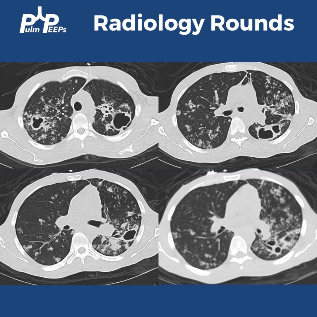
He was found to have bilateral apical cavitary disease, centrilobular nodules, and tree-in-bud opacities. He developed a productive cough with blood-tinged sputum as well as diarrhea.
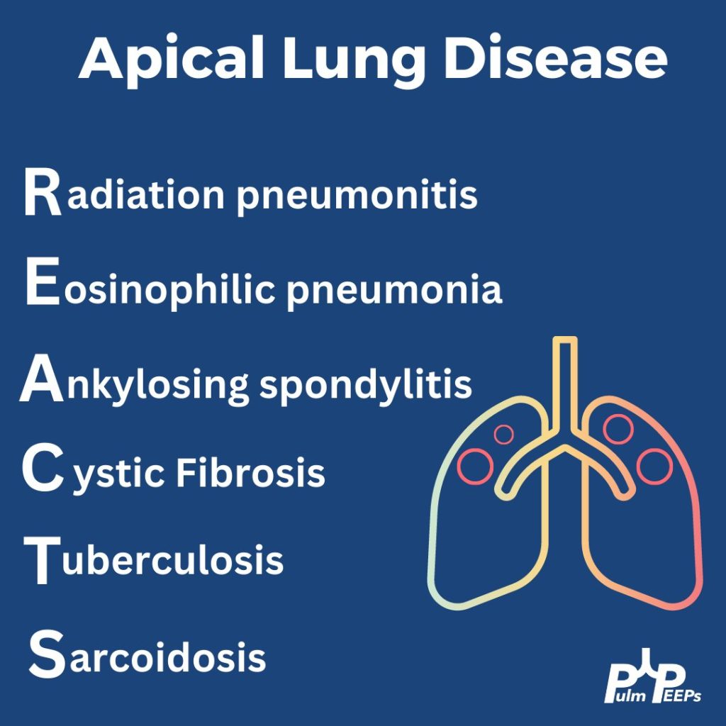
Given his apical lung disease, what is on your differential?
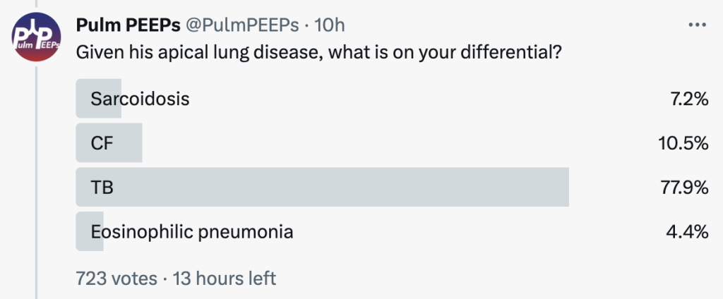
When thinking about apical lung disease, remember the mnemonic REACTS to help with your differential.
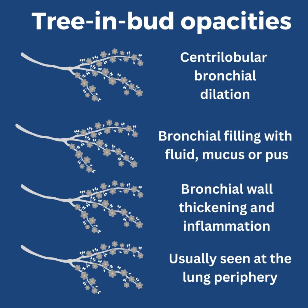
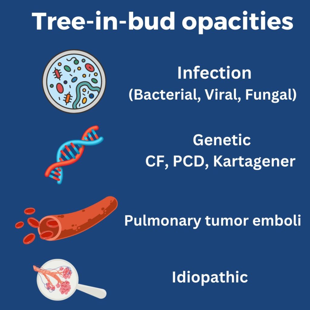
What are tree-in-bud opacities? They are findings seen on CT chest suggesting bronchial dilation, inflammation, and bronchial filling with fluid, mucus, or pus that can be caused by infections and non-infectious etiologies.
He had sputum and AFB cultures sent and his AFB smear was positive. He was ultimately diagnosed with disseminated TB and started on RIPE therapy.