107. Fellows’ Case Files: University of Kansas Medical Center KUMC

After a brief hiatus, we are excited to be back today with another Fellows’ Case Files! Today we’re virtually visiting the University of Kansas Medical Center (KUMC) to hear about a fascinating pulmonary presentation. There are some fantastic case images and key learning points. Take a listen and see if you can make the diagnosis along with us. As always, let us know your thoughts and definitely reach out if you have an interesting case you’d like to share.
Dr. Vishwajit Hegde completed his internal medicine residency at University of Kansas Medical Center where he stayed for fellowship and is currently a second year Pulmonary and Critical Care medicine fellow. 

Dr. Sahil Pandya is an Associate Professor of Medicine and Program Director of the PCCM Fellowship at KUMC.

1) Initial frame & diagnostic mindset

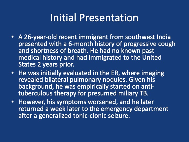
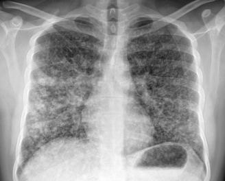
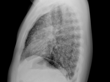
  • Young (26), subacute → chronic dyspnea/cough with diffuse pulmonary nodules; avoid premature closure on TB.
  • Use a Bayesian approach: combine pre-test probability (epidemiology, exposures, tempo) with targeted tests to decide next steps.
  • Always confirm TB when possible (micro/path + resistance testing); empiric RIPE may be reasonable but shouldn’t replace tissue when stakes are high.

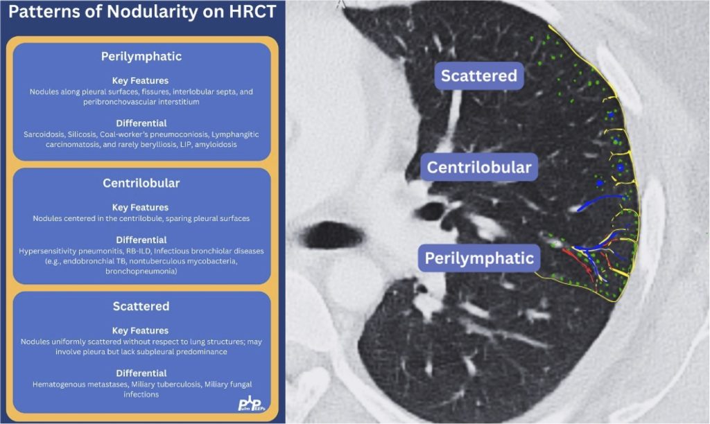
2) Imaging pearls—nodular pattern recognition

  • Ask three things: craniocaudal distribution, symmetry, central vs peripheral.
  • Centrilobular (spares pleura/fissures): airway-centered (e.g., NTM, bronchiolitis, tree-in-bud).
  • Perilymphatic (tracks fissures/pleura & septa): sarcoid, lymphangitic spread.
  • Random/diffuse (involves pleural surfaces): hematogenous spread → think miliary TB, disseminated fungal, septic emboli, metastatic disease.
  • Interval change matters: new cavitation and confluence can upweight infection or aggressive malignancy.

3) Neuro findings—ring-enhancing lesions

  • Differential: septic emboli/abscess, nocardia, fungal, TB, parasites, metastases, vasculitis, sarcoid.
  • Partner with neuroradiology for pattern nuances; treat seizures but keep searching for the unifying diagnosis.

4) Lab/serology strategy

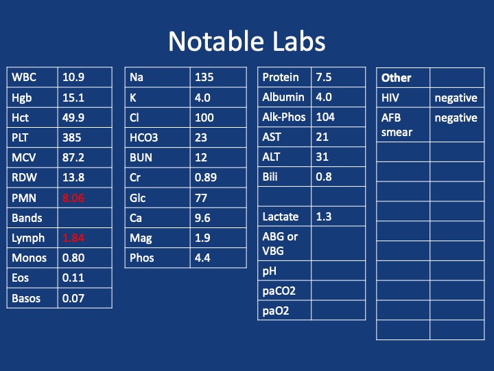
  • Broad infectious workup (AFB × multiple, fungal serologies), HIV and basic immune screen.
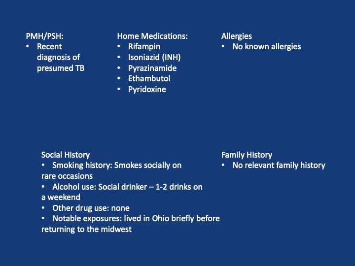
  • Negative/indeterminate tests don’t end the search—revisit history (e.g., Ohio travel → histo/blasto risk).

5) “Tissue is the issue”—choosing the procedure

  • For diffuse nodules with mediastinal adenopathy and stable patient: EBUS-TBNA + BAL, consider transbronchial or cryobiopsy.
  • Cryobiopsy pros: larger, less crush artifact, better for molecular testing; cons: ↑ bleeding/pneumothorax vs forceps.
  • VATS still best for certain ILD questions or if less invasive routes are non-diagnostic—but weigh patient preference and stage/likelihood of yield.
6) ROSE (rapid on-site evaluation) in bronchoscopy

  • Confirms adequacy in real time, steers you away from necrotic zones, helps decide when you’ve got enough for molecular studies, and when to pivot sites—reduces anesthesia time and repeat procedures.

7) Final diagnosis & management

  • Path: TTF-1+, CK7+, napsin A → pulmonary adenocarcinoma with a fusion driver.
  • Therapy: Targeted TKI (crizotinib) → dramatic radiographic response of miliary lung disease and CNS lesions.
  • Teaching point: even “miliary TB-like” lungs + CNS lesions in a 20-something can be driver-positive lung cancer—don’t let age or pattern blind you.

  1. Desai, S., Devaraj, A., Lynch, D., & Sverzellati, N. (2020). Webb, Müller and Naidich’s high-resolution CT of the lung (6th ed.). Lippincott Williams & Wilkins.
  2. Rajeswaran, G., Becker, J. L., Michailidis, C., Pozniak, A. L., & Padley, S. P. G. (2006). The radiology of IRIS (immune reconstitution inflammatory syndrome) in patients with mycobacterial tuberculosis and HIV co-infection: appearances in 11 patients. Clinical radiology, 61(10), 833-843
  3. Poletti, V., Ravaglia, C., & Tomassetti, S. (2016). Transbronchial  cryobiopsy in diffuse parenchymal lung diseases. Current opinion in pulmonary medicine, 22(3), 289-296.
  4. Norman, G. R., Monteiro, S. D., Sherbino, J., Ilgen, J. S., Schmidt, H. G., & Mamede, S. (2017). The causes of errors in clinical reasoning: cognitive biases, knowledge deficits, and dual process thinking. Academic Medicine, 92(1), 23-30.

103. Fellows’ Case Files: University of Virginia

Today, we’re virtually visiting the University of Virginia for another Fellows’ Case Files. This is a fantastic case that covers ARDS, the infectious work up of an immunosuppressed patient, and the evaluation of undifferentiated shock. Please let us know what you think of the episode and always feel free to reach out with interesting cases!


John Popovich completed his residency training and chief year at UVA and has stayed on there for his pulmonary and critical care fellowship.

Tim Scialla is an associate professor of medicine at UVA. He completed his residency and fellowship at Johns Hopkins Hospital where he was also an ACS. His clinical and research focuses are advanced airways disease. He is also the program director of the PCCM fellowship.

Matt Freedman completed his residency training at Virginia Commonwealth University and is currently a second year fellow at University of Virginia.


Patient: 52-year-old male with psoriasis, HIV/AIDS (CD4 count: 71), presenting with progressive shortness of breath, fever, non-productive cough, and weight loss.

Vital signs: Febrile (103°F), tachycardic (HR 110), hypoxemic on 6L O₂ (SpO₂ 90–92%).

Exam: Diffuse crackles, ill-appearing.

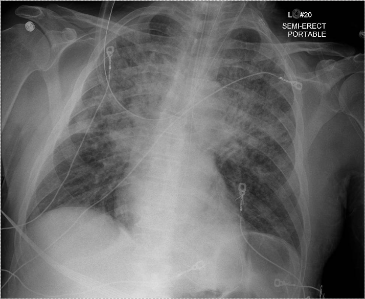
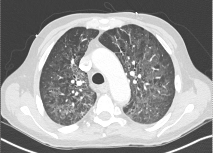
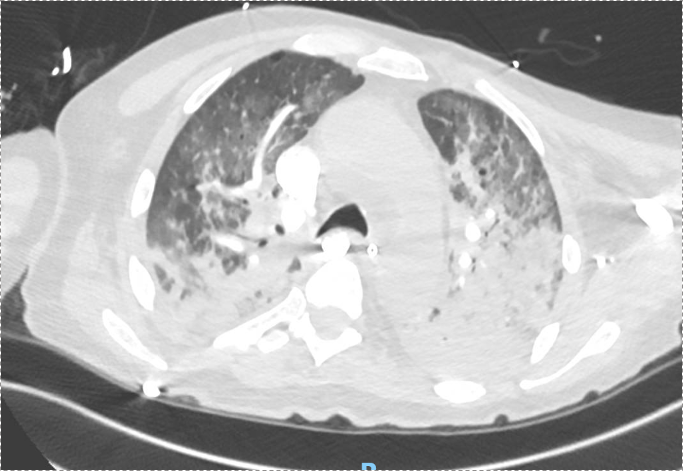
Imaging: CXR and CT showed bilateral upper lobe infiltrates, ground-glass opacities, septal thickening, and peripheral cystic changes.


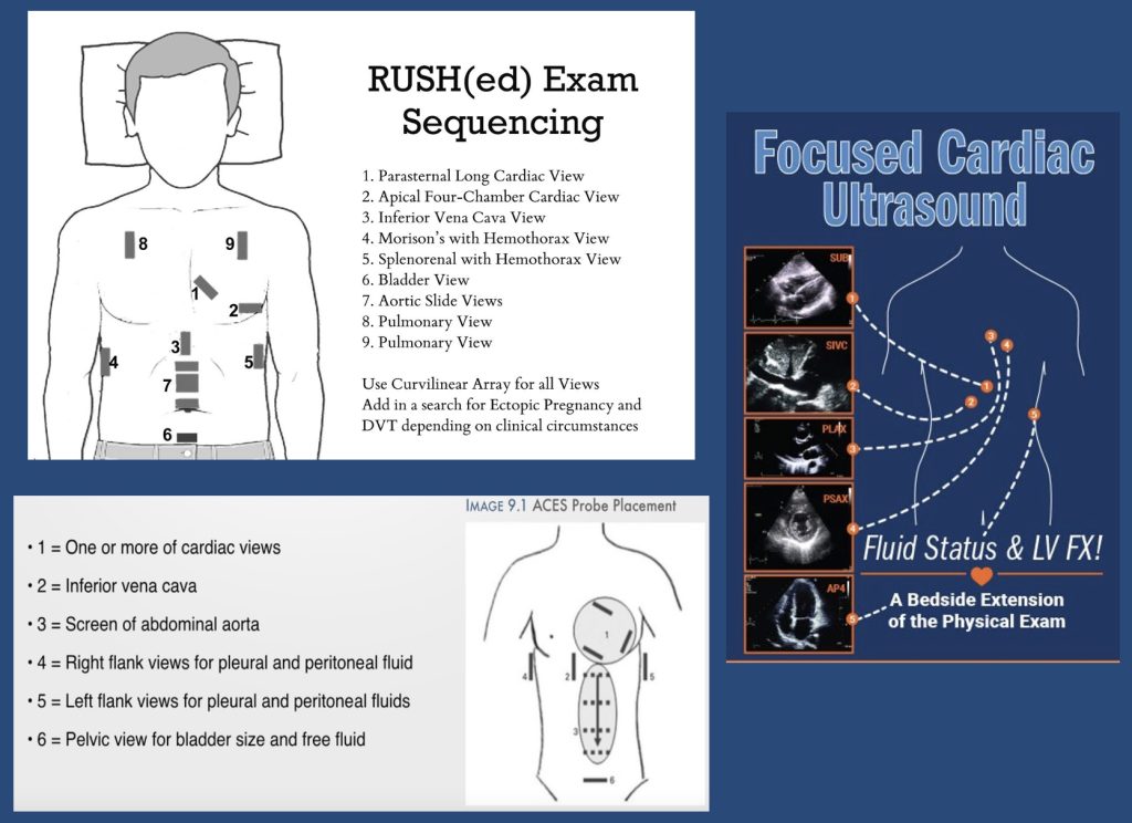
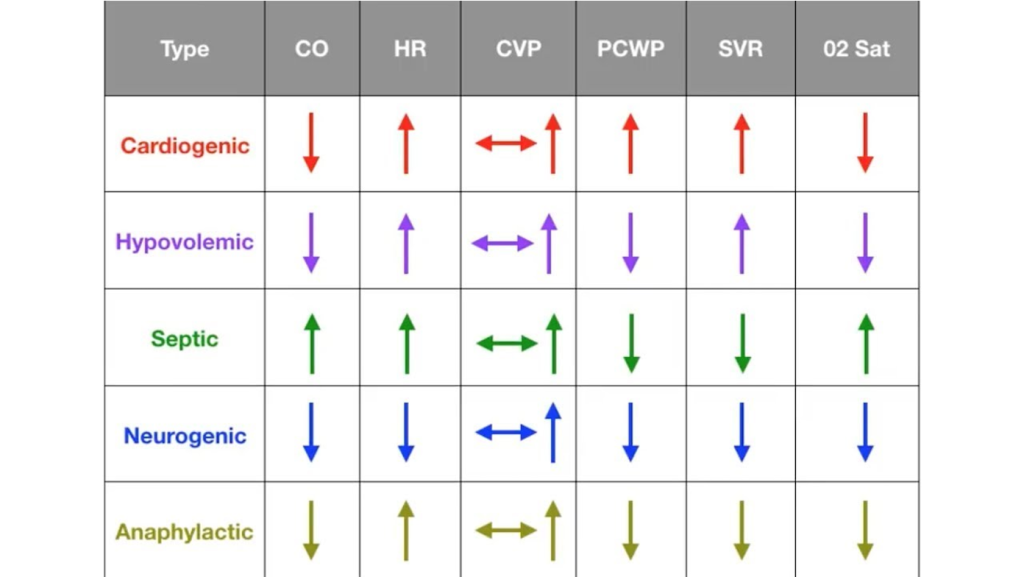
POCUS algorithms for investigating shock

Shock physiology:


Diagnostic Reasoning in Immunocompromised Hosts

  • Framework: Anchor the differential based on type of immunosuppression.
    • HIV/AIDS → T-cell dysfunction, affecting susceptibility to PCP, TB, CMV, fungi (e.g. histo/blasto), and common CAP organisms.
  • PCP considerations:
    • PCP can occur despite prophylaxis (e.g. Bactrim), especially if adherence or resistance issues exist.
    • Classic symptoms in AIDS: acute, febrile, hypoxemic respiratory failure.

Use of Serum Markers and Imaging

  • LDH: Elevated in PCP, but non-specific. High negative predictive value when normal.
  • 1,3-β-D-glucan: Elevated in PCP and other fungal infections. Very sensitive for PCP (up to 95%).
  • Imaging: Ground-glass opacities with cystic changes support PCP diagnosis.

Role of Bronchoscopy and Diagnostic Yield

  • BAL studies to obtain:
    • DFA for PCP (rapid, high specificity, lower sensitivity)
    • PCR for PCP (higher sensitivity, slower turnaround)
    • Cultures: bacterial, fungal, mycobacterial
    • Cytology, galactomannan, histo/blasto urine antigens
  • Bronch Risk-Benefit:
    • Can change management in 40–60% of cases.
    • Complication rate: ~10–15%, most often hypoxemia.
    • Heuristic for pre-bronch ABG on non-rebreather:
      • PaO₂ >150 → likely safe
      • 100–150 → ~25% risk of intubation
      • <100 → high risk of decompensation

Steroids in PCP and Severe CAP

  • Steroids indicated in PCP with significant hypoxemia (PaO₂ <70 mmHg).
  • With new CAP guidelines (Cape Cod trial), steroids may also be considered in severe bacterial CAP.

Shock Evaluation in ICU

  • Framework: Simplify into likely causes — distributive most common, but rule out cardiogenic, obstructive, hypovolemic.
  • Physical exam + POCUS essential early.
    • POCUS: cardiac views, IVC, lung US, abdominal free fluid.
    • Low EF doesn’t exclude distributive shock.
  • PA catheter (Swan) utility:
    • Useful when physiology unclear or when tracking response to therapy is critical.
    • Swan data in this patient: low CVP and wedge, high SVR → distributive shock, not cardiogenic despite low EF.

99. Fellows’ Case Files: Rutgers – Robert Wood Johnson Medical School

We’re back with another edition of Fellows’ Case Files! Today, we’re virtually visiting Rutgers University, Robert Wood Johnson Medical School to work through a fascinating pulmonary case. Enjoy, and let us know your thoughts.

Khalil El Gharib completed his residency training at Northwell at Staten Island University Hospital Program and is currently a first year fellow at Rutgers Robert Wood Johnson Medical School.

Sabiha Hussain completed her residency training at Robert Wood Johnson Medical School and her fellowship training at Columbia Presbyterian Medical Center in New York. She is currently a Professor of Medicine and the fellowship Program Director.

  • Patient: 28-year-old male with Asperger’s syndrome and IgA nephropathy.
  • Symptoms: 3-month history of progressive dry cough and dyspnea on exertion; later developed mild hemoptysis.
  • Notable exposure: Questionable black mold in the patient’s apartment.

Initial Workup and Diagnostic Reasoning

  • Vital signs: Hypoxemia (SpO₂ 91% on room air).
  • Exam: Inspiratory crackles.
  • ABG findings: Elevated A–a gradient (~50), indicating a gas exchange problem.
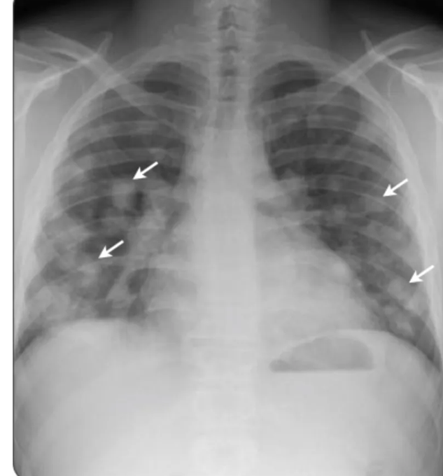
  • Chest X-ray: Bilateral, patchy infiltrates without specific lobar preference.
  • Initial management: Discharged with empiric antibiotics for presumed multifocal pneumonia.

Re-Presentation and Further Testing

  • Symptoms worsened; now with blood-tinged sputum.
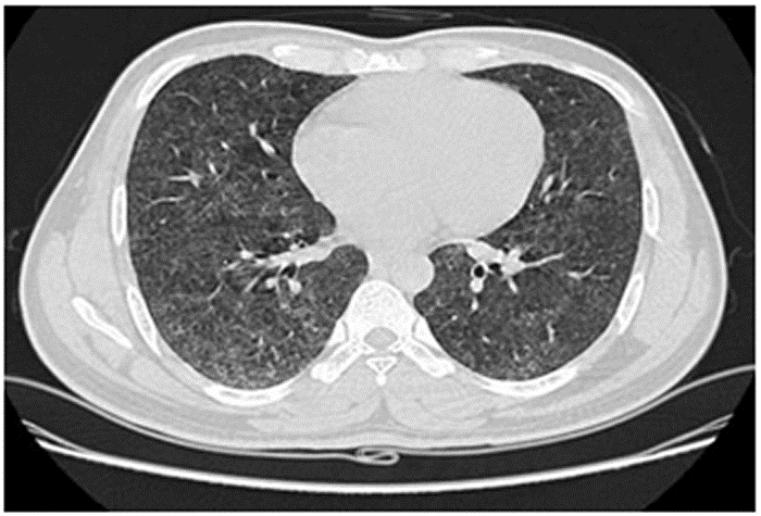
  • Chest CT: Showed diffuse ground-glass opacities (GGOs) without fibrosis, consolidation, or lymphadenopathy.

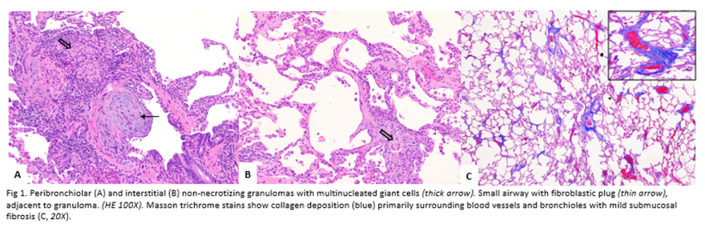
Pathology images a courtesy to Dr Isago Jerrett, pathology resident at RWJMS

Diagnostic Framework for Hypersensitivity Pneumonitis (HP)

  • New classification: Based on fibrotic vs. non-fibrotic phenotype (not acute/chronic).
  • CT features of HP:
    • GGOs with lobular air trapping.
    • “Three-density sign” (normal lung, low-density air-trapping, and ground-glass opacities).
  • BAL: Typically shows lymphocytic predominance in chronic HP, neutrophilic in early stages.
  • Serum IgG testing: Helps identify antigen exposure but doesn’t confirm disease alone.
  • Lung biopsy (VATS): Revealed poorly formed granulomas and airway-centered inflammation—consistent with HP.

Differential Diagnosis of Granulomatous Disease

  • Infectious: TB, fungal (must rule out with stains/cultures).
  • Non-infectious: Sarcoidosis, HP, granulomatosis with polyangiitis.
  • Key pathology clues for HP: Loosely formed granulomas, airway inflammation, giant cells.

Management and Outcome

  • Primary treatment: Antigen avoidance (patient moved out of mold-exposed apartment).
  • Adjunct therapy: Oral prednisone with a slow taper.
  • Outcome: Symptomatic and radiographic improvement over six months.

Teaching Pearls

  • Always take a detailed environmental and occupational exposure history.
  • Hypoxemia with an elevated A–a gradient in a young adult should trigger concern for interstitial/parenchymal lung disease.
  • CT and history are often enough to diagnose HP—biopsy is reserved for uncertain cases.
  • Remember evolving terminology: think fibrotic vs. non-fibrotic HP, not acute/chronic.

88. Fellows’ Case Files: NYU

We are joined today by two amazing educators from NYU for our latest Fellows’ Case Files Episode. Listen today as we go through a great case with some fantastic teaching points highlighted throughout the episode.

Dr. Jeremy Grossman completed his Medicine-Pediatrics residency at Stony Brook Medicine where he was also a Chief Resident. He is currently a second-year PCCM fellow at NYU.

Dr. Shari Brosnahan is an Assistant Professor of Medicine and one of the Assistant Program Directors for the NYU Langone’s Division of Pulmonary, Critical Care, and Sleep Medicine. Her clinical and research interests are focused on pulmonary embolism and thrombosis in critically ill patients.

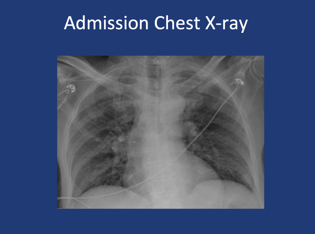
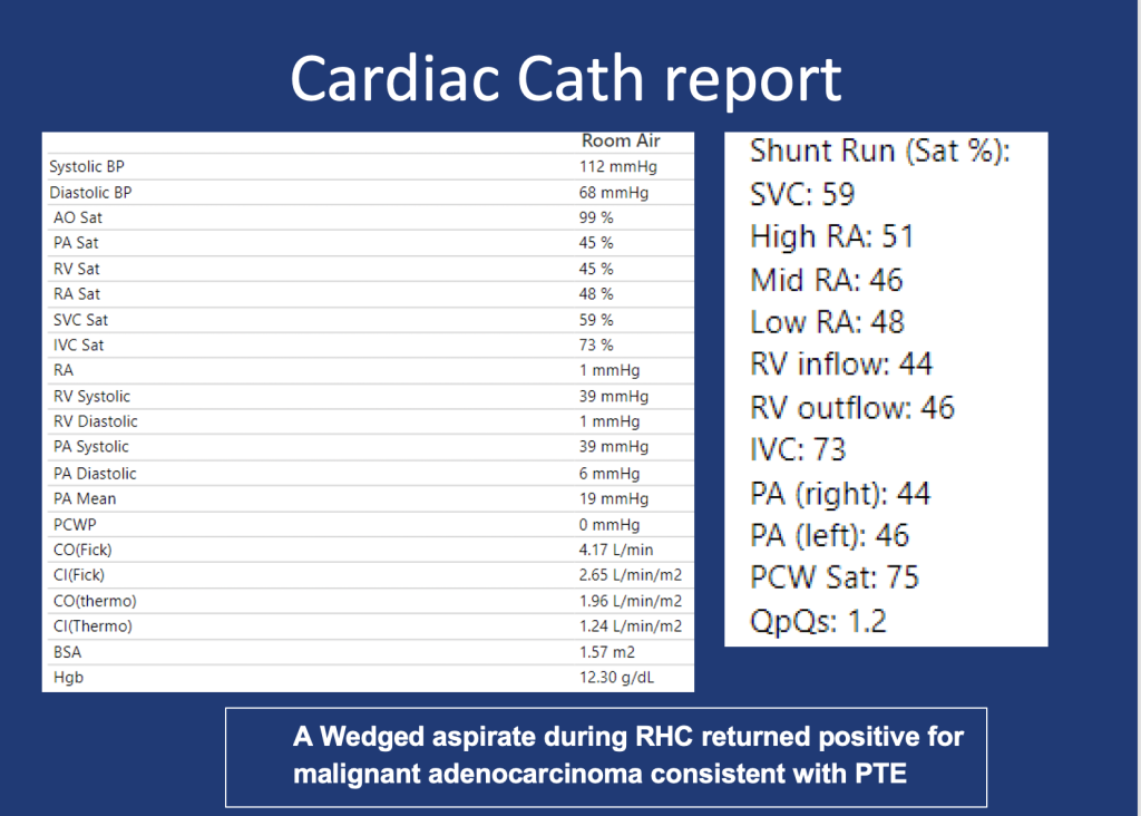
An 80-year-old male presents with shortness of breath. At home, his oxygen saturation was 82% on room air, improving only to 86% on 4L nasal cannula. Over the past month, he has experienced worsening symptoms, including a dry cough, fatigue, and difficulty speaking or ambulating due to shortness of breath at rest. He denies recent fever, sputum production, chest pain, or lower extremity swelling and presents to the ED for further evaluation.

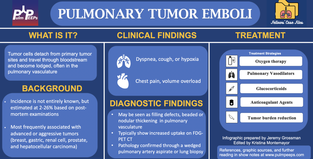
1.In any patient with a history of malignancy and hypoxia, clinicians should keep pulmonary tumor emboli (PTE) on the differential as early intervention may alter outcomes.

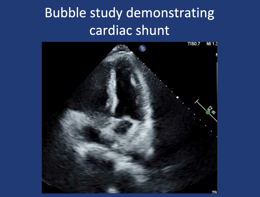
2.PTE contributes to hypoxia via mechanisms of mechanical obstruction of pulmonary arteries leading to shunting, VQ Mismatch, and in some cases pulmonary hypertension due to increased PVR.

3.A wedged aspirate can be used to diagnose PTE ante-mortem

82. Fellows’ Case Files: UMass Chan

We have another great case in our Fellows’ Case Files coming today from UMass Chan. Listen in for a great discussion about a fascinating case with interesting physical exam and radiographic findings.

Dr. Jen Kodela completed her residency training at UMass Memorial Medical Center and is currently a third year PCCM fellow at UMass Chan.

Dr. Ariel McKenna completed her residency training at Maine Medical Center and is also currently a third year PCCM fellow at UMass Chan.

Dr. Will Wong is an Assistant Professor of Medicine and is the Program Director of the PCCM fellowship at UMass Chan

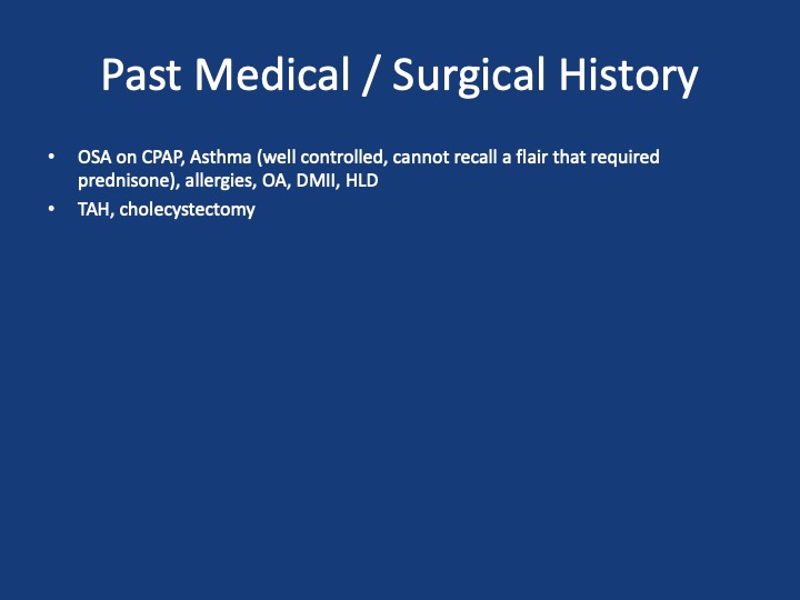
A 75 y/o F presenting with acute on chronic SOB, cough, L sided chest pain and rash. She has had ~7 months of progressive dyspnea, now a/w 2 months of productive cough, and several weeks of L sided chest pain and rash. She has been seen multiple times in the past two months for these sxs. During that time she received multiple antibiotic courses (urgent care, outpatient providers), including augmentin, azithromycin and levaquin, and asthma directed therapy (no steroids). Imaging throughout that time (CXRs, CTPE) show progression from a LLL infiltrate to bibasilar infiltrates. Despite these interventions, sxs continue to worsen. One month prior she was admitted to an OSH w/ continued worsening, vitals stable, exam nonfocal, mild leukocytosis but infectious w/u bland. Received broad spectrum abx. Bronch w/ BAL offers negative cultures, cytology, cell count w/ 66% neutrophils, 14% eosinophils. Discharged w/ dx of PNA on a 10 day course of levaquin and new exertional oxygen requirement of 2L. She then presents to Umass ~1 month later w/ continued progression of sxs

1. Formulate a differential diagnosis for non-resolving pneumonia

2. Evaluate the utility of transbronchial biopsy in the workup of undifferentiated ILD

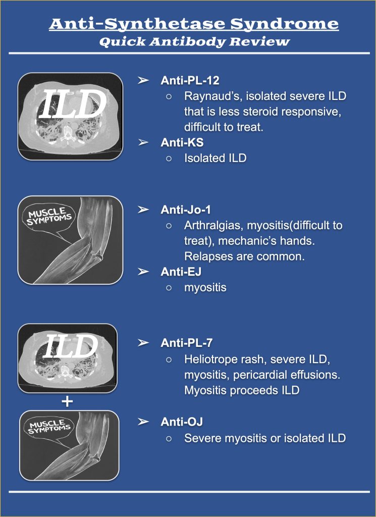
3. Describe the clinical manifestations of antisynthetase syndrome and identify the differences in presentation associated with PL-12 positivity

1. Kuru T, Lynch JP 3rd. Nonresolving or slowly resolving pneumonia. Clin Chest Med. 1999 Sep;20(3):623-51. doi: 10.1016/s0272-5231(05)70241-0. PMID: 10516909.

2. Troy LK, Grainge C, Corte TJ, Williamson JP, Vallely MP, Cooper WA, Mahar A, Myers JL, Lai S, Mulyadi E, Torzillo PJ, Phillips MJ, Jo HE, Webster SE, Lin QT, Rhodes JE, Salamonsen M, Wrobel JP, Harris B, Don G, Wu PJC, Ng BJ, Oldmeadow C, Raghu G, Lau EMT; Cryobiopsy versus Open Lung biopsy in the Diagnosis of Interstitial lung disease alliance (COLDICE) Investigators. Diagnostic accuracy of transbronchial lung cryobiopsy for interstitial lung disease diagnosis (COLDICE): a prospective, comparative study. Lancet Respir Med. 2020 Feb;8(2):171-181. doi: 10.1016/S2213-2600(19)30342-X. Epub 2019 Sep 29. PMID: 31578168.

3. Hallowell RW, Danoff SK. Diagnosis and Management of Myositis-Associated Lung Disease. Chest. 2023 Jun;163(6):1476-1491. doi: 10.1016/j.chest.2023.01.031. Epub 2023 Feb 9. PMID: 36764512.

4. Hallowell RW, Paik JJ. Myositis-associated interstitial lung disease: a comprehensive approach to diagnosis and management. Clin Exp Rheumatol. 2022 Feb;40(2):373-383. doi: 10.55563/clinexprheumatol/brvl1v. Epub 2021 Mar 25. PMID: 33769263; PMCID: PMC8855729.

5. Marie I, Josse S, Decaux O, Dominique S, Diot E, Landron C, Roblot P, Jouneau S, Hatron PY, Tiev KP, Vittecoq O, Noel D, Mouthon L, Menard JF, Jouen F. Comparison of long-term outcome between anti-Jo1- and anti-PL7/PL12 positive patients with antisynthetase syndrome. Autoimmun Rev. 2012 Aug;11(10):739-45. doi: 10.1016/j.autrev.2012.01.006. Epub 2012 Feb 3. PMID: 22326685.

76. Fellows’ Case Files: University of Rochester

Today we’re back with another stop on our Fellows’ Case Files journey and making our way to the University of Rochester. Tune in to hear about this fascinating case and learn some key teaching points along the way.

Dr. Shiv Patel completed his IM residency and a Chief year at the California Pacific Medical Center- Van Ness Campus and is currently a second-year PCCM fellow at the University of Rochester.

Dr. Mary Anne Morgan is an Associate Professor of Medicine and the Fellowship Program Director for the PCCM Fellowship at the University of Rochester. Her clinical interests range from the care of critically ill patients in the ICU to the diagnosis and management of rare lung disease in her role as Director of the University of Rochester LAM Clinic. She loves unwrapping clinical reasoning with trainees, exploring issues around communication and teamwork in the ICU, and is excited about curriculum revitalization in the growing URMC PCCM fellowship program.

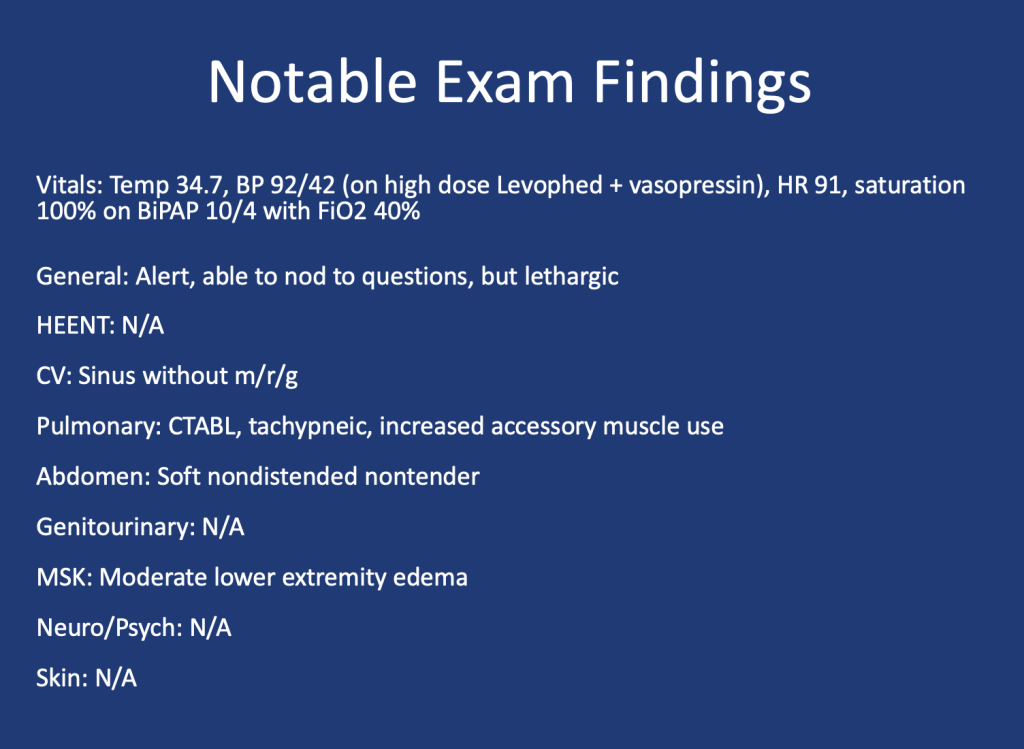
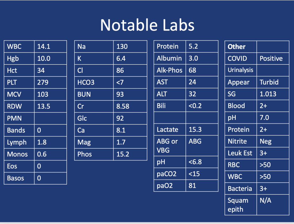
 A 75 y.o. female with a history of Hypertension, Hyperlipidemia, and Type 2 Diabetes presented for evaluation of hypoglycemia and generalized fatigue. She had felt poorly for about a week with symptoms of back pain, generalized weakness, and dyspnea, all of which acutely worsened on the day of presentation. 

She was found to be hypoglycemic with a blood glucose level in the to 40’s. Initial vital signs included a heart rate of 56, blood pressure of 70/40, respiratory rate of 30, and temperature of 28.5 degrees Celsius.

Lactic Acidosis: Type A, Type B and Type D

Type A: Typically secondary to conditions that impair oxygen delivery (respiratory failure, PE) to tissues or decrease tissue perfusion (severe anemia, shock). Patients typically present with hypotension, tachycardia, tachypnea, altered mental status, and signs of organ dysfunction.


Type B: Typically secondary to conditions that directly affect cellular metabolism or lactate clearance and characterized by the presence of hyperlactatemia without evidence of tissue hypoperfusion or hypoxia. Conditions associated include liver dysfunction (e.g., liver failure, cirrhosis), malignancies (especially hematological malignancies), medications/toxins (e.g., metformin, cyanide poisoning), inborn errors of metabolism, and mitochondrial disorders.

Type D: Less common presentation and can be seen in patients with short gut syndrome.

1.Blough B, Moreland A, Mora A Jr. Metformin-induced lactic acidosis with emphasis on the anion gap. Proc (Bayl Univ Med Cent). 2015 Jan;28(1):31-3. doi: 10.1080/08998280.2015.11929178. PMID: 25552792; PMCID: PMC4264704.

2.Callelo et al. Extracorporeal Treatment for Metformin Poisoning: Systematic Review and Recommendations From the Extracorporeal Treatments in Poisoning Workgroup. DOI: 10.1097/CCM.0000000000001002

3.Friesecke, S., Abel, P., Roser, M. et al. Outcome of severe lactic acidosis associated with metformin accumulation. Crit Care 14, R226 (2010). https://doi.org/10.1186/cc9376

4.Madias NE. Lactic acidosis. Kidney Int. 1986 Mar;29(3):752-74. doi: 10.1038/ki.1986.62. PMID: 3702227.

5. Stiller RH, Luks AM, Çoruh B. All That Raises Lactate Is Not Sepsis. ATS Sch. 2023 Jun 12;4(3):385-386. doi: 10.34197/ats-scholar.2023-0032OT.

71. Fellows’ Case Files: University of New Mexico

Today we’re visiting the University of New Mexico for another interesting entry in our Fellows’ Case Files.


Neel Vahil is a second-year internal medicine resident at the University of New Mexico. He completed medical school at New York Medical College and is planning on applying to pulmonary critical care fellowship programs.

Ishan Patel is a third year PCCM fellow at the University of New Mexico and will be pursuing a second fellowship in clinical informatics this year. He completed medical school and residency in Internal Medicine at Oregon Health & Science University. His fellowship research has focused on clinical outcomes of intensivist-led ECMO programs.

Dr. Lucie Griffin completed her internal medicine residency and PCCM fellowship at the University of New Mexico and is currently the Director of the Albuquerque VA medical intensive care unit.


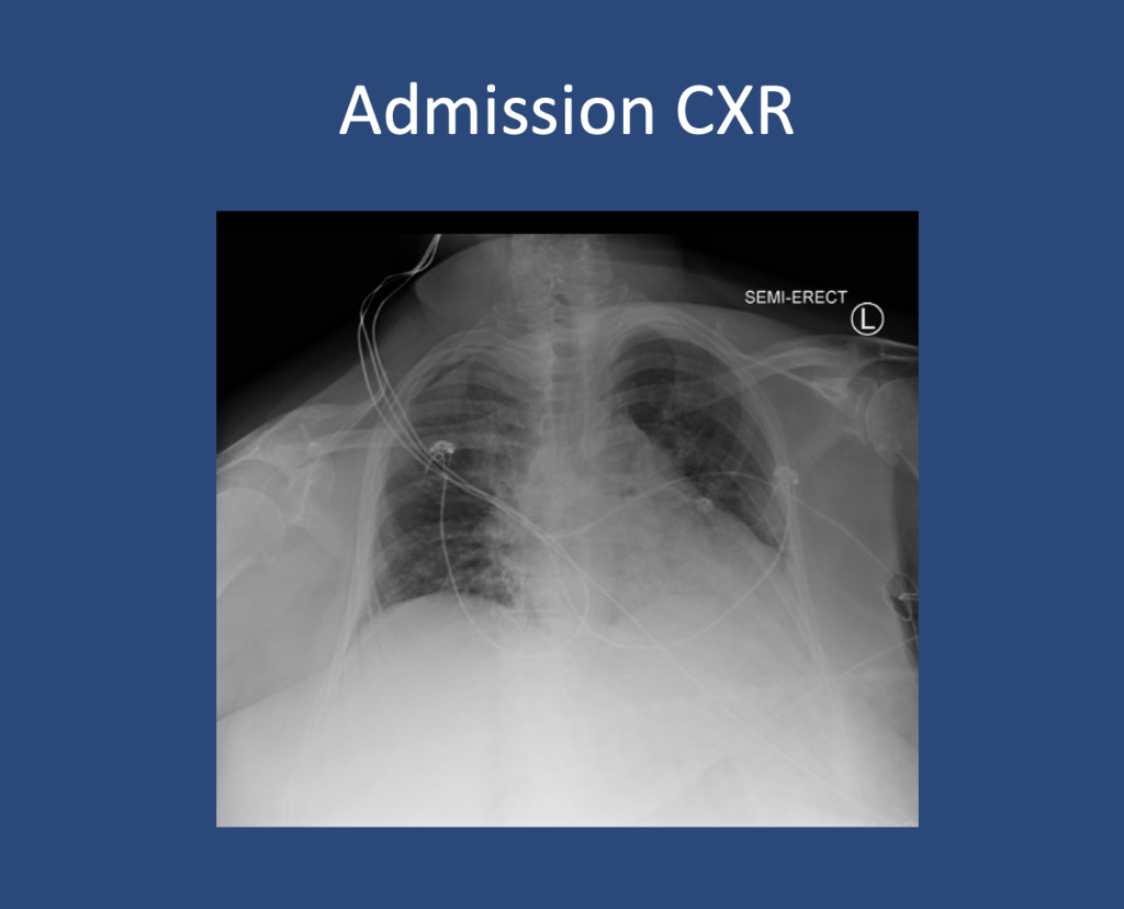
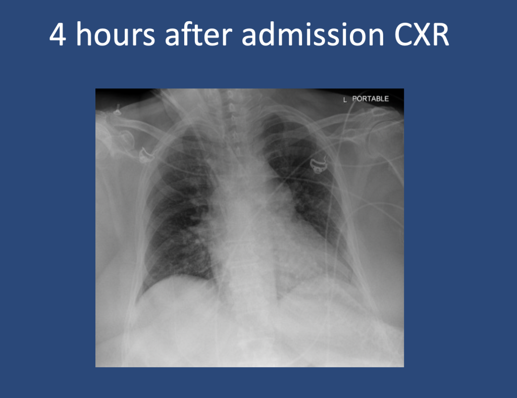
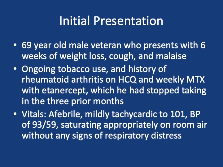
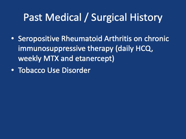
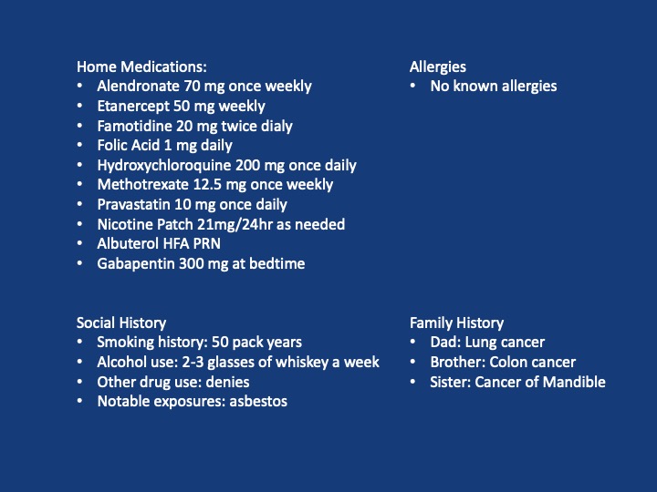
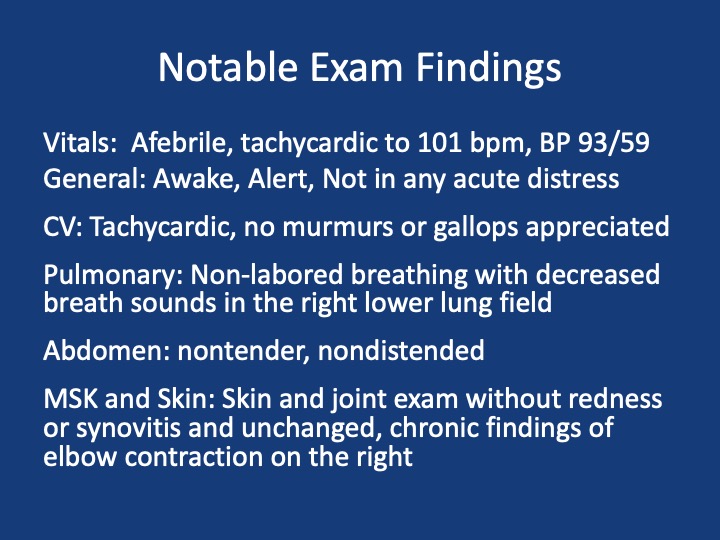
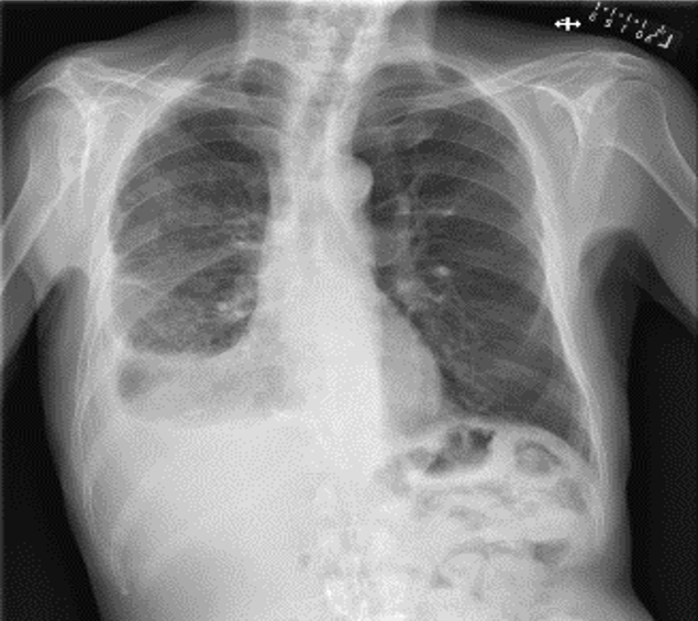
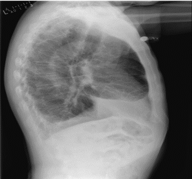
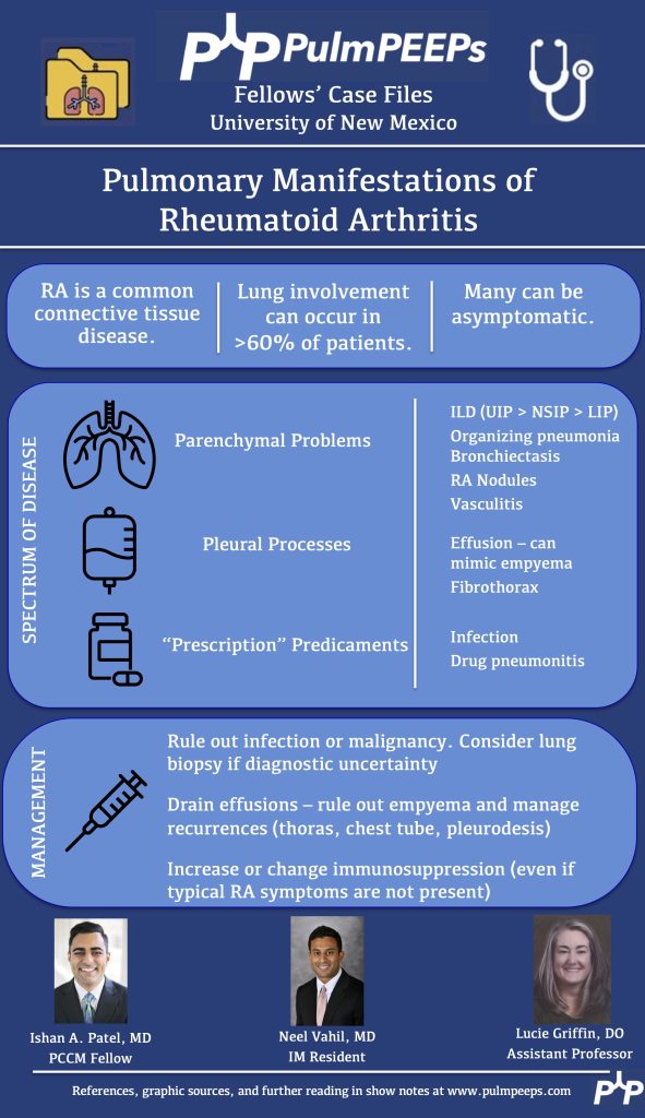
A 69 year old male veteran who presents with 6 weeks of weight loss, cough, and malaise. He has ongoing tobacco use, and history of rheumatoid arthritis on HCQ and weekly MTX with etanercept, which he had stopped taking in the three prior months. Vitals: Afebrile, mildly tachycardic to 101, BP of 93/59, saturating appropriately on room air without any signs of respiratory distress


Komarla A, Yu GH, Shahane A. Pleural effusion, pneumothorax, and lung entrapment in rheumatoid arthritis. J Clin Rheumatol. 2015;21(4):211-215.

Boddington MM, Spriggs AI, Morton JA, Mowat AG. Cytodiagnosis of rheumatoid pleural effusions. J Clin Pathol. 1971;24(2):95-106.

Balbir-Gurman A, Yigla M, Nahir AM, Braun-Moscovici Y. Rheumatoid pleural effusion. Semin Arthritis Rheum. 2006;35(6):368-378

68. Fellows’ Case Files: Mount Sinai Morningside

We’re back with another Case Files episode from Mt. Sinai Morningside. Listen in to hear another great case and some key learning points along the way.

Dr. Sara Luby is a third-year Internal Medicine resident and rising chief resident at Mt. Sinai Morningside/West and planning on applying to Pulmonary and Critical Care fellowship this upcoming year.

Dr. Javier Zulueta is the  Chief of the Division of Pulmonary, Critical Care, and Sleep Medicine at Mount Sinai Morningside. He completed residency training at St. Luke’s Medical Center/Case Western in Cleveland and fellowship in Pulmonary/Critical Care at Tufts Medical Center in Boston. His research focuses on lung cancer screening and incidental lung findings.

 Dr. Mirna Mohanraj is the Associate Program Director for the Pulmonary and Critical Care Fellowship at Mt. Sinai Morningside / Beth Israel and an associate professor of medicine and medical education at the Icahn School of Medicine at Mount Sinai. She completed residency training at University of Chicago and fellowship training at Mt. Sinai Hospital.

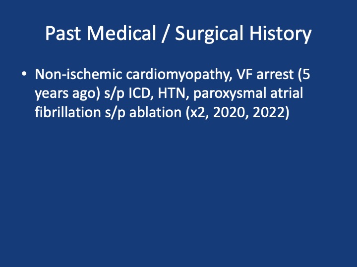
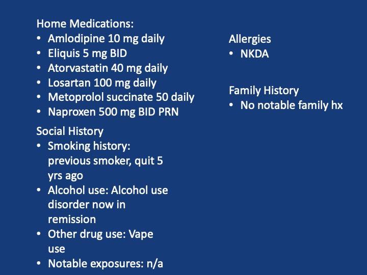
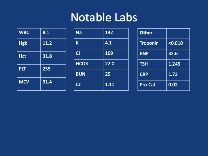
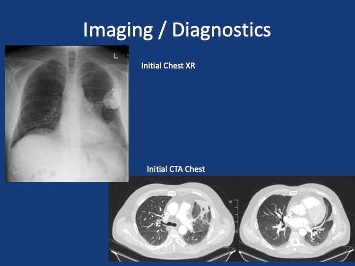
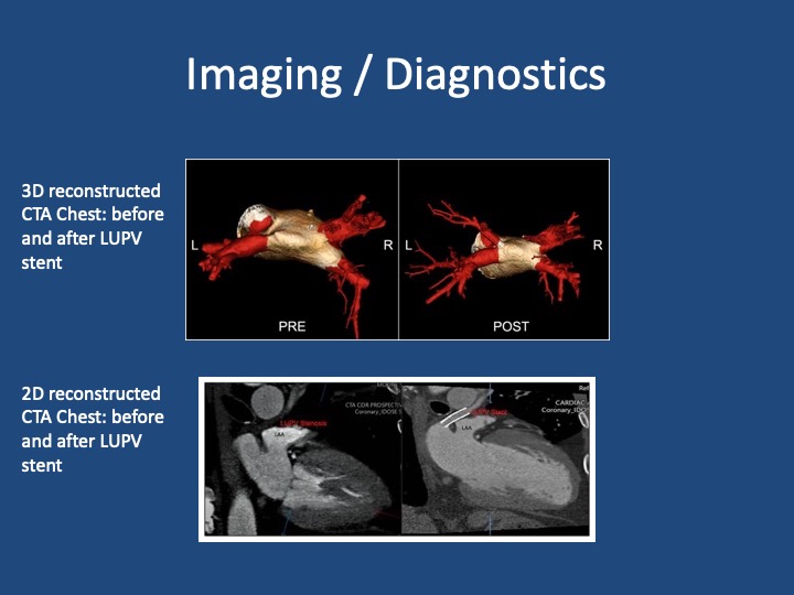
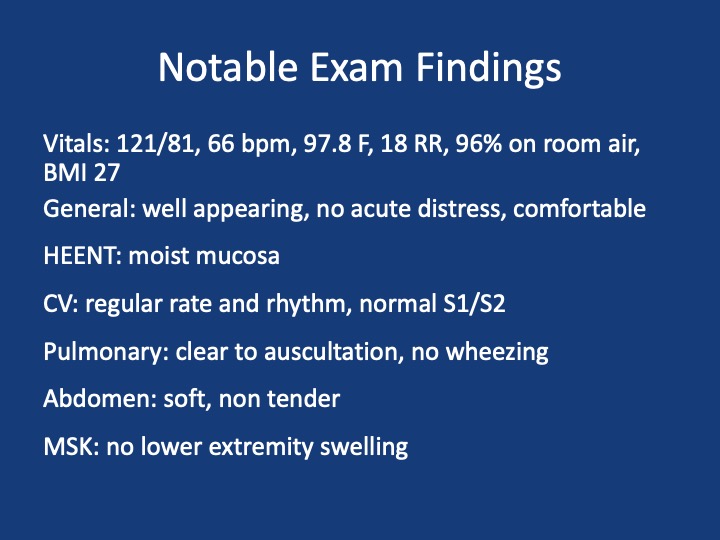
A 51 year old male presents with two days of acute on chronic chest pain and shortness of breath, worsening over the last month. His initial vitals: 143/ 100, pulse 85, temperature 36.5 °C (97.87°F), RR 16, SpO2 97 % on room air, BMI 29.8

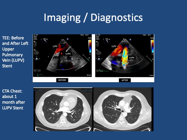
Shroff N, Choi W, Villanueva-Meyer J, Palacio DM, Bhargava P. Pulmonary vein occlusion: A delayed complication following radiofrequency ablation for atrial fibrillation. Radiol Case Rep. 2021;16(12):3666-3671. doi:10.1016/j.radcr.2021.09.015 

Fender EA, Widmer RJ, Hodge DO, et al. Assessment and Management of Pulmonary Vein Occlusion After Atrial Fibrillation Ablation. JACC: Cardiovascular Interventions. Vol 11(16); 2018. doi:10.1016/j.jcin.2018.05.020 

López-Reyes R, García-Ortega A, Torrents A, et al. Pulmonary venous thrombosis secondary to radiofrequency ablation of the pulmonary veins. Respir Med Case Rep. 2018;23:46-48. doi:10.1016/j.rmcr.2017.11.008

Mizuno A, Mauler-Wittwer S, Muller H, Noble S. Recurrent pneumonia post atrial fibrillation ablation: do not forget to look for pulmonary vein stenosis. BMJ Case Rep. 2022;15(12):e250896. doi:10.1136/bcr-2022-250896

67. Fellows’ Case Files: Northwestern University

Listen in today to another stop on our Fellows’ Case Files journey. We’re at Northwestern University for another great case presentation. Tune in, check out our associated infographic, and let us know what you think!

Meet Our Guests

Jamie Rowell is a first-year clinical fellow in the Northwestern PCCM program. She completed medical school at the Medical University of South Carolina and her internal medicine residency and Chief Residency at the University of Vermont Medical Center.

Cathy Gao is an Instructor of Medicine at Northwestern and completed her PCCM fellowship there last year. Her research focuses on using machine learning applied to ICU EHR data to characterize patient trajectories and identify potential interventions to improve outcomes.

Clara Schroedl is an Associate Professor of Medicine in Pulmonary and Critical Care and Medical Education. She is the program director of the Northwestern PCCM fellowship program, with an interest in medical education and simulation.

Case Presentation

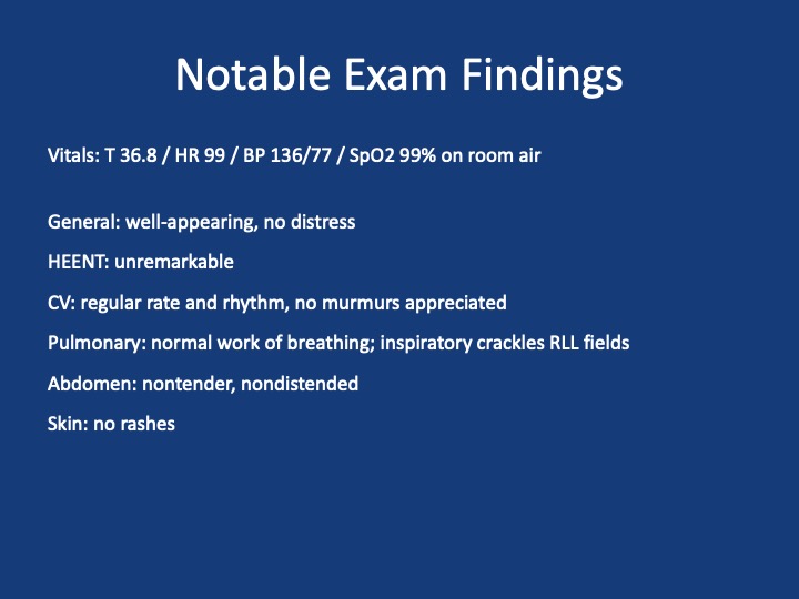
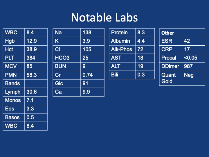
A 25-year-old previously healthy woman presents with recurrent episodes of right chest pain and cough. In October she was treated with antibiotics and felt somewhat better but in December, she presented again with chest pain, and again was treated with antibiotics. The pain improved but she still felt breathless. In February, again she had intense chest pain interfering with life, and was given NSAIDs and took high dose TID without clear benefit.

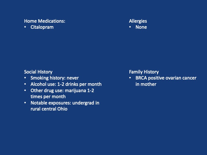
One month later, she coughed up some bloody mucus, so now she is presenting for evaluation. The chest pain is worse with deep breaths and improves in between these episodes. She only notes it on her right side. At this point, she does sometimes feel short of breath; she used to run 5 miles but is now struggling to run two miles. She denies any unusual exposures. She went to school in central rural Ohio for a while. She has no history of pulmonary infections, no exposure to mold or animals, and no history of vaping.

Key Learning Points

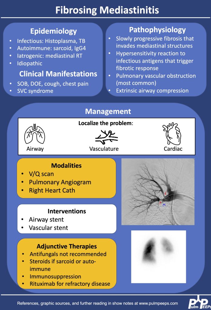
1.Making the diagnosis of Fibrosing Mediastinitis :

–Etiologies: histoplasmosis, sarcoidosis, tuberculosis, IgG4, Behcet, ANCA vasculitis

–Imaging modalities: CT chest, perfusion studies, pulmonary angiogram

–Imaging characteristics:  infiltrative, heterogeneous, fibrotic process that crosses fat planes and encroaches on nearby structures causing airway or vascular stenoses  

2. Management strategies:

–No curative therapies. Goal to relieve symptom burden

–Airway stents

–Vascular stents

–Rituximab

–Antifungals, steroids generally not considered effective

References and Further Reading

Kern et al. Bronchoscopic Management of Airway Compression due to Fibrosing Mediastinitis. Annals of the American Thoracic Society 2017. 14: 1235-1359 

Welby JP, Fender EA, Peikert T, Holmes DR Jr, Bjarnason H, Knavel-Koepsel EM. Evaluation of Outcomes Following Pulmonary Artery Stenting in Fibrosing Mediastinitis. Cardiovasc Intervent Radiol. 2021 Mar;44(3):384-391. doi: 10.1007/s00270-020-02714-z. Epub 2020 Nov 17. PMID: 33205295.

Westerly, BD Targeting B Lymphocytes in Progressive Fibrosing Mediastinitis. Am J Respir Crit Care Med. 2014 Nov 1; 190(9): 1069–1071.

https://rarediseases.org/rare-diseases/fibrosing-mediastinitis/#complete-report

https://pubmed.ncbi.nlm.nih.gov/21422386/

https://academic.oup.com/cid/article/30/4/688/421789

https://pubmed.ncbi.nlm.nih.gov/22033450/

https://www.sciencedirect.com/science/article/pii/S2352906715300087

https://www.atsjournals.org/doi/pdf/10.1513/AnnalsATS.201610-782RL

64. Fellows’ Case Files: Emory University School of Medicine

Hi everyone, we’re here with another Fellows’ Case Files. Today, we’re going virtually to Emory University School of Medicine. We’re joined by Associated Editor Luke Hedrick to dive into a critical care case. Listen in and let us know if you have any additional thoughts or questions!

Meet Our Guests

Luke Hedrick is a first-year pulmonary and critical care fellow at Emory University. He did his internal medicine residency at BIDMC in Boston. He is also one of our amazing Associate Editors here at Pulm PEEPs

Shirine Allam is an Associate Professor of Medicine at the Emory University School of Medicine where she is the Program Director of both the Pulmonary and Critical Care Medicine fellowship as well as the Critical Care Medicine fellowship. She completed her PCCM training at the Mayo Clinic in Rochester, followed by a Sleep Medicine fellowship at Stanford. She has received multiple teaching awards throughout her career

Case Presentation

A 32-year-old male is brought in by his coworkers unresponsive. He is a construction worker and was his usual self in the morning at the start of the day, but when they broke for lunch they noticed he was acting different—his arms were drooping, and while he initially was able to answer yes/no, he soon started babbling, then grunting, then vomited and became unresponsive. They laid him flat, threw cold water on him because it was 110 degrees and humid outside that day, and brought him to the ED.

When they arrive in the ED, he is unresponsive and warm to the touch. His vitals are notable for an oral temperature of 105, HR in the 160s, BP 76/34, a RR in the high 30s, and an SpO2 100% RA. His exam is relatively unremarkable other than for significant diaphoresis and both bowel and bladder incontinence.

Key Learning Points

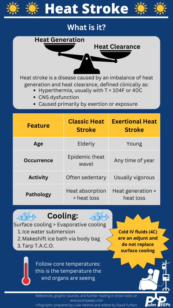
  1. Definition and recognition of heat stroke: Heat stroke is characterized by hyperthermia (>104°F or 40°C) accompanied by CNS dysfunction, primarily caused by exertion or exposure. Encephalitis without significant heat load does not constitute heat stroke.
  2. Management priorities: Rapid cooling is paramount to minimize long-term complications and organ failure. Cooling should be initiated as soon as possible, even before transportation to a hospital, particularly in cases of exertional heat stroke.
  3. Cooling methods: Surface cooling, such as immersion in ice water, is the most effective way to cool heat-stroke patients. Alternative methods include the TACO method and evaporative cooling, although they are less efficient. Refrigerated IV fluids can be used as an adjunct, but they do not replace the need for surface cooling.
  4. Monitoring and goals: Shivering during cooling should be monitored to prevent excessive heat generation. The goal is to reach a normal core body temperature (~38°C or 100.4°F). Traditional antipyretics like aspirin and acetaminophen should be avoided due to ineffectiveness and potential toxicity.
  5. Approach to endotracheal tube (ETT) exchange: ETT exchange requires preparation for potential complications. This includes ensuring the availability of airway equipment, sedation of the patient, and having additional personnel for assistance. Direct visualization using a video laryngoscope is recommended, along with measuring and marking the exchange catheter for proper insertion depth.

The following infographic can be downloaded from our website:

References and Further Reading

1.Epstein Y, Yanovich R. Heatstroke. New England Journal of Medicine. 2019;380(25):2449-2459. doi:10.1056/NEJMra1810762

2. Sorensen C, Hess J. Treatment and Prevention of Heat-Related Illness. New England Journal of Medicine. 2022;387(15):1404-1413. doi:10.1056/NEJMcp2210623