Today, Dave Furfaro, Luke Hedrick, and Robert Wharton discuss the PREDMETH trial published in The New England Journal of Medicine in 2025. This was a non-inferiority trial comparing prednisone to methotrexate for upfront therapy in treatment-naive sarcoidosis patients. Listen in for a break down of the trial, analysis, and clinically applicable pearls.
Article and Reference
Todays’ episode discusses the PREDMETH trial published in NEJM in 2025.
https://www.nejm.org/doi/full/10.1056/NEJMoa2501443
Meet Our Hosts
Luke Hedrick is an Associate Editor at Pulm PEEPs and runs the Rapid Fire Journal Club Series. He is a senior PCCM fellow at Emory, and will be starting as a pulmonary attending at Duke University next year.
Robert Wharton is a recurring guest on Pulm PEEPs as a part of our Rapid Fire Journal Club Series. He completed his internal medicine residency at Mt. Sinai in New York City, and is currently a first year pulmonary and critical care fellow at Johns Hopkins.
Key Learning Points
Clinical context
- Prednisone remains the traditional first-line treatment for pulmonary sarcoidosis when treatment is indicated, with evidence for short-term improvements in symptoms, radiographic findings, and pulmonary function—but with substantial, familiar steroid toxicities (weight gain, insomnia, HTN/DM, infection risk, etc.).
- Despite widespread use, glucocorticoids haven’t been robustly tested head-to-head against many alternatives as initial therapy, and evidence for preventing long-term decline (especially in severe disease) is limited.
- Immunosuppressants (like methotrexate) are often used as steroid-sparing agents, but guideline recommendations are generally conditional/low-quality evidence, and practice varies.
Why PREDMETH matters
- It addresses a real-world question: Can methotrexate be an initial alternative to prednisone in pulmonary sarcoidosis, rather than being reserved only for steroid-sparing later?
- It also probes a common clinical belief: MTX has slower onset than prednisone (often assumed, not well-proven).
Trial design (what to know)
- Open-label, randomized, noninferiority trial across 17 hospitals in the Netherlands.
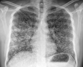
- Included patients with pulmonary sarcoidosis who had a clear pulmonary indication to start systemic therapy (moderate/severe symptoms plus objective risk features like reduced FVC/DLCO or documented decline, plus parenchymal abnormalities).
- Excluded: non–treatment-naïve patients and those whose primary indication was extrapulmonary disease.
- Treat-to-tolerability with escalation: both drugs started low and were slowly increased; switch/add-on allowed for inadequate efficacy or unacceptable side effects.
- Primary endpoint: change in FVC (with the usual caveat that FVC is “objective-ish,” but effort-dependent and not always patient-centered).
- Noninferiority margin: 5% FVC, justified as within biologic/measurement variation and “not clinically relevant.”
- Outcomes assessed at weeks 4, 16, 24; powered for ~110 patients to detect the NI margin.
Patient population (who this applies to)
- Mostly middle-aged (~40s) with mild-to-moderate physiologic impairment on average (FVC ~77% predicted; DLCO ~70% predicted).
- Netherlands-based cohort with limited Black representation (~7%), which matters for generalizability.
- Would have been helpful to know more about comorbidities (e.g., diabetes), which can strongly influence prednisone risk.
Main findings (what happened)
- Methotrexate was noninferior to prednisone at week 24 for FVC:
- Between-group difference in least-squares mean change at week 24: −1.17 percentage points (favoring prednisone) with CI −4.27 to +1.93, staying within the 5% NI margin.
- Timing mattered:
- Prednisone showed earlier benefit (notably by week 4) in FVC and across quality-of-life measures.
- By week 24, those early differences largely washed out—possibly because MTX “catches up,” and/or because crossover increased over time.
- In their reporting, MTX didn’t meet noninferiority for FVC until week 24, supporting the practical message that prednisone works faster.
Crossover and analysis nuance (important for interpretation)
- Crossover was fairly high, which complicates noninferiority interpretation:
- MTX arm: some switched to prednisone for adverse events and others had prednisone added for disease progression/persistent symptoms.
- Prednisone arm: some had MTX added.
- In noninferiority trials, heavy crossover can bias intention-to-treat analyses toward finding “no difference” (making noninferiority easier to claim). Per-protocol analyses avoid some of that but introduce other biases. They reported both.
Safety signals (what to remember clinically)
- Adverse events were very common in both arms (almost everyone), mostly mild.
- Side-effect patterns fit expectations:
- Prednisone: more insomnia (and classic steroid issues).
- MTX: more headache/cough/rash, and notably liver enzyme elevations (about 1 in 4), with a small number discontinuing.
- Serious adverse events were rare; numbers were too small to confidently separate “signal vs noise,” but overall known risk profiles apply.
Limitations (why you shouldn’t over-read it)
- Open-label design, and FVC—while objective-ish—is still effort-dependent and can be influenced by expectation/behavior.
- Small trial, limiting subgroup conclusions (e.g., severity strata, different phenotypes).
- Generalizability issues (Netherlands demographics; US populations have higher rates of obesity/metabolic syndrome, which may tilt the steroid risk-benefit equation).
- Crossover reduces precision and interpretability of between-group differences over time.
Practice implications (the “so what”)
- For many patients with pulmonary sarcoidosis needing systemic therapy, MTX is a reasonable initial alternative to prednisone when thinking long-term tolerability and steroid avoidance.
- Prednisone likely provides faster symptom/QoL relief in the first weeks—so it may be preferable when rapid improvement is important.
- The trial strengthens the case for a patient-centered discussion: short-term relief vs side-effect tradeoffs, and the possibility of early combination therapy in more severe cases (suggested, not proven).
Podcast: Play in new window | Download
Subscribe: Apple Podcasts | Spotify | Amazon Music | Android | iHeartRadio | Podcast Index | RSS