119. Guideline Series: Pulmonary Embolism

PPP Cover Images (4)

We are unbelievably excited this week to be reviewing the hot-off-the-presses 2026 Multi-Society (AHA/ACC/ACCP/ACEP/CHEST/SCAI/SHM/SIR/SVM/SVN) Pulmonary Embolism Guidelines with lead author Dr. Mark A. Creager. We will talk about key updates in these guidelines compared to prior practice, including the new risk classification model, and provide an overview from diagnosis to follow-up. Given the clinical importance and prevalence of pulmonary embolism, these guidelines are certainly going to shape practice going forward, so this episode is a can’t miss!

Watch the full video of this episode with graphics and helpful teaching visuals on our YouTube channel: https://www.youtube.com/@pulmpeeps

Dr. Mark Creager is a Professor of Medicine at Dartmouth Hitchcock Medical Center where he specializes in Cardiovascular Medicine with an emphasis on venous thromboembolic disease. He served as the lead author of the 2026 Pulmonary Embolism Guidelines.

Creager MA, Barnes GD, Giri J, Mukherjee D, Jones WS, Burnett AE, Carman T, Casanegra AI, Castellucci LA, Clark SM, Cushman M, de Wit K, Eaves JM, Fang MC, Goldberg JB, Henkin S, Johnston-Cox H, Kadavath S, Kadian-Dodov D, Keeling WB, Klein AJP, Li J, McDaniel MC, Moores LK, Piazza G, Prenger KS, Pugliese SC, Ranade M, Rosovsky RP, Russo F, Secemsky EA, Sista AK, Tefera L, Weinberg I, Westafer LM, Young MN. 2026 AHA/ACC/ACCP/ACEP/CHEST/SCAI/SHM/SIR/SVM/SVN Guideline for the Evaluation and Management of Acute Pulmonary Embolism in Adults: A Report of the American College of Cardiology/American Heart Association Joint Committee on Clinical Practice Guidelines. J Am Coll Cardiol. 2026 Feb 19:S0735-1097(25)10161-7. doi: 10.1016/j.jacc.2025.11.005. Epub ahead of print. PMID: 41712898.

Why these guidelines matter:

This is the first joint AHA/ACC clinical practice guideline specifically on acute PE, bringing together a truly multidisciplinary writing committee (cardiology, pulmonology, hematology, emergency medicine, interventional radiology, surgery, and others). Prior guidelines existed from individual societies, but nothing this comprehensive had been updated in roughly five to six years.

New PE clinical categories (A through E):

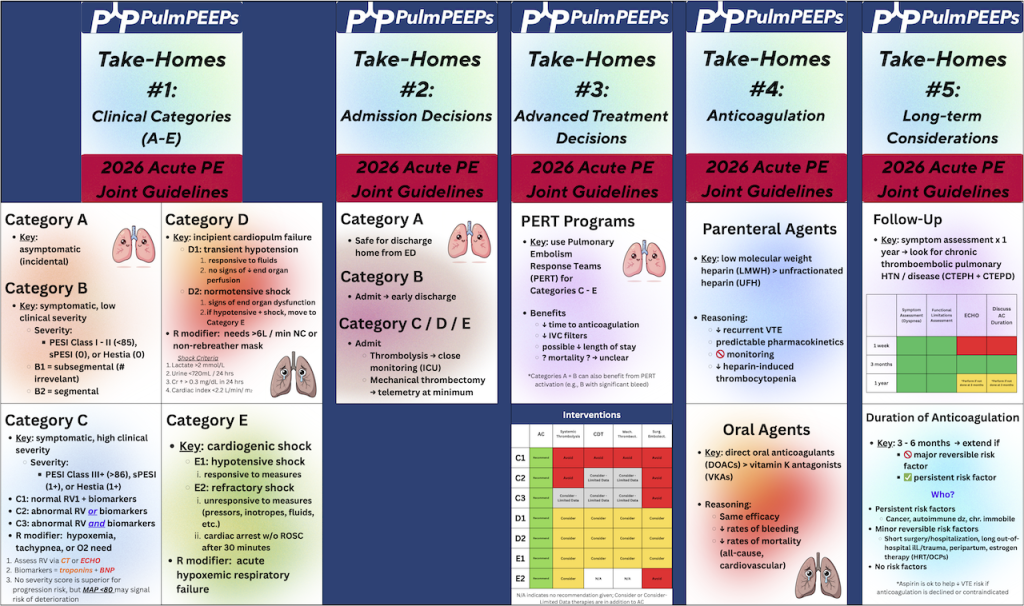
One of the most impactful changes is replacing the old “massive/submassive” and “low/intermediate/high risk” labels with five categories that form a severity continuum. Category A is subclinical (incidental PE found on imaging in asymptomatic patients). Category B covers symptomatic but low-severity patients. Category C is where much of the clinical complexity lives — symptomatic, hemodynamically stable patients subdivided into C1, C2, and C3 based on RV function and biomarkers. Category D represents incipient cardiopulmonary failure (transient hypotension, normotensive shock with end-organ dysfunction). Category E is frank cardiopulmonary failure, with E2 being the sickest — refractory or recurrent cardiac arrest. Respiratory modifiers (hypoxia requiring supplemental oxygen) layer onto C, D, and E.

Diagnostic approach:

Clinical evaluation comes first — history, exam, and validated decision tools (Wells score, revised Geneva, PERC). If clinical probability is low and D-dimer is normal, imaging can be safely avoided. If either is concerning, imaging is warranted. CTPA remains the preferred imaging modality due to superior sensitivity, specificity, wide availability, and ability to assess clot burden and alternative diagnoses. VQ scanning is still appropriate when CTPA is contraindicated, and VQ SPECT offers better reproducibility and specificity than traditional planar VQ if available. Echocardiography is not a diagnostic test for PE but is important for risk stratification — RV size, TAPSE, and tissue Doppler measures all contribute prognostic information.

Anticoagulation updates:

Anticoagulation remains the cornerstone of treatment. For patients potentially needing advanced therapies (C3, D, E), parenteral anticoagulation is started first. A notable recommendation: low molecular weight heparin is generally preferred over unfractionated heparin, based on evidence showing more effective VTE risk reduction, more predictable pharmacokinetics, no need for routine monitoring, lower rates of heparin-induced thrombocytopenia, and no increase in major bleeding. The committee acknowledged this may create discomfort for clinicians accustomed to unfractionated heparin’s easy reversibility, but the difficulty of achieving and maintaining therapeutic levels with UFH was a significant concern.

Advanced therapies:

Catheter-based thrombolysis, mechanical thrombectomy, systemic thrombolysis, and surgical embolectomy all received mostly class 2B recommendations (“can consider”) for C3 and D categories, reflecting that current evidence shows improvement in short-term surrogate measures (RV/LV ratio, hemodynamics) but lacks definitive hard outcome data on mortality. For category E1 patients, recommendations are stronger (class 2A). Multiple trials are expected soon — HI-PEITHO, PEERLESS-2, PE-TRACT, PERSEVERE, TORPEDO, and PROG — that should substantially inform future updates.

PERT teams:

Pulmonary embolism response teams are encouraged, particularly for C3, D, and E patients. They’ve been shown to reduce length of stay. For institutions without PERT capability, establishing consultation networks with larger centers is recommended.

Post-PE follow-up:

Patients shouldn’t be “left in the wilderness” after discharge. The guidelines recommend communication within the first week to ensure understanding of diagnosis and treatment, an in-person visit at or before three months to assess for persistent symptoms and discuss anticoagulation duration, ongoing surveillance for chronic thromboembolic pulmonary disease, and periodic reassessment for those on extended anticoagulation.

Infographics
116. Guidelines Series: Pulmonary Hypertension – Risk Stratification and Treatment Goals

PH 2 Face Sheet

On this week’s episode, we’re continuing our Guidelines Series exploring the 2022 ESC/ERS Guidelines for the diagnosis and treatment of Pulmonary Hypertension. If you missed our first episode in the series, give it a listen to hear about the most recent recommendations regarding Pulmonary Hypertension definitions, screening, and diagnostics. Today, we’re talking about the next steps after diagnosis. Specifically, we’ll be discussing risk stratification, establishing treatment goals, and metrics for re-evaluation. We’ll additionally introduce the mainstays of pharmacologic therapy for Pulmonary Hypertension.

Rupali Sood  grew up in Las Vegas, Nevada and made her way over to Baltimore for medical school at Johns Hopkins. She then completed her internal medicine residency training at Massachusetts General Hospital before returning back to Johns Hopkins, where she is currently a pulmonary and critical care medicine fellow. Rupali’s interests include interstitial lung disease, particularly as related to oncologic drugs, and bedside medical education.

Tom Di Vitantonio  is originally from New Jersey and attended medical school at Rutgers, New Jersey Medical School in Newark. He then completed his internal medicine residency at Weill Cornell, where he also served as a chief resident. He currently is a pulmonary and critical care medicine fellow at Johns Hopkins, and he’s passionate about caring for critically ill patients, how we approach the management of pulmonary embolism, and also about medical education of trainees to help them be more confident and patient centered.

1) Episode Roadmap

  • How to set treatment goals, assess symptom burden, and risk-stratify patients with suspected/confirmed pulmonary arterial hypertension (PAH).
  • What tools to use to re-evaluate patients on treatment
  • Intro to major PAH medication classes and how they map to pathways.

2) Case-based diagnostic reasoning

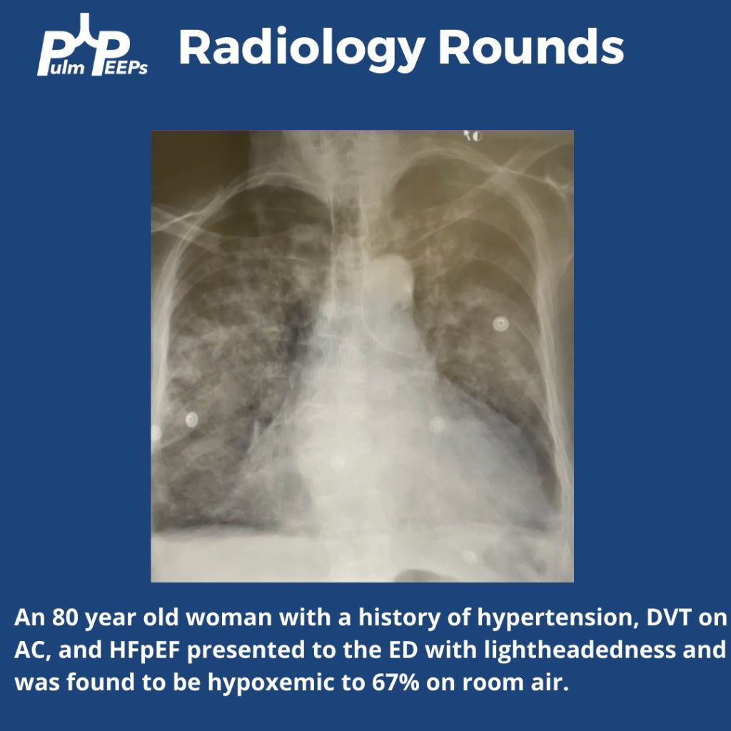
Patient: 37-year-old woman with exertional dyspnea, mild edema, abnormal echo, telangiectasias + epistaxis → raises suspicion for HHT (hereditary hemorrhagic telangiectasia) and/or early connective tissue disease.

  • Key reasoning move: start broad (Groups 2–5) and narrow using history/exam/testing.
  • In a young patient without obvious left heart or lung disease, think more about Group 1 PAH (idiopathic/heritable/associated).

HHT teaching point: HHT can cause PH in more than one way:

  • More common: high-output PH from AVMs (often hepatic/pulmonary)
  • Rare (1–2% mentioned): true PAH phenotype (vascular remodeling; associated with ALK1 in some patients), behaving like Group 1 PAH.

3) Functional class assessment

WHO Functional Class:

  • Class I: no symptoms with ordinary activity, only with exertion
  • Class II: symptoms with ordinary activity
  • Class III: symptoms with less-than-ordinary activity (can’t do usual chores/shopping without dyspnea)
  • Class IV: symptoms at rest

Practical bedside tip they give:

  • Ask if the patient can walk at their own pace or keep up with a similar-age peer/partner. If not, think Class II (or worse).

4) Risk stratification at diagnosis: why, how, and which tools

Big principle: treatment choices are driven by risk, and the goal is to move patients to low-risk quickly.

ESC/ERS approach at diagnosis (as described):

  • Use a 3-strata model predicting 1-year mortality:
    • Low: <5%
    • Intermediate: 5–20%
    • High: >20%

ESC/ERS risk assessment variables (10 domains discussed):

  • Clinical progression, signs of right heart failure, syncope
  • WHO FC
  • Biomarkers (NT-proBNP)
  • Exercise capacity (6MWD)
  • Hemodynamics
  • Imaging (echo; sometimes cardiac MRI)
  • CPET (peak VO₂; VE/VCO₂ slope)

They note: even if you don’t have everything, the calculator can still be useful with ≥3 variables.

REVEAL 2.0:

  • Builds on similar core variables but adds further patient context (demographics, renal function, BP, DLCO, etc.)

Case result: both tools put her in intermediate risk (ESC/ERS ~1.6; REVEAL 2.0 score 8), underscoring that mild symptoms can still equal meaningful mortality risk.

5) Treatment goals and follow-up philosophy

What they explicitly prioritize:

  • Help patients feel better, live longer, and stay out of the hospital
  • Use risk tools to communicate prognosis and to track improvement
  • Reassess frequently (they mention ~every 3 months early on) until low risk is achieved
  • “Time-to-low-risk” is an important treatment goal

Also emphasized:

  • The diagnosis is psychologically heavy; patients need clear counseling, reassurance about the plan, and connection to support groups.

6) Medication classes for the treatment of PAH

Nitric oxide–cGMP pathway

  • PDE5 inhibitors: sildenafil, tadalafil
  • Soluble guanylate cyclase stimulator: riociguat
  • Important safety point: don’t combine PDE5 inhibitors with riociguat (risk of significant hypotension/hemodynamic effects)

Endothelin receptor antagonists (ERAs)

  • “-sentan” drugs: bosentan (less used due to side effects/interactions), ambrisentan, macitentan
  • Teratogenicity emphasized
  • Hepatotoxicity that requires LFT monitoring
  • Can cause fluid retention and peripheral edema

Prostacyclin pathway

  • Prostacyclin analogs/agonists:
    • Epoprostenol (potent; short half-life; IV administration)
    • Treprostinil (IV/SubQ/oral/inhaled options)
    • Selexipag (oral prostacyclin receptor agonist)

7) Sotatercept (post-guidelines)

They note sotatercept wasn’t in 2022 ESC/ERS but is now “a game changer” in practice:

  • Mechanism: ligand trap affecting TGF-β signaling / remodeling biology
  • Positioned as potentially more disease-modifying than pure vasodilators
  • Still evolving: where to place it earlier vs later in regimens is an active question in the field

8) How risk category maps to initial treatment intensity

General approach they outline:

  • High risk at diagnosis: parenteral prostacyclin (IV/SubQ) strongly favored, often aggressive early
  • Intermediate risk: at least dual oral therapy (typically PDE5i + ERA); escalate if not achieving low risk
  • Low risk: at least one oral agent; many still use dual oral depending on etiology/trajectory

For the case: intermediate-risk → start dual oral therapy (they mention tadalafil + ambrisentan as a typical choice), reassess in ~3 months; add a third agent (e.g., selexipag/prostacyclin pathway) if not low risk.

Humbert M, Kovacs G, Hoeper MM, Badagliacca R, Berger RMF, Brida M, Carlsen J, Coats AJS, Escribano-Subias P, Ferrari P, Ferreira DS, Ghofrani HA, Giannakoulas G, Kiely DG, Mayer E, Meszaros G, Nagavci B, Olsson KM, Pepke-Zaba J, Quint JK, Rådegran G, Simonneau G, Sitbon O, Tonia T, Toshner M, Vachiery JL, Vonk Noordegraaf A, Delcroix M, Rosenkranz S; ESC/ERS Scientific Document Group. 2022 ESC/ERS Guidelines for the diagnosis and treatment of pulmonary hypertension. Eur Heart J. 2022 Oct 11;43(38):3618-3731. doi: 10.1093/eurheartj/ehac237. Erratum in: Eur Heart J. 2023 Apr 17;44(15):1312. doi: 10.1093/eurheartj/ehad005. PMID: 36017548.

Condon DF, Nickel NP, Anderson R, Mirza S, de Jesus Perez VA. The 6th World Symposium on Pulmonary Hypertension: what’s old is new. F1000Res. 2019 Jun 19;8:F1000 Faculty Rev-888. doi: 10.12688/f1000research.18811.1. PMID: 31249672; PMCID: PMC6584967.

Maron BA. Revised Definition of Pulmonary Hypertension and Approach to Management: A Clinical Primer. J Am Heart Assoc. 2023 Apr 18;12(8):e029024. doi: 10.1161/JAHA.122.029024. Epub 2023 Apr 7. PMID: 37026538; PMCID: PMC10227272.

Hoeper MM, Badesch DB, Ghofrani HA, Gibbs JSR, Gomberg-Maitland M, McLaughlin VV, Preston IR, Souza R, Waxman AB, Grünig E, Kopeć G, Meyer G, Olsson KM, Rosenkranz S, Xu Y, Miller B, Fowler M, Butler J, Koglin J, de Oliveira Pena J, Humbert M; STELLAR Trial Investigators. Phase 3 Trial of Sotatercept for Treatment of Pulmonary Arterial Hypertension. N Engl J Med. 2023 Apr 20;388(16):1478-1490. doi: 10.1056/NEJMoa2213558. Epub 2023 Mar 6. PMID: 36877098.

Ruopp NF, Cockrill BA. Diagnosis and Treatment of Pulmonary Arterial Hypertension: A Review. JAMA. 2022 Apr 12;327(14):1379-1391. doi: 10.1001/jama.2022.4402. Erratum in: JAMA. 2022 Sep 6;328(9):892. doi: 10.1001/jama.2022.13696. PMID: 35412560.

112. Guidelines Series: Pulmonary Hypertension – Definitions, Screening, and Diagnosis

PH 1 Guideline Series FacePage

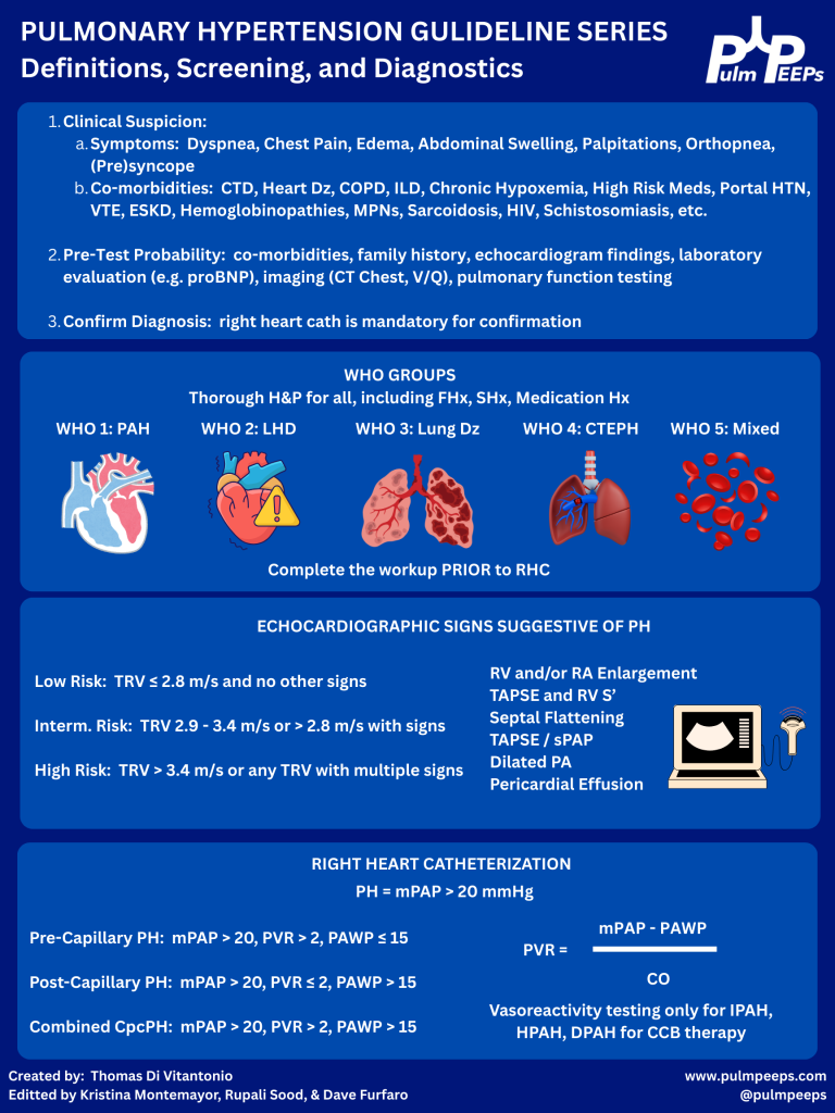
Today we’re kicking off another segment in our Guidelines Series, and doing a deep dive into the 2022 ESC/ERS Guidelines for the diagnosis and treatment of pulmonary hypertension. Over a series of episodes we’ll talk about the most recent updates to definitions around pulmonary hypertension, recognizing and diagnosing Group 1 – 5 pulmonary hypertension, risk stratification, and treatments. In this first episode, we will review the most recent definitions, including changes to the definitions that were new in 2022. We’ll then talk about recognizing and diagnosing pulmonary hypertension with tips and insights along the way.


Rupali Sood  grew up in Las Vegas, Nevada and made her way over to Baltimore for medical school at Johns Hopkins. She then completed her internal medicine residency training at Massachusetts General Hospital before returning back to Johns Hopkins, where she is currently a pulmonary and critical care medicine fellow alongside Tom. Rupali’s interests include interstitial lung disease, particularly as related to oncologic drugs. And she also loves bedside medical education.

Tom Di Vitantonio  is originally from New Jersey and attended medical school at Rutgers, New Jersey Medical School in Newark. He then completed his internal medicine residency at Weill Cornell, where he also served as a chief resident. He currently is a pulmonary and critical care medicine fellow at Johns Hopkins, and he’s passionate about caring for critically ill patients, how we approach the management of pulmonary embolism, and also about medical education of trainees to help them be more confident and patient centered in the care they have going forward.

  1. Why to have a high index of suspicion for pulmonary hypertension (PH)
  • PH often presents subtly with slowly progressive dyspnea on exertion, fatigue, lightheadedness, exertional chest pain, or syncope.
  • There’s often a delay of 1–2+ years from symptom onset to diagnosis, which is associated with worse mortality.
  • Early recognition and treatment, especially for pulmonary arterial hypertension (PAH, WHO group 1), can significantly change outcomes.


  1. When to suspect PH

Think PH when:

  • Dyspnea is out of proportion to:
    • CT parenchymal findings (relatively normal lungs)
    • Spirometry (normal FEV₁/FVC, volumes)
  • There are subtle but progressive symptoms over months:
    • Reduced exercise tolerance
    • No obvious alternative explanation (e.g., no overt HF, CAD, big ILD, etc.)
  • Physical exam may show (often late):
    • Elevated JVP, V waves (TR)
    • Peripheral edema, hepatomegaly, ascites
    • Loud P2, RV heave

In the case: a woman with systemic sclerosis + slowly progressive exertional dyspnea + relatively normal CT parenchyma and spirometry → high suspicion.


  1. WHO classification: 5 PH groups (big picture + why it matters)

Used for pathophysiology, prognosis, and treatment choices:

  1. Group 1 – PAH
    • Idiopathic, heritable (e.g., BMPR2), drug-induced (e.g., dasatinib)
    • Connective tissue disease (esp. systemic sclerosis)
    • Portal hypertension (portopulmonary HTN)
    • HIV, HHT, congenital heart disease/shunts
    • Rare: PVOD, PCH
  2. Group 2 – PH due to left heart disease
    • HFrEF, HFpEF, valvular disease
    • Most common cause worldwide.
  3. Group 3 – PH due to lung disease/hypoxia
    • COPD, ILD, combined pulmonary fibrosis–emphysema
    • OSA/obesity hypoventilation, chronic hypoxemia
  4. Group 4 – CTEPH
    • Chronic thromboembolic pulmonary hypertension
  5. Group 5 – Multifactorial/unclear
    • Sarcoidosis, myeloproliferative disorders, CKD, sickle cell, etc.

Patients can span multiple groups (e.g., systemic sclerosis: group 1 and/or group 3; sickle cell: many mechanisms).


  1. Initial workup & refining pre-test probability

Once you suspect PH, you’re trying to answer:

  1. Does this patient likely have PH?
  2. If yes, what group(s) are most likely?

Core non-invasive tests:

  • NT-proBNP (preferred over BNP)
    • Surrogate of RV strain and prognosis.
    • Normal value makes significant RV failure less likely.
  • Oxygenation & exercise
    • Resting SpO₂ plus ambulatory sats; consider 6-minute walk test.
    • Exertional desaturation is common and clinically meaningful.
  • CXR & ECG
    • Low yield but may show RV enlargement, right axis deviation, etc.
  • Pulmonary function tests
    • Full set: spirometry, volumes, DLCO.
    • Clue: isolated or disproportionately low DLCO with relatively preserved FVC suggests pulmonary vascular disease.
  • Imaging
    • High-res CT chest – parenchymal disease (ILD, emphysema).
    • V/Q scan – best screening test for CTEPH; better than CT angiography for chronic disease.
  • Sleep testing / overnight oximetry
    • When OSA/nocturnal hypoxemia suspected.


  1. Echo: estimating PH probability (not diagnosis)

TTE is the key screening tool but does not diagnose PH.

Main elements:

  1. Peak tricuspid regurgitant (TR) velocity
    • Used to estimate pulmonary artery systolic pressure (PASP).
    • Categories:
      • Low probability: TR velocity < 2.8 m/s, no other PH signs.
      • Intermediate: 2.9–3.4 m/s ± other PH signs.
      • High: > 3.4 m/s.

The presence and severity of TR ≠ TR velocity. You can have severe TR without PH.

  1. “Other signs” of PH/RV dysfunction on echo:
    • RV enlargement or systolic dysfunction (qualitative, TAPSE < ~1.7 cm, S′ ↓)
    • RA enlargement
    • Septal flattening (D-shaped LV; systolic = pressure overload, diastolic + systolic = volume + pressure)
    • Dilated PA
    • Pericardial effusion

Interpretation pattern:

  • Low pre-test probability + TR v < 2.8 + no other signs → PH unlikely.
  • Intermediate TR v (2.9–3.4) + high pre-test probability and/or other PH signs → consider RHC.
  • High TR v (>3.4) or clearly abnormal RV → strongly consider RHC if it would change management.

Also:

  • Echo is great to follow RV size/function and PASP over time once PH is diagnosed and treated.

Case echo:

  • TR velocity 3.1 m/s + mild RA enlargement + moderate RV enlargement + TAPSE 1.6 cm → intermediate probability, consistent with PH and RV involvement.


  1. Right heart cath (RHC): gold standard & updated definitions

You cannot definitively diagnose or classify PH without RHC.

Key directly measured values:

  • RA, RV, PA pressures
  • Pulmonary capillary wedge pressure (PCWP/PAWP) ≈ LVEDP
  • Oxygen saturations in chambers/vessels
  • Cardiac output (thermodilution)

Key derived values:

  • Cardiac output (Fick)
  • Pulmonary vascular resistance (PVR)

Updated hemodynamic definitions:

  1. Pulmonary hypertension (PH)
    • mPAP ≥ 20 mm Hg (lowered from ≥ 25).
  2. Pre-capillary PH (think PAH, group 1; also groups 3, 4, some 5):
    • mPAP ≥ 20
    • PAWP ≤ 15
    • PVR > 2 Wood units (new lower threshold)
  3. Isolated post-capillary PH (IpcPH) (group 2)
    • mPAP ≥ 20
    • PAWP > 15
    • PVR ≤ 2
  4. Combined pre- and post-capillary PH (CpcPH)
    • mPAP ≥ 20
    • PAWP > 15
    • PVR > 2

Rationale for the changes:

  • Normal mPAP in healthy people is < ~19; 20 is about 2 SD above normal.
  • Patients with mPAP 20–24 (esp. systemic sclerosis) already have worse outcomes than those < 20.
  • Lowering PVR cutoff from 3 → 2 WU better aligns with these new thresholds and catches earlier precapillary disease.

Practical interpretation:

  • You use mPAP + PAWP + PVR to:
    • Confirm PH.
    • Distinguish pre- vs post-capillary.
    • Identify mixed disease.
  • Echo tells you probability; RHC tells you what type and how severe.


  1. Vasoreactivity testing (acute vasodilator testing)
  • Only indicated in:
    • Idiopathic (IPAH)
    • Heritable PAH
    • Drug-induced PAH
      → Not routine for all PH patients.
  • Performed in the cath lab with short-acting vasodilator (e.g., inhaled NO).

Positive test:

  • ↓ mPAP ≥ 10 mm Hg
  • To an absolute mPAP ≤ 40 mm Hg
  • No fall in cardiac output

Why it matters:

  • Identifies a small subset who can be treated with high-dose calcium channel blockers long-term and often have better prognosis.
  • Does not predict response to other PAH therapies (ERA, PDE5i, prostacyclin, etc.).


  1. Screening high-risk populations

Some groups warrant systematic screening because of high PAH risk.

  1. a) Systemic sclerosis / systemic sclerosis spectrum
  • Annual screening if:
    • Disease duration ≥ 3 years
    • FVC ≥ 40% predicted
    • DLCO < 60% predicted
  • DETECT algorithm (2-step):
    • Step 1: uses labs and simple tests (FVC/DLCO ratio, NT-proBNP, autoantibodies, right axis deviation on ECG, telangiectasias).
    • If positive → Step 2: adds echo (TR velocity, RA size).
    • If high risk after Step 2 → RHC.
  • Goal: catch early PAH before symptoms are severe.
  1. b) Other high-risk groups

Annual screening (usually with echo ± NT-proBNP, PFTs) for:

  • Known heritable PAH mutations (e.g., BMPR2)
  • Portal hypertension (esp. considering liver transplant or TIPS)
  • HIV

Always layer this on top of clinical symptoms and progression.


  1. Big practical takeaways (what to apply on Monday)
  1. Don’t label “pulmonary hypertension” off CT or echo alone.
    • Enlarged PA on CT or elevated PASP on echo ≠ diagnosis.
    • RHC is required.
  2. Think PH early when:
    • Dyspnea is out of proportion to imaging and spirometry.
    • There is a relevant risk factor (systemic sclerosis, portal HTN, HIV, prior PE, congenital heart disease, etc.).
  3. Use the WHO groups to structure your differential and workup:
    • Group 1 vs 2 vs 3 vs 4 vs 5 → drives what tests you order and what treatments you eventually consider.
  4. Echo = probability. RHC = truth.
    • Echo gives you low / intermediate / high PH probability.
    • RHC gives you pre- vs post-capillary, PVR, and hemodynamics needed for therapy.
  5. Know the new numbers:
    • mPAP ≥ 20 = PH
    • PAWP cutoff = 15
    • PVR > 2 WU = precapillary component
  6. Don’t forget NT-proBNP, DLCO, V/Q scan, and high-risk screening (especially in systemic sclerosis and BMPR2 carriers).


Humbert M, Kovacs G, Hoeper MM, Badagliacca R, Berger RMF, Brida M, Carlsen J, Coats AJS, Escribano-Subias P, Ferrari P, Ferreira DS, Ghofrani HA, Giannakoulas G, Kiely DG, Mayer E, Meszaros G, Nagavci B, Olsson KM, Pepke-Zaba J, Quint JK, Rådegran G, Simonneau G, Sitbon O, Tonia T, Toshner M, Vachiery JL, Vonk Noordegraaf A, Delcroix M, Rosenkranz S; ESC/ERS Scientific Document Group. 2022 ESC/ERS Guidelines for the diagnosis and treatment of pulmonary hypertension. Eur Heart J. 2022 Oct 11;43(38):3618-3731. doi: 10.1093/eurheartj/ehac237. Erratum in: Eur Heart J. 2023 Apr 17;44(15):1312. doi: 10.1093/eurheartj/ehad005. PMID: 36017548.

Condon DF, Nickel NP, Anderson R, Mirza S, de Jesus Perez VA. The 6th World Symposium on Pulmonary Hypertension: what’s old is new. F1000Res. 2019 Jun 19;8:F1000 Faculty Rev-888. doi: 10.12688/f1000research.18811.1. PMID: 31249672; PMCID: PMC6584967.

Maron BA. Revised Definition of Pulmonary Hypertension and Approach to Management: A Clinical Primer. J Am Heart Assoc. 2023 Apr 18;12(8):e029024. doi: 10.1161/JAHA.122.029024. Epub 2023 Apr 7. PMID: 37026538; PMCID: PMC10227272.

101. RFJC – NAVIGATOR

Logo

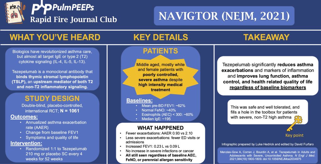
We’re back with another Rapid Fire Journal Club. Luke Hedrick and Dave Furfaro discuss the NAVIGATOR trial published in NEJM in 2021 evaluating tezepelumab for adults with asthma.

Article and Reference

We are talking today about the NAVIGATOR trial evaluating the use of tezepelumab in adults with asthma.

Menzies-Gow A, Corren J, Bourdin A, Chupp G, Israel E, Wechsler ME, Brightling CE, Griffiths JM, Hellqvist Å, Bowen K, Kaur P, Almqvist G, Ponnarambil S, Colice G. Tezepelumab in Adults and Adolescents with Severe, Uncontrolled Asthma. N Engl J Med. 2021 May 13;384(19):1800-1809. doi: 10.1056/NEJMoa2034975. PMID: 33979488.

https://www.nejm.org/doi/full/10.1056/NEJMoa2034975

Key Learning Points

Background & Rationale

  • Asthma biologics already exist, targeting IgE and type 2 cytokines (IL-4, IL-5, IL-13), but there’s an unmet need for patients with non-allergic or non-eosinophilic phenotypes.
  • Tezepelumab is a monoclonal antibody targeting TSLP (thymic stromal lymphopoietin), an upstream mediator of both T2 and non-T2 inflammation, offering a potentially broader therapeutic effect.


📌 Study Design (Navigator Trial)

  • Phase 3, double-blind, placebo-controlled RCT
  • Conducted in 18 countries from 2017-2020
  • N = 1,061 patients, aged 12-80 with moderate to severe asthma
  • All were on medium/high-dose ICS + controller med
  • Required ≥2 exacerbations in prior year


📌 Outcomes

  • Primary Outcome: Annualized rate of asthma exacerbations (events per patient-year)
  • Secondary Outcomes:
    • Change in pre-bronchodilator FEV₁
    • Symptoms & quality of life (with predefined MCIDs)
    • Subgroup analyses by eosinophil count, FeNO, and perennial allergen sensitivity


📌 Key Inclusion/Exclusion

  • Inclusion: 12-80 years, guideline-based therapy, ≥2 exacerbations
  • Exclusion: recent biologic use, mild/asymptomatic asthma, no reversibility on spirometry


📌 Patient Population (Table 1 Summary)

  • Middle-aged, predominantly white, female
  • Poorly controlled severe asthma despite high-intensity therapy
  • ~75% on high-dose ICS, ~10% on oral steroids
  • ~40% had normal FeNO
  • ~60% had eosinophils <300
  • Median IgE ~195


 Results

Efficacy:

  • Annualized exacerbation rate:
    • 0.93 (tezepelumab) vs. 2.1 (placebo)
    • Rate ratio: 0.44, p<0.001 (very positive)
  • In eosinophils <300 group: rate ratio 0.59, still effective
  • FEV₁ improved by ~+0.25 L (vs. +0.09 L placebo), significant & sustained from week 2 onward
  • Quality of life: statistically improved but did not meet MCID, so unclear clinical impact
  • Severity of exacerbations reduced: fewer hospitalizations & ED visits in the treatment arm
  • ~40% of treated patients still had some exacerbations → not a cure, but improves severity

Safety:

  • Very well tolerated
  • 77% reported adverse events (more common in placebo)
  • No anaphylaxis, no GBS, no cancer signal
  • Most common AEs: URTI, headache, nasopharyngitis
  • Injection site reactions: 3.6%
  • Serious AEs were lower in drug arm than placebo


Overall Takeaway

  • Tezepelumab significantly reduces asthma exacerbations (including in patients with low eosinophils), improves lung function, and is safe and well tolerated.
  • Provides a broad-acting biologic option even for patients who may not be eligible for existing T2-high biologics.
  • Now widely used as part of the asthma biologic armamentarium for poorly controlled asthma despite maximal inhaled therapy.

Infographic:


88. Fellows’ Case Files: NYU

PulmPEEPs NYU Case Files

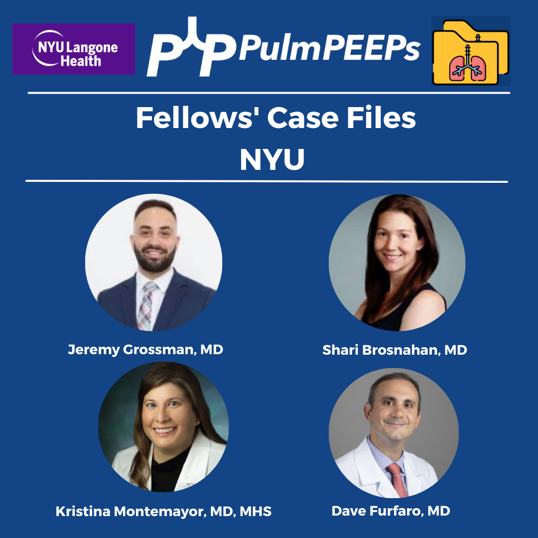
We are joined today by two amazing educators from NYU for our latest Fellows’ Case Files Episode. Listen today as we go through a great case with some fantastic teaching points highlighted throughout the episode.

Dr. Jeremy Grossman completed his Medicine-Pediatrics residency at Stony Brook Medicine where he was also a Chief Resident. He is currently a second-year PCCM fellow at NYU.

Dr. Shari Brosnahan is an Assistant Professor of Medicine and one of the Assistant Program Directors for the NYU Langone’s Division of Pulmonary, Critical Care, and Sleep Medicine. Her clinical and research interests are focused on pulmonary embolism and thrombosis in critically ill patients.

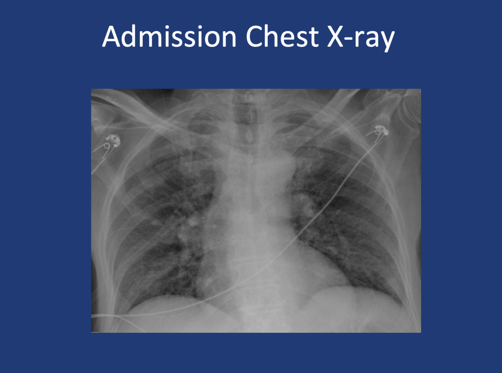
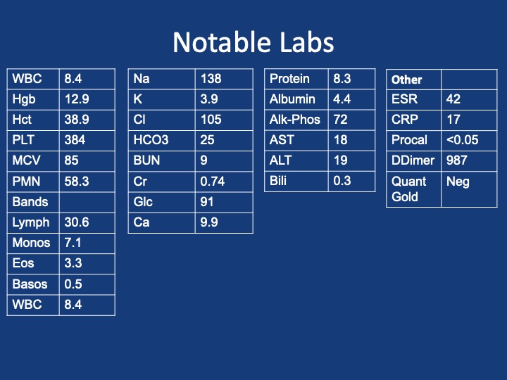
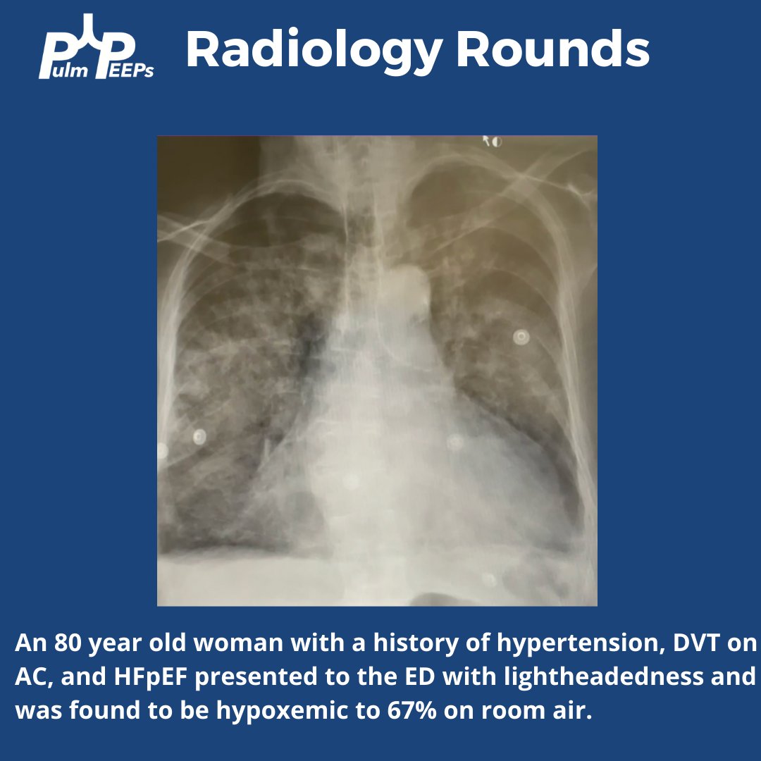
An 80-year-old male presents with shortness of breath. At home, his oxygen saturation was 82% on room air, improving only to 86% on 4L nasal cannula. Over the past month, he has experienced worsening symptoms, including a dry cough, fatigue, and difficulty speaking or ambulating due to shortness of breath at rest. He denies recent fever, sputum production, chest pain, or lower extremity swelling and presents to the ED for further evaluation.

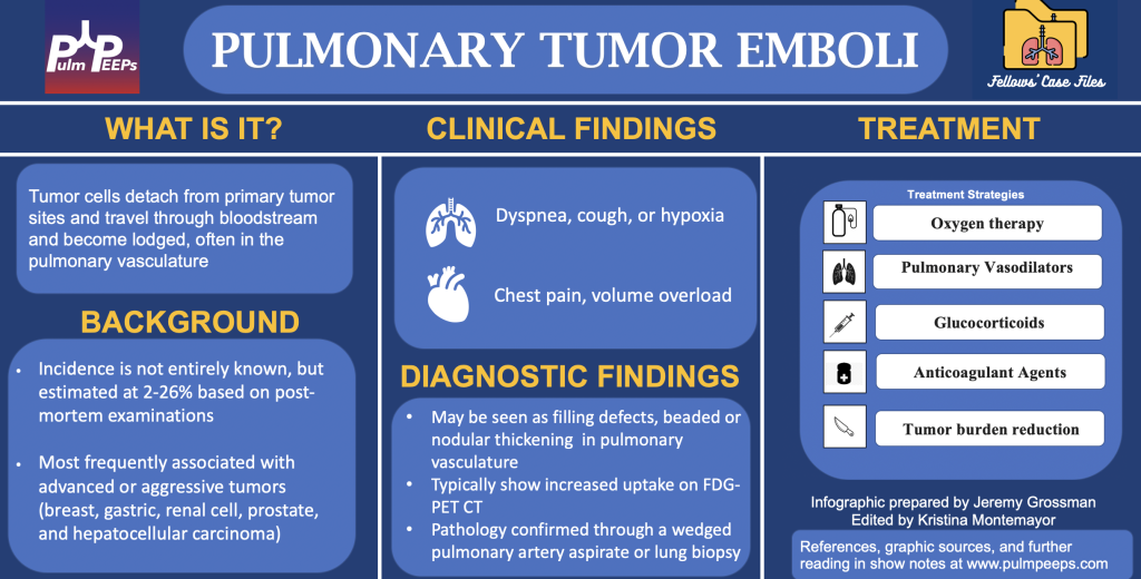
1.In any patient with a history of malignancy and hypoxia, clinicians should keep pulmonary tumor emboli (PTE) on the differential as early intervention may alter outcomes.

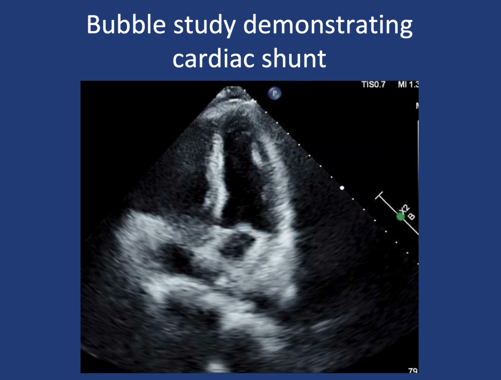
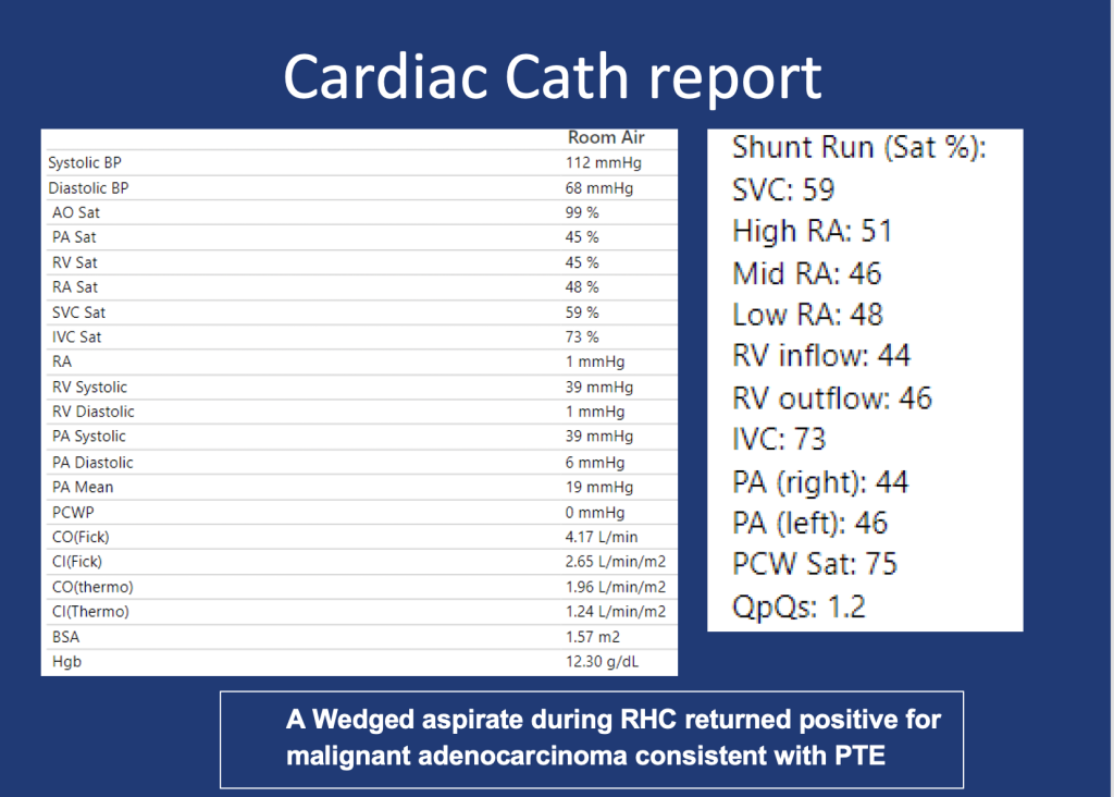
2.PTE contributes to hypoxia via mechanisms of mechanical obstruction of pulmonary arteries leading to shunting, VQ Mismatch, and in some cases pulmonary hypertension due to increased PVR.

3.A wedged aspirate can be used to diagnose PTE ante-mortem

75. Rapid Fire Journal Club 8 – STELLAR

Case Files

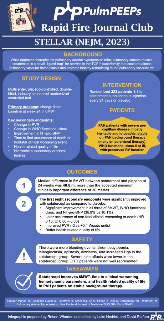
We’re back with our Rapid Fire Journal Club, and talking about the NEJM 2023 STELLAR Trial of Sotatercept in Pulmonary Arterial Hypertension. This is a landmark trial that is actively changing the face of PAH treatment today. Listen to hear the details of the trial and how its findings can be utilized to help patients.

Article and Reference

We’re looking at the STELLAR Trial today which is a Phase 3 trial of Sotatercept in Pulmonary Arterial Hypertension.

Reference: Hoeper MM, Badesch DB, Ghofrani HA, Gibbs JSR, Gomberg-Maitland M, McLaughlin VV, Preston IR, Souza R, Waxman AB, Grünig E, Kopeć G, Meyer G, Olsson KM, Rosenkranz S, Xu Y, Miller B, Fowler M, Butler J, Koglin J, de Oliveira Pena J, Humbert M; STELLAR Trial Investigators. Phase 3 Trial of Sotatercept for Treatment of Pulmonary Arterial Hypertension. N Engl J Med. 2023 Apr 20;388(16):1478-1490. doi: 10.1056/NEJMoa2213558. Epub 2023 Mar 6. PMID: 36877098.

Infographic

68. Fellows’ Case Files: Mount Sinai Morningside

Case Files

We’re back with another Case Files episode from Mt. Sinai Morningside. Listen in to hear another great case and some key learning points along the way.

Dr. Sara Luby is a third-year Internal Medicine resident and rising chief resident at Mt. Sinai Morningside/West and planning on applying to Pulmonary and Critical Care fellowship this upcoming year.

Dr. Javier Zulueta is the  Chief of the Division of Pulmonary, Critical Care, and Sleep Medicine at Mount Sinai Morningside. He completed residency training at St. Luke’s Medical Center/Case Western in Cleveland and fellowship in Pulmonary/Critical Care at Tufts Medical Center in Boston. His research focuses on lung cancer screening and incidental lung findings.

 Dr. Mirna Mohanraj is the Associate Program Director for the Pulmonary and Critical Care Fellowship at Mt. Sinai Morningside / Beth Israel and an associate professor of medicine and medical education at the Icahn School of Medicine at Mount Sinai. She completed residency training at University of Chicago and fellowship training at Mt. Sinai Hospital.

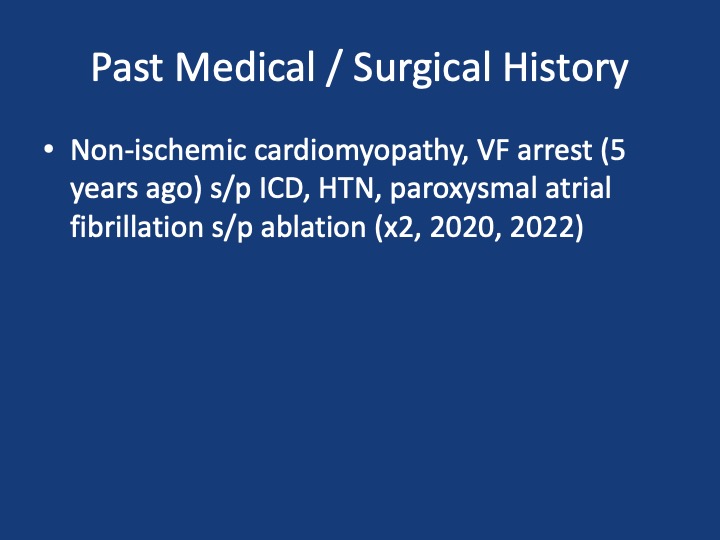
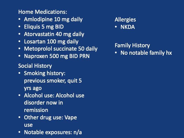
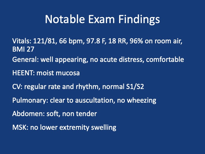
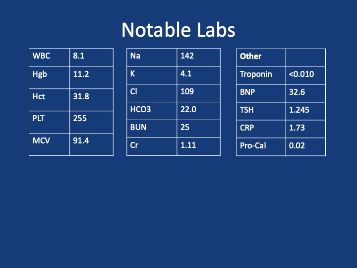
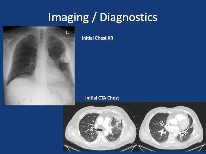
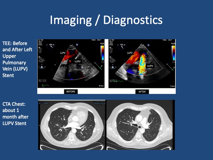
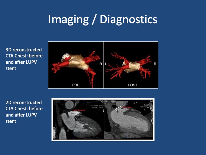
A 51 year old male presents with two days of acute on chronic chest pain and shortness of breath, worsening over the last month. His initial vitals: 143/ 100, pulse 85, temperature 36.5 °C (97.87°F), RR 16, SpO2 97 % on room air, BMI 29.8

Shroff N, Choi W, Villanueva-Meyer J, Palacio DM, Bhargava P. Pulmonary vein occlusion: A delayed complication following radiofrequency ablation for atrial fibrillation. Radiol Case Rep. 2021;16(12):3666-3671. doi:10.1016/j.radcr.2021.09.015 

Fender EA, Widmer RJ, Hodge DO, et al. Assessment and Management of Pulmonary Vein Occlusion After Atrial Fibrillation Ablation. JACC: Cardiovascular Interventions. Vol 11(16); 2018. doi:10.1016/j.jcin.2018.05.020 

López-Reyes R, García-Ortega A, Torrents A, et al. Pulmonary venous thrombosis secondary to radiofrequency ablation of the pulmonary veins. Respir Med Case Rep. 2018;23:46-48. doi:10.1016/j.rmcr.2017.11.008

Mizuno A, Mauler-Wittwer S, Muller H, Noble S. Recurrent pneumonia post atrial fibrillation ablation: do not forget to look for pulmonary vein stenosis. BMJ Case Rep. 2022;15(12):e250896. doi:10.1136/bcr-2022-250896

67. Fellows’ Case Files: Northwestern University

Case Files

Listen in today to another stop on our Fellows’ Case Files journey. We’re at Northwestern University for another great case presentation. Tune in, check out our associated infographic, and let us know what you think!

Meet Our Guests

Jamie Rowell is a first-year clinical fellow in the Northwestern PCCM program. She completed medical school at the Medical University of South Carolina and her internal medicine residency and Chief Residency at the University of Vermont Medical Center.

Cathy Gao is an Instructor of Medicine at Northwestern and completed her PCCM fellowship there last year. Her research focuses on using machine learning applied to ICU EHR data to characterize patient trajectories and identify potential interventions to improve outcomes.

Clara Schroedl is an Associate Professor of Medicine in Pulmonary and Critical Care and Medical Education. She is the program director of the Northwestern PCCM fellowship program, with an interest in medical education and simulation.

Case Presentation

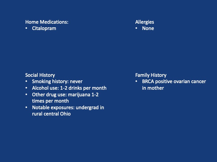
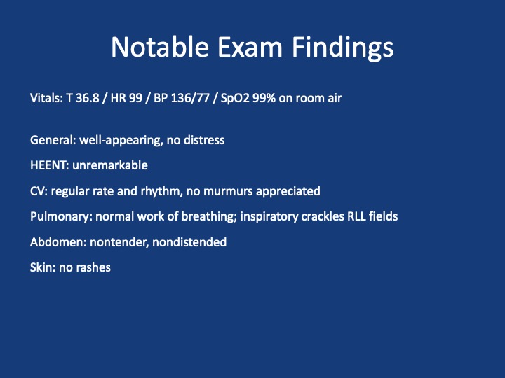
A 25-year-old previously healthy woman presents with recurrent episodes of right chest pain and cough. In October she was treated with antibiotics and felt somewhat better but in December, she presented again with chest pain, and again was treated with antibiotics. The pain improved but she still felt breathless. In February, again she had intense chest pain interfering with life, and was given NSAIDs and took high dose TID without clear benefit.

One month later, she coughed up some bloody mucus, so now she is presenting for evaluation. The chest pain is worse with deep breaths and improves in between these episodes. She only notes it on her right side. At this point, she does sometimes feel short of breath; she used to run 5 miles but is now struggling to run two miles. She denies any unusual exposures. She went to school in central rural Ohio for a while. She has no history of pulmonary infections, no exposure to mold or animals, and no history of vaping.

Key Learning Points

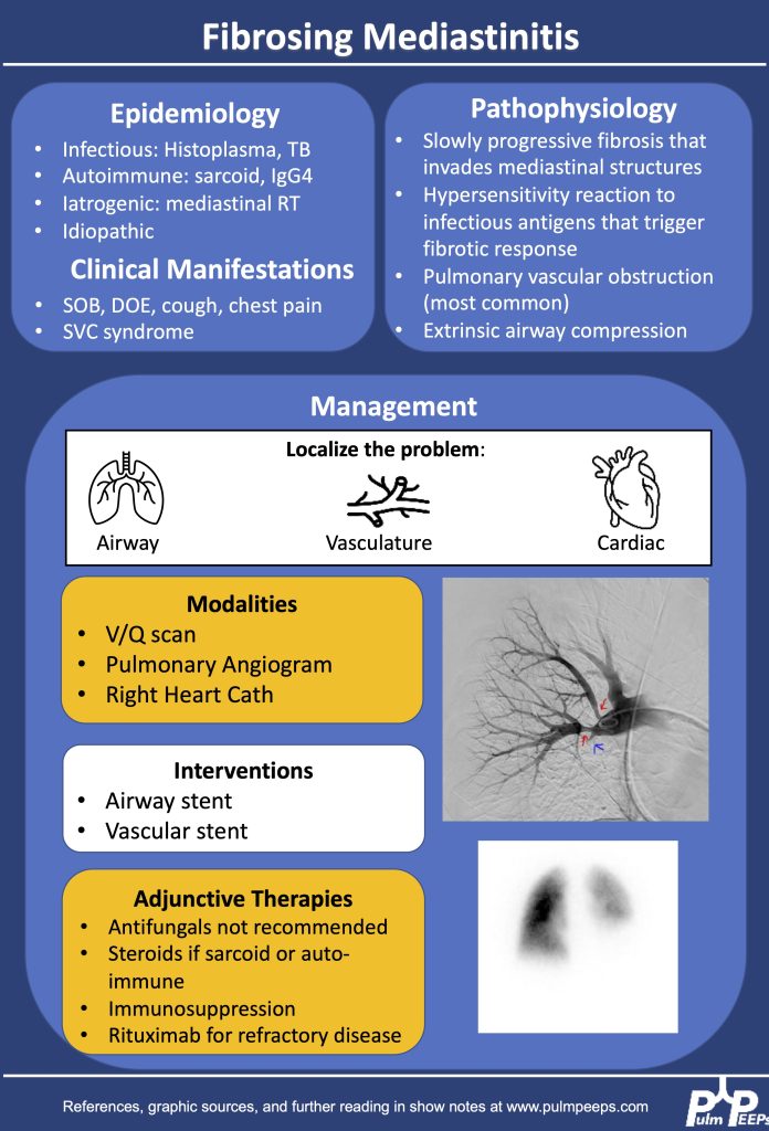
1.Making the diagnosis of Fibrosing Mediastinitis :

–Etiologies: histoplasmosis, sarcoidosis, tuberculosis, IgG4, Behcet, ANCA vasculitis

–Imaging modalities: CT chest, perfusion studies, pulmonary angiogram

–Imaging characteristics:  infiltrative, heterogeneous, fibrotic process that crosses fat planes and encroaches on nearby structures causing airway or vascular stenoses  

2. Management strategies:

–No curative therapies. Goal to relieve symptom burden

–Airway stents

–Vascular stents

–Rituximab

–Antifungals, steroids generally not considered effective

References and Further Reading

Kern et al. Bronchoscopic Management of Airway Compression due to Fibrosing Mediastinitis. Annals of the American Thoracic Society 2017. 14: 1235-1359 

Welby JP, Fender EA, Peikert T, Holmes DR Jr, Bjarnason H, Knavel-Koepsel EM. Evaluation of Outcomes Following Pulmonary Artery Stenting in Fibrosing Mediastinitis. Cardiovasc Intervent Radiol. 2021 Mar;44(3):384-391. doi: 10.1007/s00270-020-02714-z. Epub 2020 Nov 17. PMID: 33205295.

Westerly, BD Targeting B Lymphocytes in Progressive Fibrosing Mediastinitis. Am J Respir Crit Care Med. 2014 Nov 1; 190(9): 1069–1071.

https://rarediseases.org/rare-diseases/fibrosing-mediastinitis/#complete-report

https://pubmed.ncbi.nlm.nih.gov/21422386/

https://academic.oup.com/cid/article/30/4/688/421789

https://pubmed.ncbi.nlm.nih.gov/22033450/

https://www.sciencedirect.com/science/article/pii/S2352906715300087

https://www.atsjournals.org/doi/pdf/10.1513/AnnalsATS.201610-782RL

59. Top Consults: Lung Transplant 101

Case Files

We’re back with our Top Consults series to talk about Lung Transplant! This is a topic that every pulmonologist should have background knowledge about since it impacts the care of patients with end-stage lung disease of any cause. We will talk about the indications for referral and transplant, how to advise patients and some unique considerations for evaluation. Enjoy, rate and review us, and share your thoughts about the episode!

Meet Our Guests

Dr. Meghan Aversa is an Assistant Professor of Medicine at the University of Toronto and her expertise involves patients with end stage lung disease and lung transplant.

Dr. Hannah Mannem is an Associate Professor of Medicine at the University of Virginia Health. Hannah joined faculty at UVA in 2016 and she has expertise in ILD and Lung Transplant.

Learning Points

Trends in lung transplant:

  1. Global Increase in Lung Transplants: Over the past three decades, there has been a gradual worldwide increase in lung transplants, with approximately 4,500 performed annually. North America conducts over half of these transplants, and the growth is particularly notable in double lung transplants.
  2. Indications and Disease Trends: Interstitial lung disease (ILD) has seen a significant rise in lung transplant indications, surpassing COPD as the leading cause. ILD, especially idiopathic pulmonary fibrosis (IPF), constitutes a substantial portion (40%) of all transplants. However, the trend is primarily observed in North America.
  3. Decline in Cystic Fibrosis Cases: While Cystic Fibrosis is still a significant indication for lung transplant, its percentage has been declining, likely due to improvements in drugs and CFTR modulators.
  4. Evolution of Lung Transplant Candidates: Over the past five years, lung transplant candidates have become sicker, with higher listing scores and increased hospitalization rates at the time of transplant. More patients have antibodies affecting match difficulty. The average age of patients has increased, with 35% being over 65, a demographic that was previously considered contraindicated.
  5. Impact of COVID-19: The COVID-19 pandemic has influenced lung transplant trends. In 2020, UNOS added COVID-19-related ARDS and pulmonary fibrosis as indications. In 2021, these indications constituted about 10% of lung transplants, making it the third most common indication. Two-thirds were due to COVID-19 ARDS, and one-third due to pulmonary fibrosis. The long-term impact, especially with evolving vaccine dynamics, is still uncertain.

Indications for transplant referral:

  1. ISHLT Consensus Document Update (2021): The ISHLT consensus document for lung transplant candidate selection was updated in 2021. It is available on the ISHLT website and serves as a valuable guideline for pulmonologists considering referrals for lung transplant assessment.
  2. General Rule of Thumb for Chronic Lung Diseases: According to the consensus document, a general rule of thumb for all patients with chronic and stage lung diseases is to consider lung transplant if there is a high (more than 50%) risk of death from the lung disease within the next two years. Prognostic markers vary based on the underlying lung disease.
  3. Disease-Specific Recommendations: The consensus document provides disease-specific recommendations. The key diseases highlighted are COPD, ILD, CF, and PH.
    • COPD: Referral is recommended when the BODE index is in the range of 5 to 6, with additional factors that increase mortality, such as frequent exacerbations, low FEV1 (20-25%), or rapidly increasing BODE. Referral is also advised for clinically deteriorating patients or those with an unacceptably low quality of life despite maximal medical therapy.
    • ILD (Particularly IPF): Early referral is suggested, ideally at the time of diagnosis. For any pulmonary fibrosis, referral is recommended if FEC is less than 80% or declining by 10% in two years, or DLCO is less than 40% or declining by 15% in two years. Other factors for referral include radiographic progression or a need for supplemental oxygen.
    • Cystic Fibrosis (CF): Referral is encouraged for those with FEV1 less than 30%, and even 40% if there’s reduced walk distance, hypercapnia, PH, frequent exacerbations, or rapid decline.
    • Pulmonary Hypertension (PH): Referral criteria include a REVEAL score of eight, significant RV dysfunction, progressive disease on therapy, need for IV prostacyclin therapy, and specific conditions like PVOD, PCH, scleroderma pulmonary artery aneurysms, which should be referred early due to their rapid progression.

Transplant evaluation process

  1. Phases of Lung Transplant Evaluation:
    • Referral and Initial Visit: The process begins with a referral, often from a primary pulmonologist. Patients can also self-refer. The initial phase involves insurance authorization and confirming the underlying diagnosis while ensuring all other treatment options are exhausted.
    • Assessment of Disease Severity: The severity of end-stage lung disease is assessed to determine the timing of the workup, which varies depending on the patient’s condition and the center’s protocols.
    • Diagnostic Steps: A thorough diagnostic workup follows the initial visit, including various tests, imaging, and meetings with multidisciplinary teams to assess medical and social factors influencing transplant success.
    • Follow-Up Appointments: Patients typically have multiple follow-up appointments to track the evolution of the disease and ensure health maintenance and vaccinations are up to date.
    • Selection Committee: The final phase involves a selection committee that determines if the patient is a candidate. If so, there may be conditional requirements before officially listing the patient.
  2. Multidisciplinary Approach: Lung transplant evaluation involves collaboration with various specialists, including social work, finance, nutrition, pharmacy, physical therapy, and potentially other consult services. The efficiency of this process is optimized for both the patient and the medical team.
  3. Diagnostic Workup:
    • Medical Testing: Involves blood work, cardiac testing (echo, left and right heart cath), and imaging, including abdominal imaging, VQ scans, DEXA scans, and 24-hour urine analysis.
    • Multidisciplinary Meetings: Patients meet with members of the multidisciplinary team, addressing medical comorbidities as well as social and psychological factors.
    • Follow-Up Appointments: Multiple appointments allow for tracking disease progression and ensuring overall health maintenance.
  4. Selection Committee Decision: The patient receives a decision from the selection committee, determining candidacy. Sometimes, patients are considered candidates with conditions (e.g., completing vaccinations or losing weight). Timing of listing is also discussed to ensure optimal candidacy.
  5. Patient Involvement: Patients play an active role, and the process may involve self-referral, understanding and completing requirements, and active participation in follow-up appointments.
  6. Efficiency and Individualization: The evaluation process is tailored to the patient’s condition, and centers aim to efficiently organize diagnostic workup and multidisciplinary meetings to optimize patient care.

Timing of transplant listing for candidates

  • COPD Patients: For COPD patients, listing is likely when the Bode index is around 7, the FEV1 is under 20%, there is at least moderate pulmonary hypertension (PH), chronic hypercapnia, or severe exacerbations.
  • ILD Patients: Patients with interstitial lung disease (ILD) are likely to be listed when showing signs of progression or decline in forced expiratory capacity (FEC), diffusing capacity of the lungs for carbon monoxide (DLCO), or six-minute walk distance. Other indicators include hypoxemia, secondary pulmonary hypertension, or hospitalization for complications.
  • CF Patients: Cystic fibrosis (CF) patients are considered for listing when FEV1 is below 25% or is rapidly declining, and if they experience frequent hospitalizations. Listing criteria also include the presence of pulmonary hypertension, chronic hypoxemia, or hypercapnia.
  • Pulmonary Hypertension Patients: Those with primary pulmonary hypertension may be listed when the reveal score is above 10 on intravenous therapy, there is progressive hypoxemia, or if there are renal or liver dysfunctions associated with pulmonary hypertension (PH).

Changes from the LAS system to the CAS system

  1. Transition to Composite Allocation Score (CAS):
    • Background and Timing: In March 2023, the lung allocation system (LAS) transitioned to the composite allocation score (CAS), a major change in the allocation of lung transplants.
    • Reasoning Behind the Change: The change aimed to improve organ matching, prioritize sick candidates, enhance long-term survival, promote equity, increase transplant opportunities for specific patient groups (especially pediatric patients), and manage geographical variation in organ placement.
    • Components of CAS:
      • Medical Urgency: Based on waitlist mortality at one year without a transplant and the likelihood of survival post-transplant, now assessed at greater than five years, with equal weighting.
      • Recipient Variables: Includes factors like height discrepancy, blood type matching, sensitization (immune system matching), and other recipient variations.
      • Candidate Biology: Focuses on pediatric patients (less than 18 years old) and individuals are a prior living donor.
      • Donor Variables: Addresses donor characteristics, emphasizing proximity and travel distance from the organ hospital.
    • Early Data and Observations: The initial three-month monitoring period has shown changes in O blood type scores, prompting adjustments. Notable outcomes include a 16% increase in the number of lung transplants, a decrease in waitlist deaths and removals, and changes in median distance between donor hospital and transplant center.
    • Exception Scores: The number of exception scores has increased, allowing for adjustments when the assigned score may not reflect the patient’s true medical urgency.
    • Caution and Early Analysis: Early data, while promising, is subject to caution as centers were aware of the upcoming change. The impact on different age groups and the reasons for exceptions are being closely monitored and may evolve as more data becomes available.
  2. Ongoing Monitoring and Potential Evolution: The data is being closely tracked by medical directors, and further changes to the scoring system may occur based on ongoing analysis and experience with the CAS. The impact on patient outcomes and allocation efficiency will continue to be studied and refined.

Advising patients on what to expect in terms of prognosis and survival after lung transplant

  1. Survival Statistics:
    • Overall three is approximately 50 percent survival at five years, and the median survival time is approximately six and a half years.
    • Significant variations based on factors such as diagnosis, age, and comorbidities.
    • Survival outcomes differ for specific groups, e.g., cystic fibrosis (CF) patients, those older than 65, and individuals with interstitial lung disease (ILD).
  2. Quality of Life Emphasis:
    • Shift in focus from survival alone to the patient’s goals and quality of life.
    • Highlighting the importance of understanding and aligning with the patient’s individual quality of life expectations.
  3. Investment in Healthcare Team and Lifestyle Change:
    • Emphasis on the long-term commitment and involvement with the healthcare team post-transplant.
    • A substantial investment in healthcare post-transplant, including regular visits, extensive blood work, and medication management.
    • Cultural shift for patients to adapt to a new routine of frequent medical visits even when otherwise healthy.
  4. Complications and Side Effects:
    • Acknowledgment of potential complications within the first year, making the initial post-transplant period a full-time job.
    • Discussion of various complications and medication side effects, ensuring patients are informed.
    • Multidisciplinary approach involving nutritionists, physical therapists, and other specialists to address complications and enhance the patient’s quality of life.
  5. Individualized Patient Approach:
    • Recognition of the patient’s fight, spirit, and motivation as crucial factors for successful transplantation.
    • Encouraging patients to set goals for their post-transplant life.
    • Ethical considerations regarding transplanting older patients, with the importance of assessing overall well-being, motivation, and mental health.
  6. Acknowledgment of Averages and Unpredictability:
    • Communication of averages, but a reminder of the inherent unpredictability in the post-transplant course.
    • Preparing patients for potential complications and the need to adapt to unforeseen challenges.
    • Managing expectations by highlighting the unpredictability of individual transplant journeys.
  7. Quality of Life Improvement:
    • Despite complications and side effects, lung transplant often results in a significant improvement in the patient’s quality of life.
    • Patients generally experience increased satisfaction and happiness post-transplant, outweighing the challenges associated with the procedure and subsequent care.

References for further reading

  1. Leard LE, Holm AM, Valapour M, Glanville AR, Attawar S, Aversa M, Campos SV, Christon LM, Cypel M, Dellgren G, Hartwig MG, Kapnadak SG, Kolaitis NA, Kotloff RM, Patterson CM, Shlobin OA, Smith PJ, Solé A, Solomon M, Weill D, Wijsenbeek MS, Willemse BWM, Arcasoy SM, Ramos KJ. Consensus document for the selection of lung transplant candidates: An update from the International Society for Heart and Lung Transplantation. J Heart Lung Transplant. 2021 Nov;40(11):1349-1379. doi: 10.1016/j.healun.2021.07.005. Epub 2021 Jul 24. PMID: 34419372; PMCID: PMC8979471.
  2. van der Mark SC, Hoek RAS, Hellemons ME. Developments in lung transplantation over the past decade. Eur Respir Rev. 2020 Jul 21;29(157):190132. doi: 10.1183/16000617.0132-2019. PMID: 32699023; PMCID: PMC9489139.
  3. Valapour M, Lehr CJ, Wey A, Skeans MA, Miller J, Lease ED. Expected effect of the lung Composite Allocation Score system on US lung transplantation. Am J Transplant. 2022 Dec;22(12):2971-2980. doi: 10.1111/ajt.17160. Epub 2022 Aug 9. PMID: 35870119.
  4. Arcasoy SM, Kotloff RM. Lung transplantation. N Engl J Med. 1999 Apr 8;340(14):1081-91. doi: 10.1056/NEJM199904083401406. PMID: 10194239.