122. Pulm PEEPs Pearls: Steroids in Sepsis

Today we have another Pulm PEEPs Pearls episode about a core critical care topic. Furf and Monty will be giving a high level overview of the use of steroids in sepsis including a review of the relevant literature and recent guidelines, and pragmatic bedside points.

This episode was prepared with research by Pulm PEEPs Associate Editor George Doumat.

Dustin Latimer, another Pulm PEEPs Associate Editor, assisted with audio and video editing.

Why Steroids in Sepsis?

Steroids do not treat the infection — antimicrobials are always first and remain the cornerstone. The goal is addressing critical illness–related corticosteroid insufficiency (CIRCI), where cortisol production cannot keep up with the overwhelming inflammatory demand of septic shock.

Hydrocortisone helps in two main ways:

  • Blunts the dysregulated inflammatory response — tempers the excessive vasodilation and febrile response that drive harm beyond the infection itself.
  • Restores vascular sensitivity to catecholamines — sepsis downregulates adrenergic receptors; steroids turn that responsiveness back on.

Clinical takeaway: The first thing you notice is vasopressor weaning (or a bend in the escalation curve) — not a rapid improvement in fever or white count.

Caveat: These trials predate modern sepsis phenotyping. None distinguish hyperinflammatory vs. hypoinflammatory responders — they treat all comers.

The Evidence: Four Landmark Trials

Every IM resident and critical care fellow will eventually journal-club these four. The most consistent signal across all of them is faster shock reversal and reduced vasopressor use; the mortality question remains unsettled.

Trial (Year)NRegimenKey Finding
Annane (2002)~300Hydrocortisone + fludrocortisoneMortality benefit in ACTH non-responders; criticized methodology and messy cortisol-response testing; not cleanly replicated.
CORTICUS (2008)~500Hydrocortisone aloneFaster shock reversal but no mortality benefit, regardless of cortisol responsiveness. Raised (later allayed) superinfection concern. Cornerstone for abandoning routine cort-stim testing.
ADRENAL (2018)~3,800Hydrocortisone aloneFaster vasopressor weaning; no 90-day mortality benefit.
APROCCHSS (2018)~1,200Hydrocortisone + fludrocortisoneMortality benefit at 90 days.

Bottom line: Faster shock reversal is consistent. Mortality benefit appears in 2 of 4 trials (both used fludrocortisone) but not the others. A 2026 meta-analysis showed benefit for hydrocortisone + fludrocortisone vs. placebo, but

not for hydrocortisone + fludrocortisone vs. hydrocortisone alone — suggesting hydrocortisone drives the main effect.

Who Gets Steroids, and When?

  • 2021 Surviving Sepsis: Consider steroids for norepinephrine or epinephrine ≥ 0.25 mcg/kg/min for ≥ 4 hours despite adequate resuscitation — a reasonable bedside trigger.
  • Early 2026 update: Moved away from a specific numeric trigger — consider steroids when a septic patient is not responding well to vasopressors or has escalating requirements. Make a clinical decision. (Quality of evidence: low to moderate.)
  • Go faster than the threshold when: Known/suspected adrenal insufficiency or home steroids, or florid pressor-requiring shock on arrival.

A practical escalation sequence: escalating norepinephrine → add vasopressin (per VASST) → then add steroids if requirements keep climbing.

Do NOT wait for an ACTH stimulation test. It does not reliably predict who responds and only delays treatment. Sepsis is an elevated-cortisol state but can dissociate ACTH and cortisol, and cortisol-binding globulin is depleted — the test is too messy to guide care.

What to Give: The Regimen

  • Standard dose: Hydrocortisone 200 mg/day, typically 50 mg IV Q6H. (Original trials often used continuous infusions, rarely used in the U.S.) Some start with a 100 mg bolus to gain control.
  • Higher dose: If chronically on steroids / adrenally insufficient, consider ~300 mg/day (e.g., 100 mg Q8H).
  • Fludrocortisone: Unsettled. The two mortality-benefit trials added it (50 mcg PO/NG/OG daily), but hydrocortisone already has mineralocorticoid activity and meta-analyses don’t show added benefit over hydrocortisone alone. Most clinicians omit it — adding it is reasonable and safe, just be honest about the uncertainty.

Duration & Tapering

  • Typical course: ~7 days is most common. Trial practices varied (ADRENAL ~7 days; VANISH used a taper after 6 days; some continue until pressors are off).
  • No taper needed. You do not need to taper for adrenal insufficiency after a short course — just stop. If pressors dramatically rebound, you can restart, but most patients have gained the benefit they’ll get by day 7.

Pitfalls & Safety

  • Hyperglycemia: Expected and must be managed (monitor closely; insulin drip if needed). No signal for major DKA / severe complications in the trials.
  • Superinfection / fungal infection: The most-quoted concern, but the overall literature does not show a convincing, statistically significant increase. Be disciplined about stopping on schedule.
  • Muscle weakness: Steroids can worsen critical illness myopathy; a short 7-day course likely has limited effect, but be aware.
  • Other: GI bleeding (follow general PPI prophylaxis guidance) and sodium disturbances (watch for hyper-/hyponatremia).

Two things we know: (1) steroids shorten duration of vasopressor support, and (2) they are relatively safe in sepsis. Whether they improve mortality — and in whom — remains open.

The Five Pulm PEEPs Pearls

  1. Mechanism: Steroids restore catecholamine vascular sensitivity and blunt dysregulated inflammation. The clinical target is vasopressor weaning, not infection treatment.
  2. Evidence: Faster shock reversal is the most consistent finding. Mortality benefit is seen in 2 of 4 trials but not the others — still controversial. Some patients likely benefit; we don’t yet know who.
  3. Trigger: A practical 2021 threshold is levo/epi ≥ 0.25 mcg/kg/min for ≥ 4 hours. Newer guidance drops the strict number — make a clinical decision based on poor pressor response or escalation.
  4. Dose: Hydrocortisone 200 mg/day (e.g., 50 mg Q6H). Adding fludrocortisone mirrors two trials, but meta-analyses find no benefit over hydrocortisone alone.
  5. Safety: Steroids appear safe in sepsis. Monitor and treat hyperglycemia; no marked increase in superinfection.

Annane, Djillali et al. “Effect of treatment with low doses of hydrocortisone and fludrocortisone on mortality in patients with septic shock.” JAMA vol. 288,7 (2002): 862-71. doi:10.1001/jama.288.7.862

Sprung, Charles L et al. “Hydrocortisone therapy for patients with septic shock.” The New England journal of medicine vol. 358,2 (2008): 111-24. doi:10.1056/NEJMoa071366

Venkatesh, Balasubramanian et al. “Adjunctive Glucocorticoid Therapy in Patients with Septic Shock.” The New England journal of medicine vol. 378,9 (2018): 797-808. doi:10.1056/NEJMoa1705835

Annane, Djillali et al. “Hydrocortisone plus Fludrocortisone for Adults with Septic Shock.” The New England journal of medicine vol. 378,9 (2018): 809-818. doi:10.1056/NEJMoa1705716

Sun, Alin et al. “Correction: Hydrocortisone combined with fludrocortisone for treatment of adults with septic shock: an updated meta-analysis and systematic review.” Frontiers in medicine vol. 13 1811616. 2 Mar. 2026, doi:10.3389/fmed.2026.1811616

Prescott, Hallie C et al. “Executive Summary: Surviving Sepsis Campaign: International Guidelines for Management of Sepsis and Septic Shock 2026.” Critical care medicine vol. 54,4 (2026): 715-724. doi:10.1097/CCM.0000000000007089

118. Pulm PEEPs Pearls: Methacholine Challenge

Furf and Monty are back with another Pulm PEEPs Pearls episode. The topic of today’s discussion is an often discussed, but often misunderstood, test; the methacholine challenge. They’ll review when to utilize this test, how it should be performed, and the appropriate interpretation.

This episode was prepared with research by Pulm PEEPs Associate Editor George Doumat.

Dustin Latimer, another Pulm PEEPs Associate Editor, assisted with audio and video editing.

What the Test Measures

  • Methacholine challenge is a direct bronchial provocation test of airway hyperresponsiveness (AHR), a core physiologic feature of asthma.
  • Anyone will bronchoconstrict at high enough concentrations — the test looks for an abnormal threshold.
  • The key endpoint is the PC20: the methacholine concentration causing a 20% fall in FEV1.
    • Abnormal in adults: PC20 ≤ 8–16 mg/mL

Test Performance

  • Meta-analyses: pooled sensitivity ~60%, specificity ~90%.
  • Real-world cohorts: sensitivity 55–62%, specificity 56–100% (varies by population, protocol, and threshold used).
  • Not a standalone yes/no test — best used as part of a broader diagnostic pathway.

Where It Fits in the Asthma Workup

The test belongs in a stepwise approach:

  1. Step 1: Spirometry + bronchodilator response
  2. Step 2: Add FeNO and/or peak flow variability (if available)
  3. Step 3: If the picture is still unclear → methacholine challenge

It is most useful for symptomatic patients with normal spirometry and no bronchodilator reversibility. Given its cost, mild risk, and discomfort, it should not be a first-line test — most asthma diagnoses do not require it.

Technique and Medication Prep

Technique

  • ERS guidelines favor tidal breathing over deep inspiratory maneuvers.
  • Deep breaths can be bronchoprotective and blunt the response, reducing sensitivity — especially in mild or well-controlled asthma.

Medication Washout (to Avoid False Negatives)

Medication ClassWashout Period
Short-acting beta-agonists (SABA)≥ 6 hours
Long-acting beta-agonists (LABA)~24 hours
Ultra-long-acting beta-agonists~48 hours
Short-acting anticholinergics (e.g., ipratropium)~12 hours
Long-acting muscarinic antagonists (LAMA, e.g., tiotropium)7 days
  • Inhaled corticosteroids, leukotriene blockers, and antihistamines do not significantly affect the test acutely — continue these. Withdrawing ICS also carries its own risk for asthma patients.
  • Practical tip: Spell out exactly what to hold and when — for both the patient and the PFT lab — at the time the test is ordered.

Interpreting Results

Negative Test (PC20 > 16 mg/mL)

  • Very high negative predictive value in symptomatic adults.
  • Makes current asthma quite unlikely (assuming proper test conduct).
  • This is the test’s greatest strength: it is an excellent rule-out test.

Positive Test (PC20 ≤ 8–16 mg/mL)

  • More nuanced — airway hyperresponsiveness is not unique to asthma.
  • Can be positive in: chronic cough, allergic rhinitis, COPD, and even some healthy asymptomatic individuals.
  • A positive result raises probability but must be interpreted alongside the clinical story, variable respiratory symptoms, peak flow variability, FeNO, and ICS response.

Safety and Risks

  • Overall, the test is quite safe; significant adverse effects are rare.
  • Temporary breathing discomfort is expected (bronchoconstriction is being induced).
  • Severe bronchospasm is possible:
    • A trained clinician should be available; SABA inhaler/nebulizer must be immediately on hand; a physician should be reachable in the facility.
  • Contraindications / cautions:
    • Avoid if FEV1 < 70% predicted or < 1–1.5 L (baseline obstruction greatly increases risk).
    • Avoid within 3 months of an acute cardiac event (rare risk of cardiac events with unstable cardiac disease).

Five Pearls — Quick Recap

  1. What it tests: Methacholine challenge is a direct test of AHR with high specificity but variable sensitivity — it belongs inside a diagnostic pathway, not as a standalone asthma test.
  2. When to use it: Most useful for symptomatic patients with normal spirometry and no bronchodilator response, after FeNO and peak flow variability have been considered.
  3. Technique and meds matter: Use tidal breathing protocol; respect washout intervals — especially the 7-day LAMA washout and 24–48 hour LABA window — to avoid false negatives.
  4. Safety: Generally safe, but can induce significant bronchoconstriction. Have a SABA available and avoid the test in patients with FEV1 < 70% predicted.
  5. Interpretation: A negative test (PC20 > 16 mg/mL) strongly argues against current asthma. A positive test raises probability but is not specific — interpret alongside the full clinical picture.
  1. Coates AL, Wanger J, Cockcroft DW, Culver BH; Bronchoprovocation Testing Task Force: Kai-Håkon Carlsen; Diamant Z, Gauvreau G, Hall GL, Hallstrand TS, Horvath I, de Jongh FHC, Joos G, Kaminsky DA, Laube BL, Leuppi JD, Sterk PJ. ERS technical standard on bronchial challenge testing: general considerations and performance of methacholine challenge tests. Eur Respir J. 2017 May 1;49(5):1601526. doi: 10.1183/13993003.01526-2016. PMID: 28461290.
  2. Lee, J., & Song, J. U. (2021). Diagnostic comparison of methacholine and mannitol bronchial challenge tests for identifying bronchial hyperresponsiveness in asthma: a systematic review and meta-analysis. Journal of Asthma58(7), 883–891. https://doi.org/10.1080/02770903.2020.1739704
  3. Davis BE, Blais CM, Cockcroft DW. Methacholine challenge testing: comparative pharmacology. J Asthma Allergy. 2018 May 14;11:89-99. doi: 10.2147/JAA.S160607. PMID: 29785128; PMCID: PMC5957064.

117. Pulm PEEPs Pearls: Spontaneous Breathing Trials

This week’s Pulm PEEPs Pearls episode is all about spontaneous breathing trials (SBTs). SBTs are a standard part of the daily practice in the intensive care unit, but the exact methods vary across ICUs and institutions. Listen in to hear about the most common methods of SBTs, the physiology of each method, and what the evidence says.

This episode was prepared with research by Pulm PEEPs Associate Editor George Doumat.

Dustin Latimer, another Pulm PEEPs Associate Editor, assisted with audio and video editing.

  1. What an SBT is really testing
  • An SBT is a stress test for post-extubation work of breathing, not just a ventilator check.
  • The goal is to balance sensitivity and specificity:
    • Too hard → unnecessary failures and delayed extubation
    • Too easy → false positives and higher risk of reintubation
  1. Common SBT modalities and how they compare
  • T-piece
    • No inspiratory support and no PEEP
    • Highest work of breathing
    • Most “physiologic” but often too strict
  • Pressure support (PS) + PEEP (e.g., 5/5 or 8/5)
    • Offsets ETT resistance and provides modest assistance
    • Easier to pass than T-piece
  • CPAP (0/5)
    • No inspiratory help, but provides PEEP to counter ETT resistance
    • Sits between PS and T-piece in difficulty
  1. Evidence favors pressure-supported SBTs for most patients
  • Large meta-analysis (~6,000 patients, >40 RCTs):
    • Pressure-supported SBTs increase successful extubation (~7% absolute benefit)
    • No increase in reintubation rates
  • Trials (e.g., FAST trial):
    • Patients pass SBTs earlier
    • Leads to earlier extubation and fewer ventilator-associated risks
  • Bottom line: A 30-minute PS 5/5 SBT is evidence-based and appropriate for most stable ICU patients
  1. When a T-piece still makes sense

T-piece SBTs are useful when:

  • Cost of reintubation is high
    • Difficult airway
    • Prior failed extubation
  • Pretest probability of success is low
    • Prolonged or difficult weaning
    • Tracheostomy vs extubation decisions
  • Need to mimic physiology without positive pressure
    • In LV dysfunction or pulmonary edema even small amounts PEEP may significantly improve physiology
  • Some centers use a hybrid approach: PS SBT → short confirmatory T-piece before extubation
  1. CPAP as a middle ground
  • Rationale:
    • Allows full patient effort while compensating for ETT resistance
  • Evidence:
    • Fewer and smaller trials
    • Possible modest improvement in extubation success
    • No clear mortality or LOS benefit
  • Reasonable option based on patient physiology, institutional protocols, and clinician comfort
  1. No single “perfect” SBT mode
  • Across PS, T-piece, CPAP, and newer methods (e.g., high-flow via ETT) there are no consistent differences in mortality or length of stay
  • What matters most:
    • Daily protocolized screening
    • Thoughtful bedside clinical judgment
    • Matching SBT difficulty to patient-specific risk
  1. Institutional variation is normal—and acceptable
  • Examples:
    • PS 10/5 in postoperative surgical ICU patients
    • PS 5/0 as an intermediate difficulty option
  • Key question clinicians should ask: What does passing or failing this specific SBT tell me about this patient’s likelihood of post-extubation success?
  1. Take-home pearls
  1. SBTs are stress tests of post-extubation physiology.
  2. PS 5/5 for 30 minutes is a strong default for most ICU patients.
  3. T-piece trials are valuable when false positives are costly or physiology demands it.
  4. CPAP is reasonable but supported by less robust data.
  5. Consistency, daily screening, and judgment matter more than the exact mode.

  • Burns KEA, Khan J, Phoophiboon V, Trivedi V, Gomez-Builes JC, Giammarioli B, Lewis K, Chaudhuri D, Desai K, Friedrich JO. Spontaneous Breathing Trial Techniques for Extubating Adults and Children Who Are Critically Ill: A Systematic Review and Meta-Analysis. JAMA Netw Open. 2024 Feb 5;7(2):e2356794. doi: 10.1001/jamanetworkopen.2023.56794. PMID: 38393729; PMCID: PMC10891471.
  • Burns KEA, Sadeghirad B, Ghadimi M, Khan J, Phoophiboon V, Trivedi V, Gomez Builes C, Giammarioli B, Lewis K, Chaudhuri D, Desai K, Friedrich JO. Comparative effectiveness of alternative spontaneous breathing trial techniques: a systematic review and network meta-analysis of randomized trials. Crit Care. 2024 Jun 8;28(1):194. doi: 10.1186/s13054-024-04958-4. PMID: 38849936; PMCID: PMC11162018.
  • Subirà C, Hernández G, Vázquez A, Rodríguez-García R, González-Castro A, García C, Rubio O, Ventura L, López A, de la Torre MC, Keough E, Arauzo V, Hermosa C, Sánchez C, Tizón A, Tenza E, Laborda C, Cabañes S, Lacueva V, Del Mar Fernández M, Arnau A, Fernández R. Effect of Pressure Support vs T-Piece Ventilation Strategies During Spontaneous Breathing Trials on Successful Extubation Among Patients Receiving Mechanical Ventilation: A Randomized Clinical Trial. JAMA. 2019 Jun 11;321(22):2175-2182. doi: 10.1001/jama.2019.7234. Erratum in: JAMA. 2019 Aug 20;322(7):696. doi: 10.1001/jama.2019.11119. PMID: 31184740; PMCID: PMC6563557.
  • Burns KEA, Wong J, Rizvi L, Lafreniere-Roula M, Thorpe K, Devlin JW, Cook DJ, Seely A, Dodek PM, Tanios M, Piraino T, Gouskos A, Kiedrowski KC, Kay P, Mitchell S, Merner GW, Mayette M, D’Aragon F, Lamontagne F, Rochwerg B, Turgeon A, Sia YT, Charbonney E, Aslanian P, Criner GJ, Hyzy RC, Beitler JR, Kassis EB, Kutsogiannis DJ, Meade MO, Liebler J, Iyer-Kumar S, Tsang J, Cirone R, Shanholtz C, Hill NS; Canadian Critical Care Trials Group. Frequency of Screening and Spontaneous Breathing Trial Techniques: A Randomized Clinical Trial. JAMA. 2024 Dec 3;332(21):1808-1821. doi: 10.1001/jama.2024.20631. PMID: 39382222; PMCID: PMC11581551.
  • Mahul M, Jung B, Galia F, Molinari N, de Jong A, Coisel Y, Vaschetto R, Matecki S, Chanques G, Brochard L, Jaber S. Spontaneous breathing trial and post-extubation work of breathing in morbidly obese critically ill patients. Crit Care. 2016 Oct 27;20(1):346. doi: 10.1186/s13054-016-1457-4. PMID: 27784322; PMCID: PMC5081985.
  • Yi LJ, Tian X, Chen M, Lei JM, Xiao N, Jiménez-Herrera MF. Comparative Efficacy and Safety of Four Different Spontaneous Breathing Trials for Weaning From Mechanical Ventilation: A Systematic Review and Network Meta-Analysis. Front Med (Lausanne). 2021 Nov 22;8:731196. doi: 10.3389/fmed.2021.731196. PMID: 34881255; PMCID: PMC8647911.​

114. Pulm PEEPs Pearls: Airway Clearance Techniques in Non-CF Bronchiectasis

This week’s Pulm PEEPs Pearls episode is a focused discussion between Furf and Monty about non-pharmacologic techniques for airway clearance in the non-Cystic Fibrosis bronchiectasis population. This is a focused, high-yield discussion of the key points about airway clearance, including practical tips and a discussion of the evidence.

This episode was prepared in conjunction with George Doumat MD. Goerge is an internal medicine resident at UT Southwestern and joined us for a Pulm PEEPs – BMJ Thorax journal club episode. He is now acting as a Pulm PEEPs Editor for the Pulm PEEPs Pearls series.

1) Why airway clearance matters in non-CF bronchiectasis

  • Non-CF bronchiectasis is defined by irreversible bronchial dilation with impaired mucociliary clearance, leading to mucus retention.
  • Retained sputum drives the classic vicious cycle: mucus → infection → neutrophilic inflammation → airway damage → worse clearance.
  • Airway clearance techniques (ACTs) are meant to interrupt this cycle, primarily by improving mucus mobilization and symptom control.

2) What ACTs are trying to achieve clinically

  • Main benefits are:
    • More effective sputum clearance
    • Reduced cough/dyspnea burden
    • Improved activity tolerance and quality of life
  • Effects on spirometry are usually small.
  • Exacerbation reduction is possible, but evidence is mixed—some longer-term data suggest benefit for specific techniques.

3) The main ACT “families” and when to use them

Breathing-based techniques (device-free, flexible)

  • ACBT (Active Cycle of Breathing Technique): breath control → deep breaths with holds → huffing.
    • Pros: portable, adaptable, good first-line option.
    • Key requirement: teaching/coaching to get technique right.
  • Autogenic drainage: controlled breathing at different lung volumes to move mucus from peripheral → central airways.
    • Pros: no device, can work well once learned.
    • Cons: more technically demanding, needs training and practice.

PEP / Oscillatory PEP (stents airways + “vibrates” mucus loose)

  • PEP: back-pressure helps prevent small airway collapse during exhalation; often paired with huff/cough.
  • Oscillatory PEP (Flutter/Acapella/Aerobika): adds oscillation that many patients find easy and satisfying to use.
    • Good fit for: people who benefit from airway stenting, want something portable, and prefer a device.

Mechanical/manual techniques (help when patient can’t self-clear well)

  • HFCWO (“the vest”): external chest wall oscillation; helpful for high sputum volumes, dexterity limits, or difficulty coordinating breathing maneuvers.
  • Postural drainage/percussion/vibration: caregiver/therapist-assisted options; still useful but consider:
    • GERD/reflux risk with certain positions
    • Hemoptysis risk with vigorous techniques

4) How to choose the “right” technique (the practical framework)

There is no one-size-fits-all. Match the tool to the patient:

  • Sputum burden (volume/viscosity)
  • Strength, coordination, cognition, dexterity
  • Comorbidities (GERD, hemoptysis history, severe obstruction/airway collapse)
  • Lifestyle + portability (what they’ll actually do)
  • Cost/access and availability of respiratory therapy/physio support

A key mindset from the script: this is not a lifetime contract—reassess and adjust over time with shared decision-making.

5) Evidence takeaways (what improves, what doesn’t)

  • ACTs reliably improve sputum expectoration and often symptoms/QoL.
  • QoL/cough scores (e.g., SGRQ, LCQ) tend to improve modestly, particularly with oscillatory PEP and some vest studies.
  • Lung function: typically minimal change; occasional short-term FEV₁ benefit is reported in some vest trials.
  • Exacerbations: mixed overall; the script highlights a longer-term RCT of ELTGOL showing fewer exacerbations at 12 months vs placebo exercises.
  • Safety: generally excellent; main cautions are hemoptysis and reflux (depending on technique/positioning).

6) Special population pearls

  • Hemoptysis / fragile airways: start with gentle breathing-based ACTs (ACBT, controlled huffing); avoid overly vigorous oscillatory/manual methods if concerned.
  • Severe obstruction or early airway collapse: PEP/oscillatory PEP can help by keeping small airways open on exhalation.
  • Mobility/coordination barriers: consider HFCWO vest or simple oscillatory PEP devices to enable daily adherence.
  • During exacerbations: keep it simple—1–2 reliable techniques, prioritize daily consistency, and re-check technique.

7) The “real” bottom line

  • Start with simple, self-manageable options (often ACBT ± PEP).
  • The “best” ACT is the one the patient will do consistently.
  • Reassess technique and fit over time; education and demonstration are part of the therapy.

 Lee AL et al., “Airway clearance techniques for bronchiectasis,” Cochrane Database Syst Rev. 2015; PMC7175838. PMID: 26591003.

Athanazio RA et al., “Airway Clearance Techniques in Bronchiectasis,” Front Med (Lausanne). 2020; PMC7674976. PMID: 33251032.

Iacono R et al., “Mucociliary clearance techniques for treating non-cystic fibrosis bronchiectasis,” Eur Rev Med Pharmacol Sci. 2015; PMID: 26078380.

Polverino E et al., “European Respiratory Society statement on airway clearance techniques in bronchiectasis,” Eur Respir J. 2023; PMID: 37142337.

Doumat G, Aksamit TR, Kanj AN. Bronchiectasis: A clinical review of inflammation. Respir Med. 2025 Aug;244:108179. doi: 10.1016/j.rmed.2025.108179. Epub 2025 May 25. PMID: 40425105.

111. Pulm PEEPs Pearls: Methylene Blue

Furf and Monty are back today with another Pulm PEEPs Pearls episode, and discussing the use of methylene blue for patients with septic shock. They review the clinical scenarios when this comes up, the mechanism, some key data, and some take aways, all in 15 minutes! Let us know any other topics you’d like covered on the show and make sure to like, give us 5 stars, and subscribe wherever you’re listening to this podcast.

This episode was prepared in conjunction with George Doumat MD. Goerge is an internal medicine resident at UT Southwestern and joined us for a Pulm PEEPs – BMJ Thorax journal club episode. He is now acting as a Pulm PEEPs Editor for the Pulm PEEPs Pearls series.

  1. Clinical context: when does methylene blue even come up?
  • This is not a first-line sepsis drug.
  • It’s considered in catecholamine-refractory vasoplegic septic shock, typically when:
    • Norepinephrine is at high dose
    • Vasopressin is on board
    • Often a 3rd or 4th vasopressor is being used (e.g., phenylephrine, angiotensin II)
  • The phenotype is strongly vasodilatory/vasoplegic (warm, distributive shock) rather than primarily cardiogenic.
  1. Mechanism of action (why it might help)
  • Methylene blue:
    • Inhibits inducible nitric oxide synthase and guanylate cyclase.
    • Blunts excess nitric oxide and cyclic GMP–mediated vasodilation, which are key in vasoplegic sepsis.
  • Practical translation:
    • It restores vascular tone and can make the vasculature more responsive to catecholamines.
  • It’s also used in post-CPB vasoplegia (e.g., after cardiac surgery, especially in patients on ACE inhibitors) and has migrated from that world into ICU sepsis practice.
  1. Typical dosing strategy (as described in the episode)
  • Common approach:
    • 1–3 mg/kg IV bolus, then
    • Reassess hemodynamics (MAP, dynamic perfusion markers).
    • If there’s a response, consider a continuous infusion or repeat bolus.
  • Key nuance: unlike other pressors that start as drips, methylene blue is often trialed as a bolus first to see if it’s doing anything.
  1. What does the evidence suggest?

Most data are from small, single-center, heterogeneous studies, so evidence quality is low. Meta-analyses and systematic reviews (through ~2024–25) suggest:

  • Hemodynamics
    • Can increase MAP (roughly 1–10 mmHg across studies).
    • May shorten total vasopressor duration (one meta-analysis ~30 hours less, though this is not definitive).
  • Secondary physiologic effects
    • Some small improvements in PaO₂/FiO₂ (P/F) ratio in certain studies.
  • Clinical outcomes
    • Possible reduction in hospital length of stay (≈ up to 2 days in some pooled analyses).
    • Some signal toward lower short-term mortality, but:
      • Studies are small
      • Heterogeneous
      • Evidence is very low certainty
  • Bottom line:
    • There’s a repeatable signal that methylene blue:
      • Raises MAP
      • Helps reduce catecholamine requirements
    • But hard clinical outcomes (mortality, LOS, ventilator days) remain uncertain.
  1. Safety profile & important adverse effects

Things to watch for:

  • Methemoglobinemia
  • Serotonin syndrome
    • Especially in patients on SSRIs, though in life-threatening refractory shock the hosts still lean toward using it with caution.
  • Pulse oximeter artifact
    • Can distort SpO₂ readings.
  • Urine discoloration
    • Blue/green urine—benign but striking.

Notably:

  • Methylene blue is both a treatment for and a potential cause of methemoglobinemia, depending on context and dosing.
  1. Guidelines & where it fits in practice
  • Surviving Sepsis Campaign 2021:
    • Does not recommend methylene blue for routine use in septic shock.
  • No major critical care society includes it in standard septic shock bundles or protocols.
  • The hosts frame methylene blue as:
    • A rescue therapy, not guideline therapy.
    • Something to consider only in refractory vasoplegic shock, ideally with:
      • Multidisciplinary discussion (intensivist, pharmacist, etc.).
      • Clear documentation that this is off-guideline, salvage use.
  1. Practical bedside framing (“2 a.m. in the ICU”)

They emphasize three pillars of practice:

  1. Physiology – mechanism makes sense (NO / cGMP / vasodilation).
  2. Empiric evidence – small studies and meta-analyses show a signal but low-quality data.
  3. Bedside reality – at 2 a.m., with a patient in multi-pressor, refractory vasoplegic shock, you sometimes reach for imperfect tools.

So, the practical take:

  • You should NOT:
    • Use methylene blue early.
    • Treat it as part of standard sepsis care.
  • You may consider it when:
    • Shock is clearly vasoplegic and refractory.
    • Norepi + vasopressin + at least one more vasopressor are maxed.
    • Team agrees this is salvage therapy and understands the limited evidence and side effects.

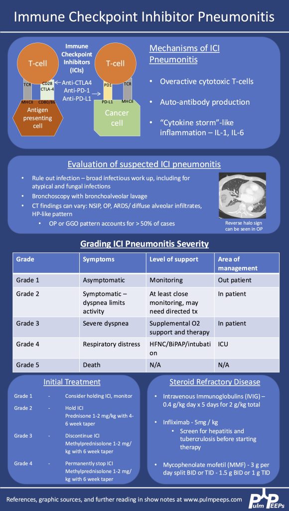
106. Pulm PEEPs Pearls: ICI Pneumonitis

We are so excited to be launching a new series here at Pulm PEEPs! We’ll be talking about high yield topics in 15 minutes or less. In this series, Furf and Monty will tackle core points and provide an overview, key points, and further reading. We’re starting with a key point review of Immune Checkpoint Inhibitor Pneumonitis. Let us know if there are other topics you want to hear about!

  1. Epidemiology & Pathophysiology
    • Increasingly common as immunotherapy use grows in oncology.
    • Caused by immune activation from PD-1, PD-L1, or CTLA-4 inhibitors.
    • Mechanisms:
      1. Overactive T cells
      2. Autoantibody production
      3. Cytokine-mediated inflammation (e.g., ↑IL-1, ↑IL-6)
  1. Clinical Suspicion & Diagnosis
    • Any new respiratory symptoms in a patient currently or previously on ICI → consider ICI pneumonitis.
    • CT findings are variable: can mimic organizing pneumonia, NSIP, ARDS, or diffuse ground glass opacities. Imaging pattern does not determine severity grade.
    • Diagnosis is of exclusion — infection and malignancy progression must be ruled out first.
    • Workup:
      • Broad infectious evaluation (cultures, viral panel, fungal markers).
      • Early bronchoscopy with BAL if feasible — typically lymphocyte-predominant in ICI pneumonitis.
      • Screen for TB and hepatitis early (in case infliximab is needed).
  1. Severity Grading (Symptom- & O₂-based, not imaging-based)
    • Grade 1: Asymptomatic → monitor, may hold ICI.
    • Grade 2: Symptomatic but not hypoxic → prednisone 1 mg/kg/day PO.
    • Grade 3–4: Hypoxemia or ICU-level care → methylprednisolone 1–2 mg/kg/day IV. Usually hold or permanently stop ICI.
  1. Steroid Management
    • Typical taper: over 6 weeks for grade ≥3.
      • Week 1: 1–2 mg/kg/day
      • Gradual step-down to 0.25 mg/kg/day by week 5, then stop week 6.
    • Chronic/recurrent cases may need slower tapers over months.
    • Add GI prophylaxis and PJP prophylaxis during prolonged steroid use.
  1. If Steroids Fail (no improvement after 48–72 hrs)
    • Consider adding:
      • IVIG (2 g/kg over 5 days)
      • Infliximab (TNF-α inhibitor — requires TB/hepatitis screening)
      • Mycophenolate mofetil (1–1.5 g/day BID or TID, start at effective dose quickly)
    • IVIG may have lower mortality in some series but comes with risks (volume overload, thrombosis, infusion reactions).
  1. Emerging Therapies
    • JAK inhibitors are under investigation as possible future options.
  1. Multidisciplinary Care
    • ICU management is a team sport — coordinate with oncology, critical care, infectious disease, and pharmacy.


  1. Managing Immune Checkpoint Inhibitor Pneumonitis in the ICU. Montemayor, Kristina et al.CHEST Critical Care, Volume 3, Issue 1, 100126
  2. Lavalle S, Masiello E, Valerio MR, Aliprandi A, Scandurra G, Gebbia V, Sambataro D. Immune checkpoint inhibitor therapy‑related pneumonitis: How, when and why to diagnose and manage (Review). Exp Ther Med. 2024 Jul 30;28(4):381. doi: 10.3892/etm.2024.12670. PMID: 39113908; PMCID: PMC11304171.
  3. Delaunay M, Prévot G, Collot S, Guilleminault L, Didier A, Mazières J. Management of pulmonary toxicity associated with immune checkpoint inhibitors. Eur Respir Rev. 2019 Nov 6;28(154):190012. doi: 10.1183/16000617.0012-2019. PMID: 31694838; PMCID: PMC9488507.