52. Fellows’ Case Files: University of Pittsburgh

We’re excited to be back with another Fellows’ Case Files. Today, we’re visiting the University of Pittsburgh to meet a fantastic fellow and a dedicated educator, and to hear about a fascinating case. Let us know if you’ve ever had a similar case, and share your interesting cases with us!
Meet Our Guests

Rachel Wojcik obtained her B.S. in Biology from Mercyhurst University and a Master’s in Liberal Studies from the University of Denver in Global Affairs with a focus on Healthcare. She completed her MD at the University of Colorado before completing her residency and chief resident year at the University of Pittsburgh and has continued her training at Pitt for PCCM fellowship.

Dr. Stephanie Maximous is an Assistant Professor of  Medicine at the University of Pittsburgh School of Medicine and is the Clinical Education APD for the Pulmonary and Critical Care Fellowship program. She completed her fellowship at Pitt in addition to obtaining a Master’s Degree in Medical Education there. She teaches in and directs courses throughout the medical school, residency, and fellowship and was recently awarded the 2023 Outstanding Subspecialty Teaching Attending Award from the housestaff.

Case Presentation

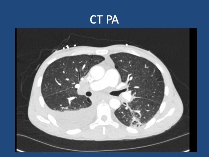
Patient: A 70-year-old male with a history of idiopathic thrombocytopenia on chronic prednisone and a history of tobacco use disorder.

Presentation: Came to the hospital with 2-3 days of right-sided weakness and slurred speech.

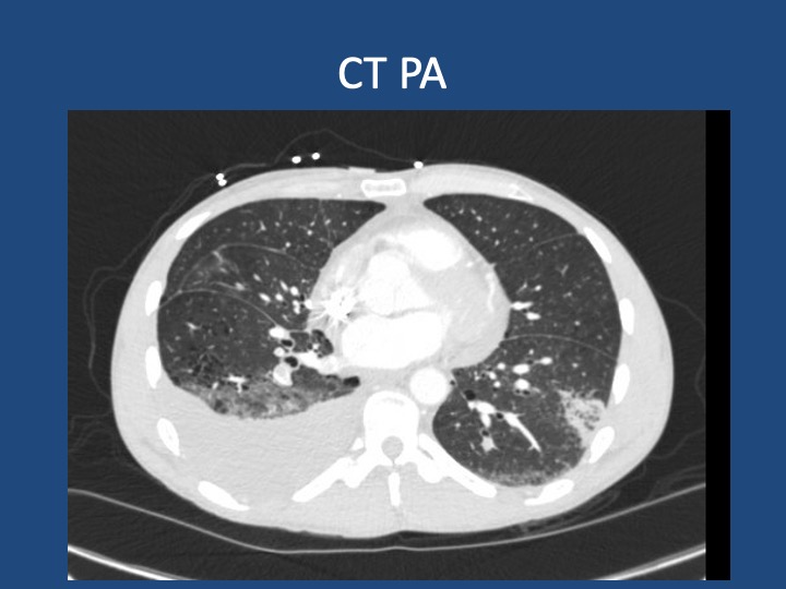
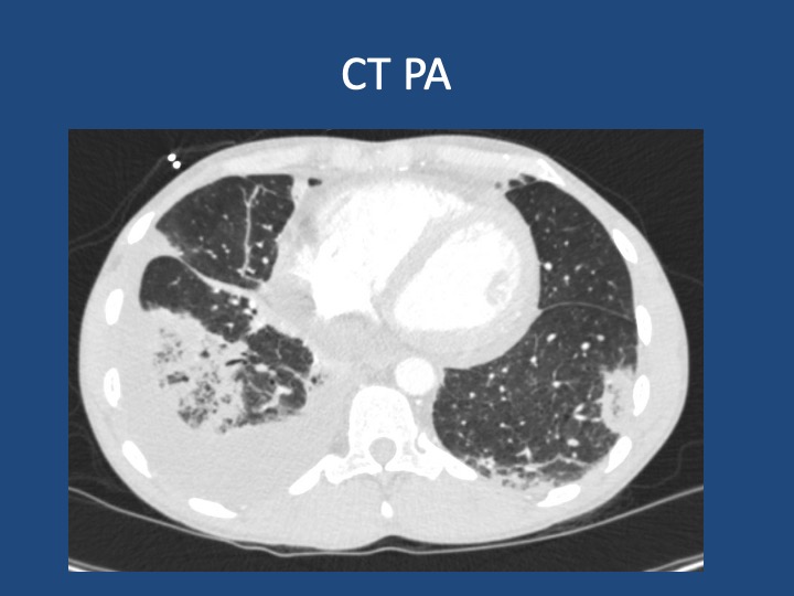
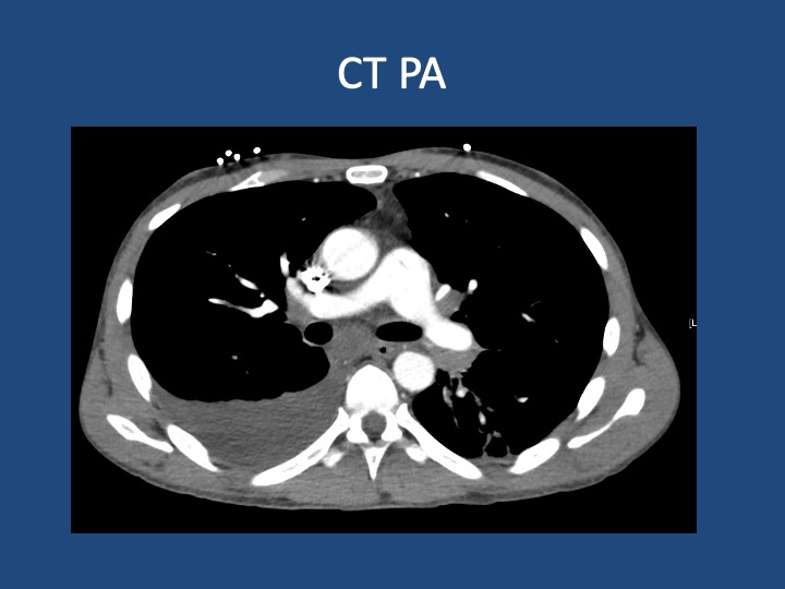
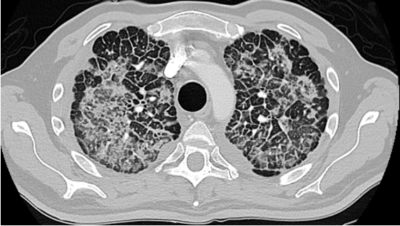
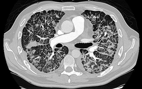
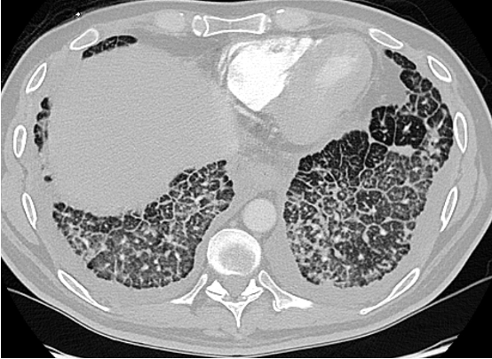
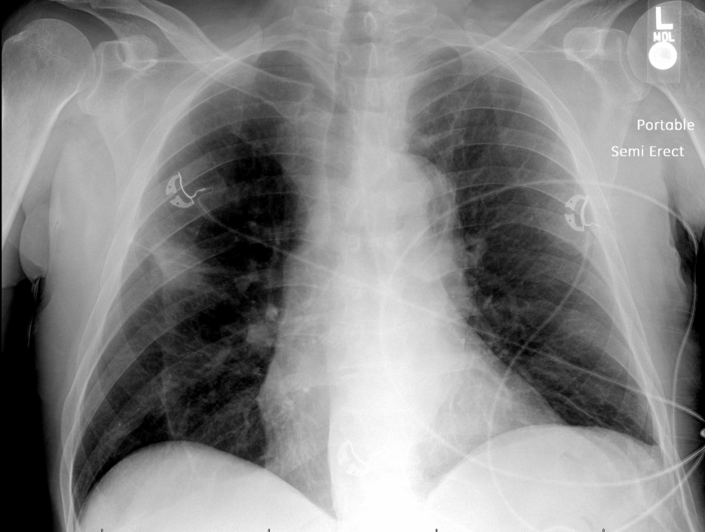
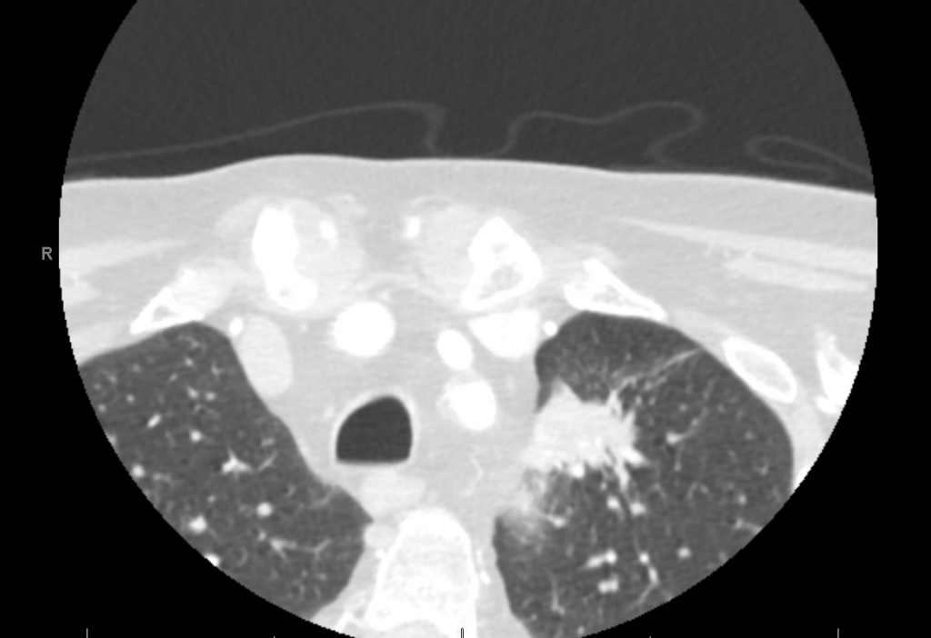
Findings: MRI showed a moderate-sized left pontine stroke. A CT angiogram of the neck showed no evidence of an occlusion, but a spiculated two-centimeter nodule at the apex of the left lung was found.

Additional Information: He requires a walker for mobility and needs help with activities like taking a shower and dressing. He had an unintentional 20-pound weight loss over six months, increased fatigue, and malaise.

Previous Investigations: A chest x-ray ordered two months prior by his hematologist was unremarkable, and a CT of the abdomen and pelvis showed no masses.
Key Learning Points

Bronchoscopy in Decision Making:

  • The decision to perform bronchoscopy in patients depends on a myriad of factors, including the location of any lesions, accessibility, potential risks, and the potential diagnostic yield.
  • Fiber optic bronchoscopy with BAL can rule out infections, and if no diagnosis is reached, more invasive methods like surgical biopsy might be necessary.
  • Consider the location of consolidated masses; navigational bronchoscopy might be needed for lesions without a clear airway leading into them.

Nocardia Insights:

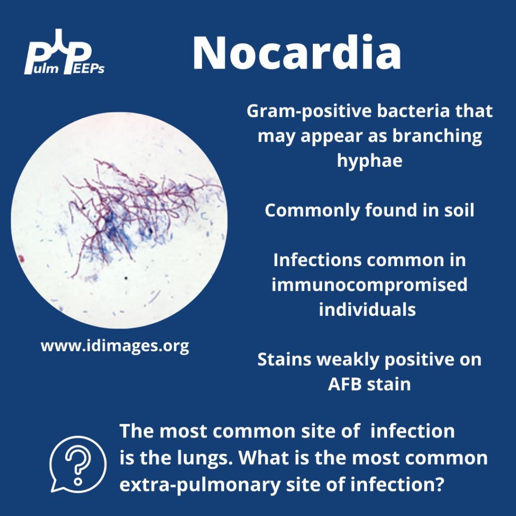
  • Nocardia is a gram-positive bacterium that stains weakly acid-fast.
  • It can be found in soil and certain water sources and can infect through the skin or by inhalation.
  • Two-thirds of patients with Nocardia are immunocompromised.
  • The dosage of Bactrim given for PJP prophylaxis doesn’t prevent Nocardia infections in immunocompromised individuals.
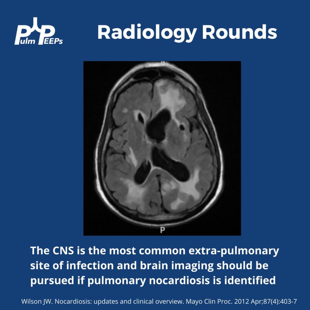
  • While the lungs are the most common infection site, Nocardia can manifest elsewhere, like the skin or CNS.

Treatment Approach:

  • Bactrim is the mainstay of treatment for Nocardia. If someone is allergic, desensitizing them can be crucial.
  • IV induction phases vary in length depending on the severity of the disease.
  • The overall treatment duration is protracted to prevent relapse.

Takeaway Points:

  • Bactrim for PJP prophylaxis doesn’t necessarily prevent Nocardia infections in immunocompromised individuals.
  • If someone is allergic to Bactrim, consider desensitizing them due to its importance in treating Nocardia.
References and Further Reading:

Menéndez R, Cordero PJ, Santos M, Gobernado M, Marco V. Pulmonary infection with Nocardia species: a report of 10 cases and review. Eur Respir J. 1997 Jul;10(7):1542-6. doi: 10.1183/09031936.97.10071542. PMID: 9230244.

Zia K, Nafees T, Faizan M, Salam O, Asad SI, Khan YA, Altaf A. Ten Year Review of Pulmonary Nocardiosis: A Series of 55 Cases. Cureus. 2019 May 26;11(5):e4759. doi: 10.7759/cureus.4759. PMID: 31363440; PMCID: PMC6663111.

48. Fellows’ Case Files: Boston University

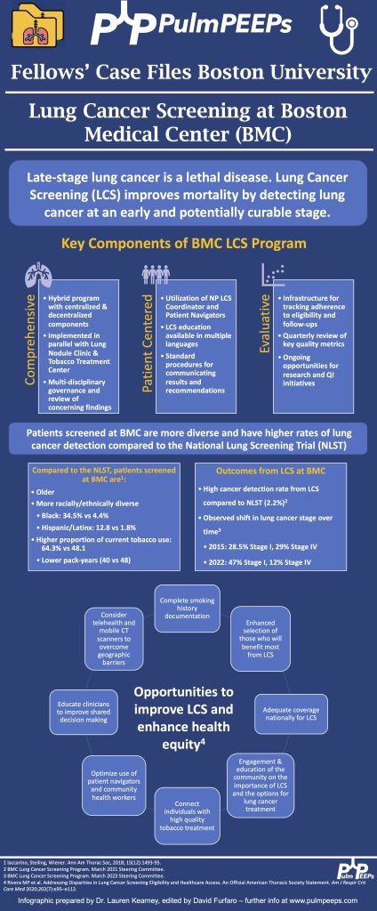
Today we’re back with another stop on our Fellows’ Case Files journey. We’re at Boston University and Boston Medical Center with an outstanding fellow, the program director, and an expert guest to dive into a new case. Tune in to hear about this fascinating case and learn some key pulmonary points along the way.

Meet Our Guests

Lauren Kearney is a research fellow at Boston University. She completed her internal medicine residency and chief residency at BU. She is pursuing a research career in health equity and community-based participatory research to improve outcomes for patients with lung cancer and other pulmonary conditions.

Chris Reardon is a Clinical Professor of Medicine at Boston University, where she is also the Fellowship Training Program Director for Pulmonary and Critical Care Medicine. She is additionally the Director of Respiratory Care Services at Pappas Rehab Hospital for Children.

Katie Steiling is an Assistant Professor of Medicine at Boston University and a Member of the Bioinformatics Graduate Program. She founded the Lung Nodule Clinic at Boston Medical Center and co-chairs the Lung Cancer Screening Steering Committee. She is dedicated to improving the equitable treatment of patients with and at risk for lung cancer.

Case Presentation

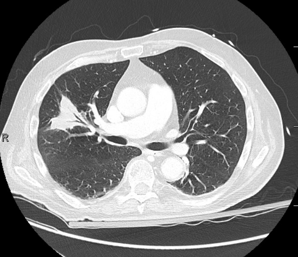
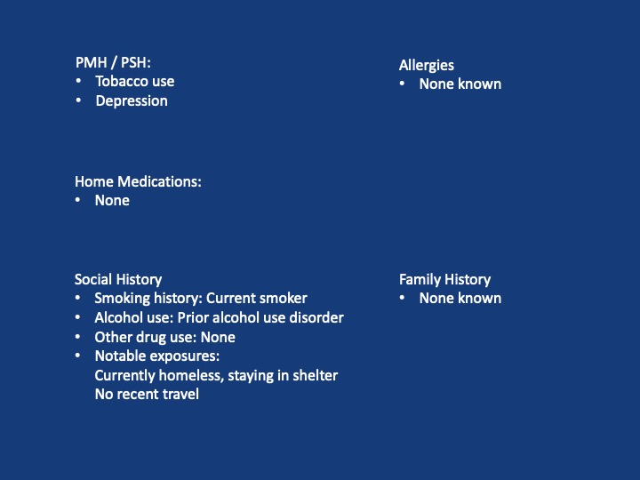
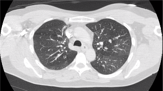
A 44-year-old man who is undomiciled and a current smoker presents with three days of intermittent, progressively worsening pleuritic chest pain with a cough productive of blood-tinged sputum. This is in the setting of 1-2 months of fatigue and anorexia, and 2 weeks of bilateral calf swelling.

Key Learning Points

  1. Factors that may lead to initial hypercoagulability testing in first unprovoked PE: young age, thrombosis in multiple or unusual vascular beds, history of warfarin-induced skin necrosis, arterial thrombosis.
  2. When lung cancer is suspected, identification of metastases to identify the highest radiologic stage and optimal biopsy site is essential.
  3. Given that pleural fluid sensitivity is only ~46%, negative cytology should not assuage further malignancy work-up in a high-risk patient.
  4. Black individuals in the US are at higher risk of developing and dying of lung cancer- the reasons for which are complex & multifactorial. Yet, gaps remain in the targeted identification of these patients, adequate lung cancer screening, and connection to diagnosis & treatment.
39. Fellows’ Case Files: Indiana University

Join us as we head to Indiana University! Listen in as we discuss another great case and hear teaching points from our amazing guests.

Meet our Guests


Parth Savsani is currently an internal medicine resident at Indiana University School of Medicine. He received his undergraduate degree from the University of Wisconsin-Madison and his medical degree from the University of Illinois College of Medicine. He enjoys medical education and was selected to be the VA chief resident next year.

Maria Srour is a Pulmonary and Critical Care Fellow at Indiana University School of Medicine. She completed her internal medicine residency at Saint Louis University where she was also a chief resident, and received her medical degree from IU. She works in global health to improve care for sepsis patients in low resources settings, and is currently pursuing her MPH.

Laura Hinkle is a Indiana University die hard and has been there from her since medical school through residency and fellowship, and is now an Assistant Professor of Clinical  Medicine and the Associate Program Director for the Pulmonary and Critical Care Medicine Fellowship. She will be taking over as the Program Director July 1, 2023. She is a dedicated educator and is the Key Clinical Educator for Pulmonary and Critical Care, and the Director of the Clinical Transitions Curriculum.  Additionally, she is working on a Master’s Degree in Education through the University of Cincinnati. 

Case Presentation

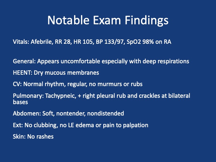
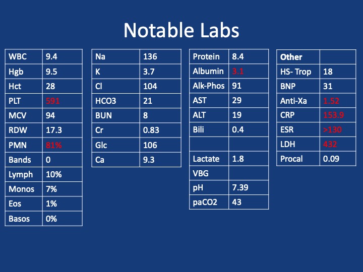
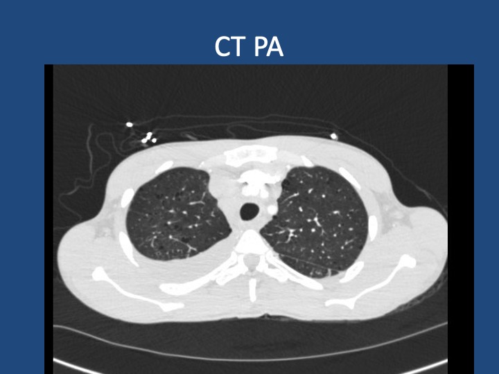
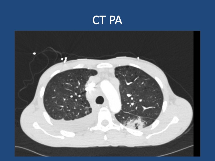
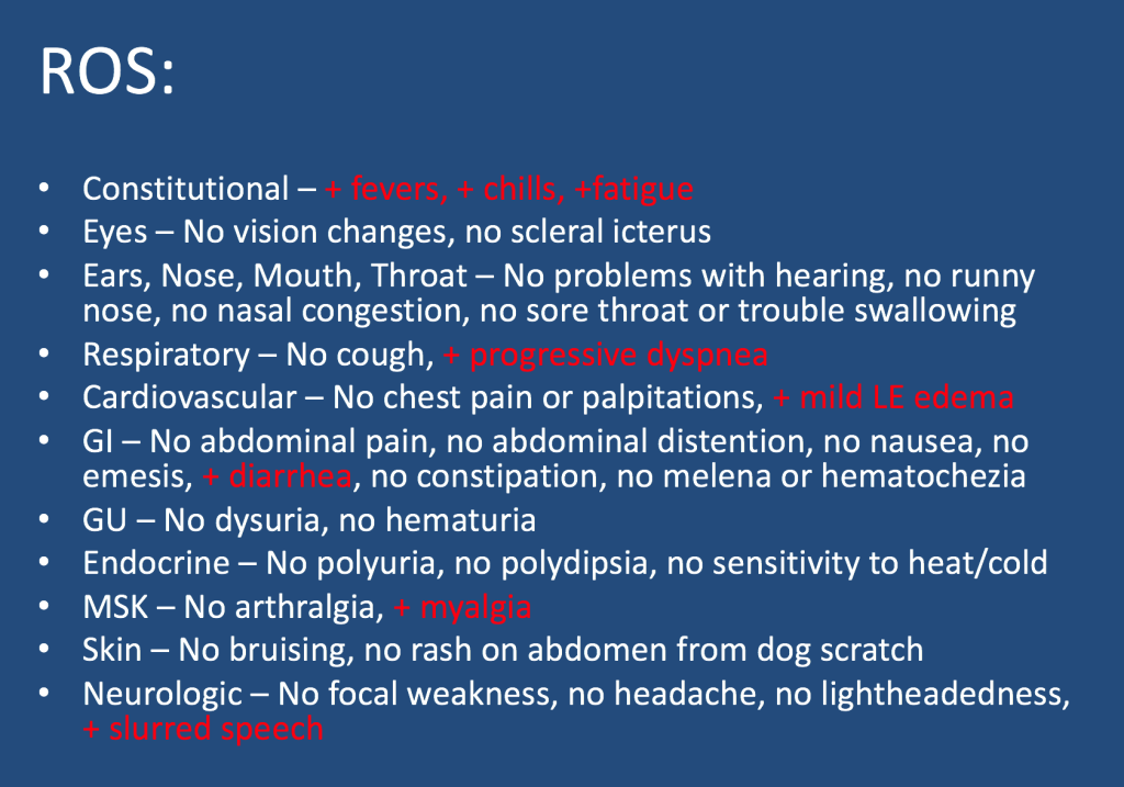
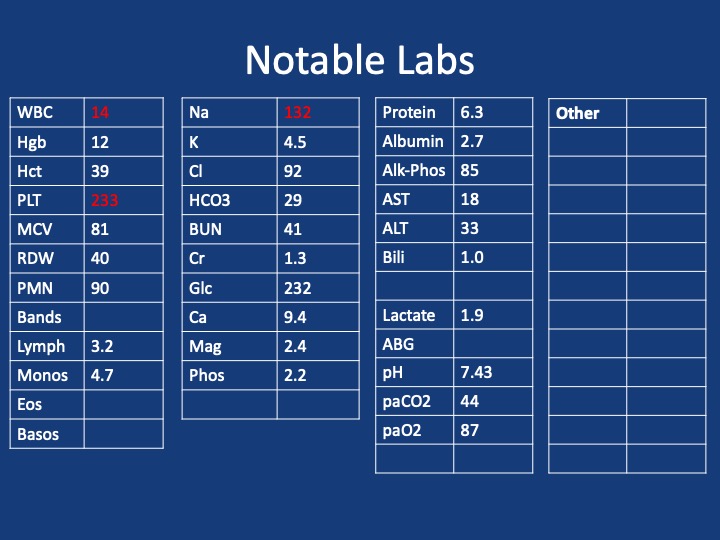
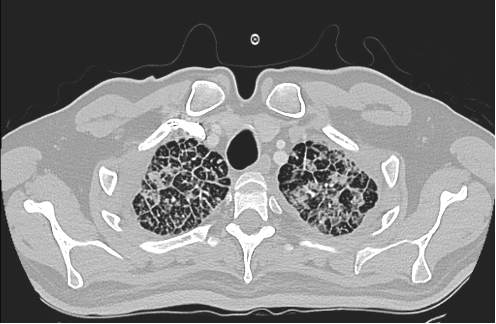
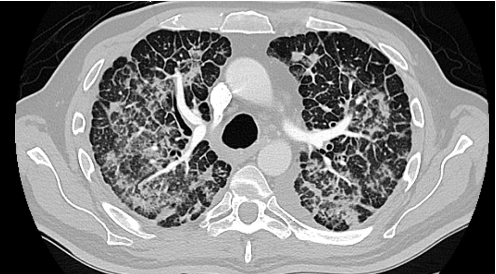
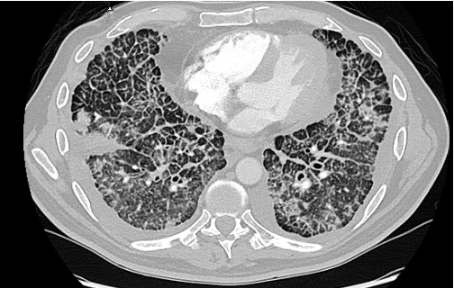
A male in her early 60s is transferred from a neighboring facility with a 1 week history of fatigue and lethargy. Three days prior to presentation he developed dyspnea and increased weakness with a near fall at home. HIs family also reported recent fevers, chills, dyspnea, and diarrhea. On his way to seek evaluation, he developed slurred speech without any other focal abnormalities.

Additional information is summarized as below:

Follow along our episode to hear the final diagnosis and key teaching points from the case!

38. Fellows’ Case Files: Houston Methodist

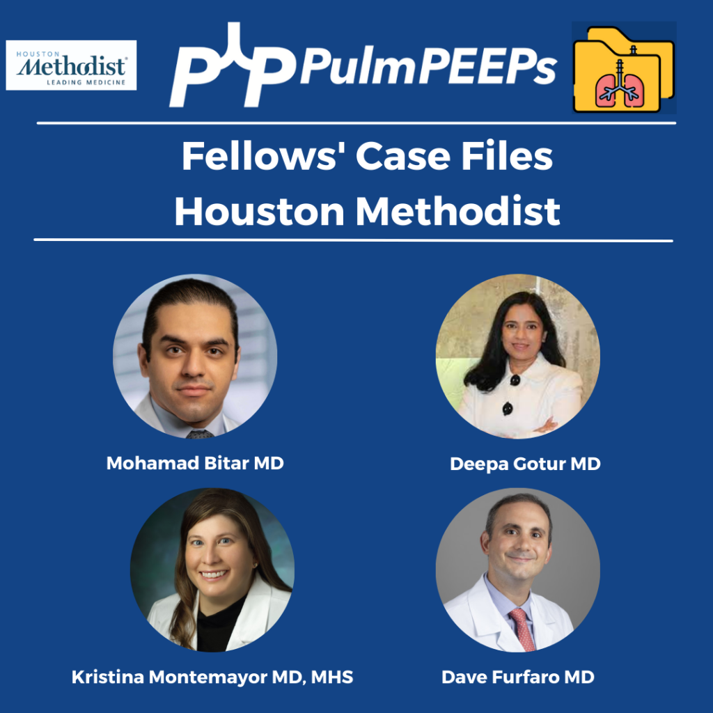
Join us as we head to the Texas Medical Center and are joined by Dr. Bitar and Dr. Gotur from Houston Methodist Pulmonary and Critical Care Medicine.

Meet our Guests

Dr. Mohamad Bitar is a Pulmonary and Critical Care Fellow at the Houston Methodist Pulmonary and Critical Care Program in Houston, Texas. Dr. Bitar completed his medical school at Misr University for Science and Technology and his Internal Medicine residency at Good Samaritan Hospital.

Dr. Deepa Gotur is an Associate Professor of Clinical Medicine at Weill Cornell Medical College and an Adjunct Associate professor at Texas A&M. She is also the Pulmonary and Critical Care Program Director at Houston Methodist. She is a sepsis and ARDS researcher, as well as a dedicated educator.

Case Presentation

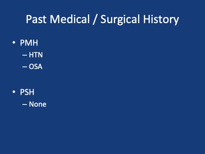
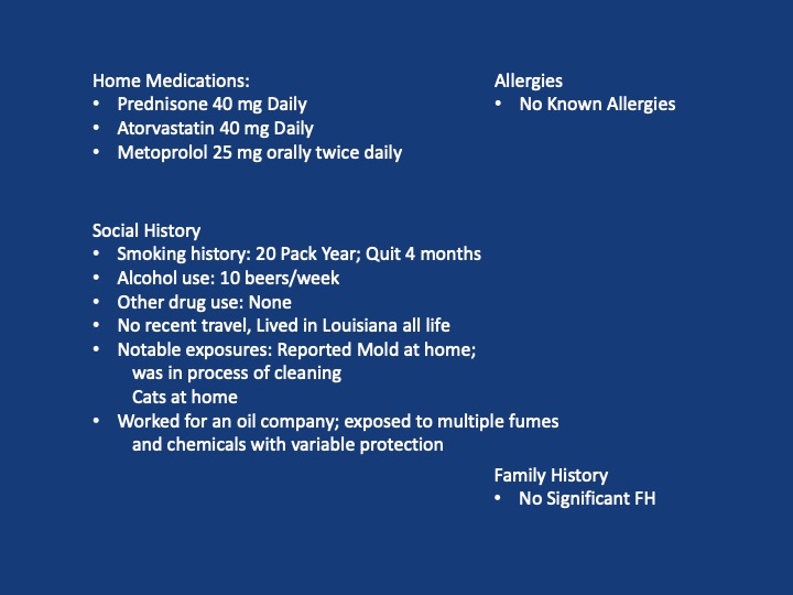
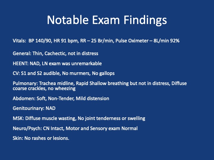
A 40s-year-old male presents with 3 months of progressive shortness of breath. He was diagnosed with COVID and given treatment with steroids, and antibiotics with no response.

He presented to the hospital 3 months later after not being able to carry out daily activities and was found to have low oxygen saturation

Admitted, initial CT Chest – showed “Non-specific patchy ground-glass bilateral pulmonary infiltrates with Coarsened interstitial markings that could be related to COVID-19/atypical infection”

References and further reading

1. Silva, C. Isabela S. MD, PhD; Müller, Nestor L. MD, PhD. Idiopathic Interstitial Pneumonias. Journal of Thoracic Imaging 24(4):p 260-273, November 2009.

2.Gruden JF, Naidich DP, Machnicki SC, Cohen SL, Girvin F, Raoof S. An Algorithmic Approach to the Interpretation of Diffuse Lung Disease on Chest CT Imaging: A Theory of Almost Everything. Chest. 2020 Mar;157(3):612-635.

3. Ryu JH, Daniels CE, Hartman TE, Yi ES. Diagnosis of interstitial lung diseases. Mayo Clin Proc. 2007 Aug;82(8):976-86.

34. Fellows’ Case Files: The Ohio State University College of Medicine

Welcome back to Pulm PEEPs Fellows’ Case Files series. We are traveling to the midwest to visit The Ohio State University College of Medicine and hear about another great pulmonary case.

Meet Our Guests

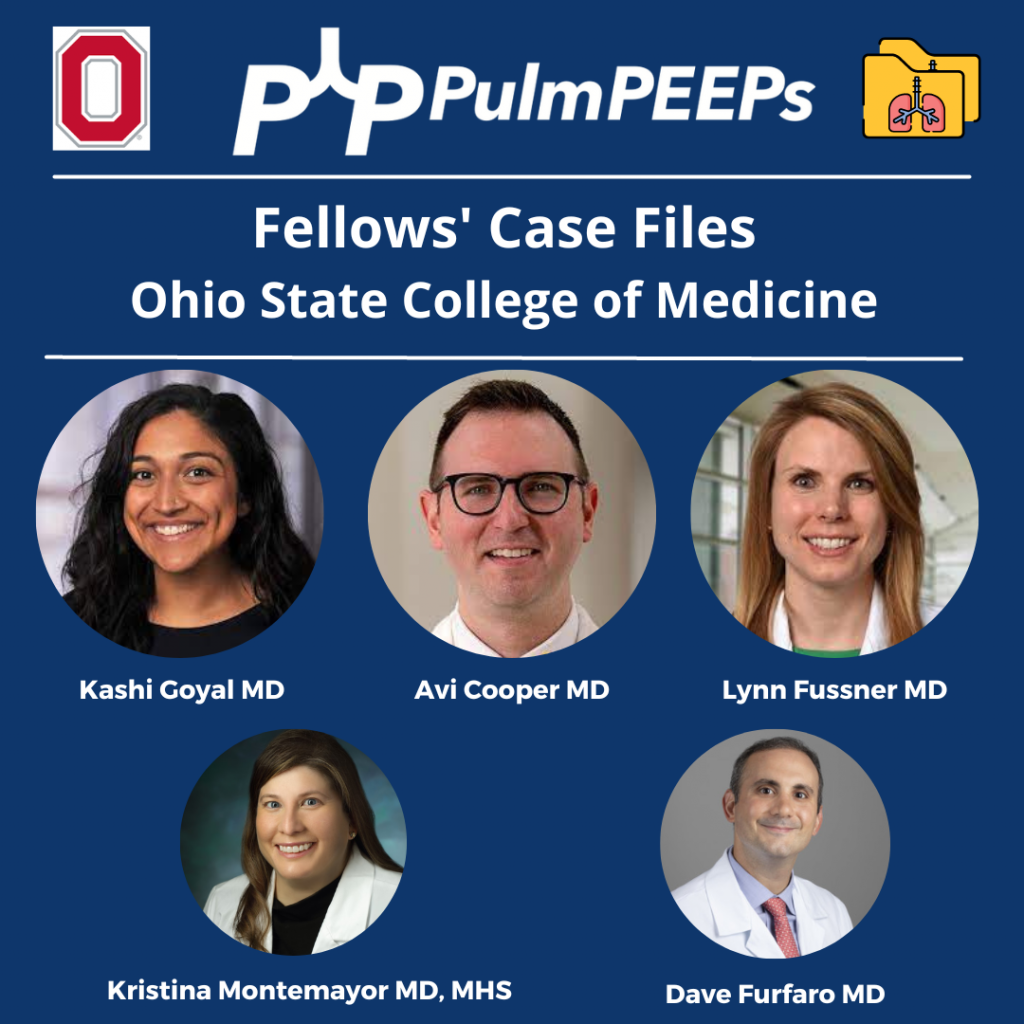
Kashi Goyal is a second-year Pulmonary and Critical Care Fellow at The Ohio State University Wexner Medical Center. She obtained her MD at OSU, and then completed her Internal Medicine residency at Beth Israel Deaconess Medical Center. She worked as a hospitalist and educator before going back to fellowship and remains passionate about medical education.

Lynn Fussner is an Associate Professor of Internal Medicine at OSU and has been there since completing her fellowship and Post-doctorate at Mayo Clinic. In addition to her clinical work in the multidisciplinary vasculitis clinic, she is a translational researcher with a focus on inflammatory pulmonary disorders and vasculitis.

Avi Cooper is an Assistant Professor of Medicine at Ohio State University College of Medicine and the Program Director of the Pulmonary and Critical Care Fellowship. He is an Associate Editor at the Journal of Graduate Medical Education. Last but not least, he co-hosts the Curious Clinician Podcast, one of the most popular medical education podcasts.

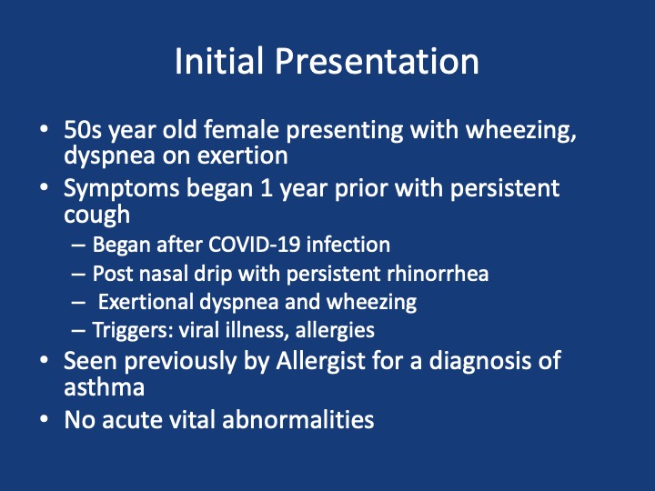
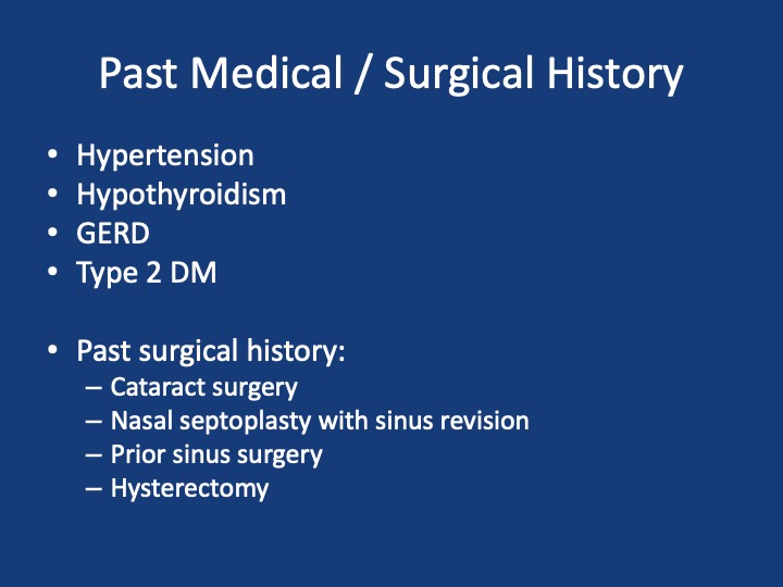
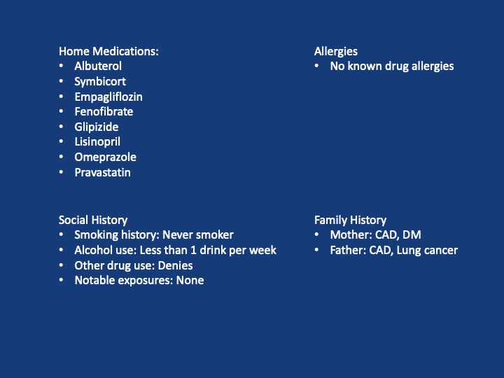
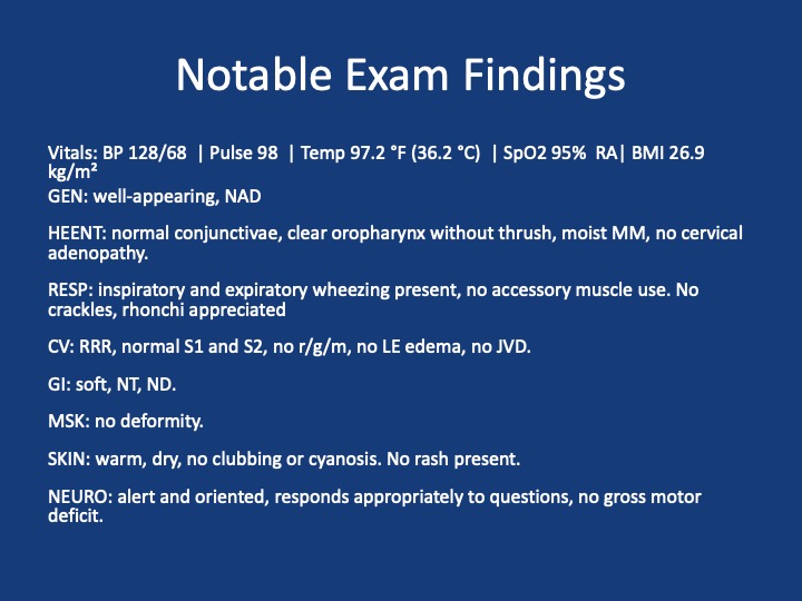
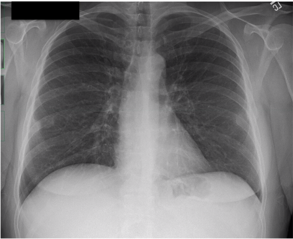
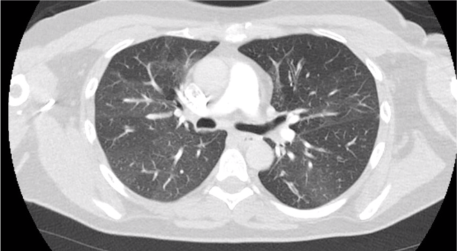
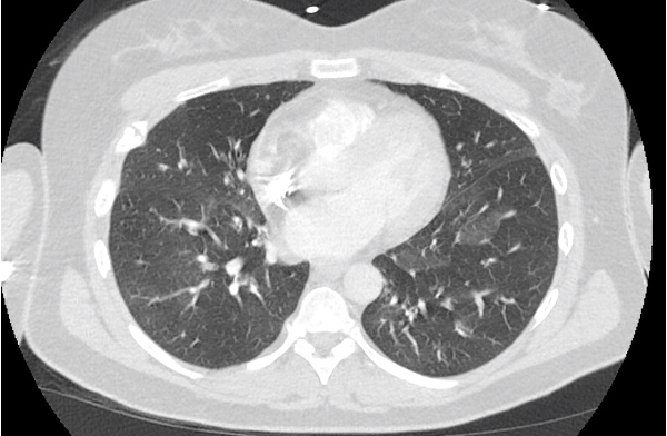
Patient Presentation

Key Learning Points

**Spoilers Ahead** If you want to think through the case on your own we advise listening to the episode first before looking at these points.

  • The three most common causes of cough in adults in the USA are cough variant asthma, GERD, and post-nasal drip
  • A post-viral cough can last for 8-12 weeks and still be within normal
  • Sinus symptoms in a chronic cough can just be sinusitis and post-nasal drip, but should consider eosinophilic granulomatosis with polyangiitis (EGPA), aspirin exacerbated respiratory disease (AERD), cystic fibrosis, or ciliary dyskinesia.
  • Examination of a wheeze
    • Fixed sound vs variable
    • Pitch: larger central airways vs lower peripheral airways
    • Is it throughout the cycle or at a certain phase?
    • Ask the patient to cough before listening and ask them to breathe out through their mouth
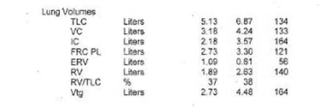
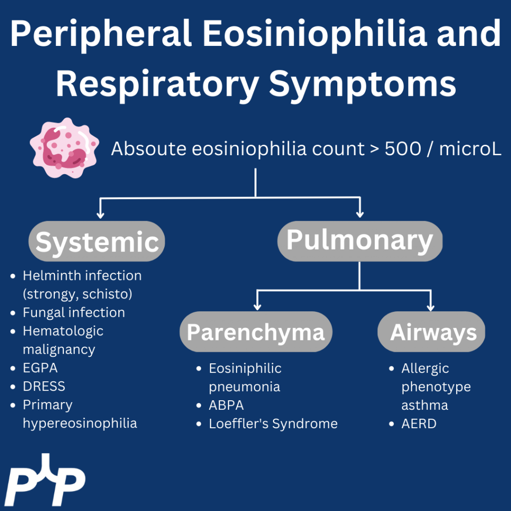
  • Approach to eosinophilia in a patient with cough and dyspnea
    •  Multi-system involvement vs lungs
      • Multi-system involvement
        • Vasculitis
        • Parasitic infection
        • Hematologic malignancy
        • Medication side effect
        • Primary hypereosinophilic syndromes
      • Within the lungs:
        • Parenchymal disease
          • Loeffler’s syndrome
          • Eosinophilic pneumonia
        • Airway disease
          • Asthma
          • ABPA
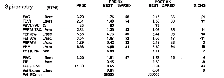
  • If you have a high suspicion for airways disease, PFTs should be requested with bronchodilator testing regardless of the degree of obstruction on baseline spirometry
  • Asthma alone should not cause ground glass opacities, so if see these in a patient with asthma we think about:
    • Infection, especially atypical infections
    • EGPA
    • Vasculitis with DAH
    • ABPA
    • Hypogammaglobulinemia or other immunodeficiency
  • EGPA diagnosis
    • ANCA testing is only positive in 60% of patients with EGPA so a negative test doesn’t rule it out by any means
    • It is easiest to make a diagnosis when there is a clear small vessel manifestation
      • Alveolar hemorrhage
      • Mononeuritis multiplex
      • Glomerulonephritis
    • Many patients with asthma, nasal polyposis, and high peripheral eosinophilia have EGPA but don’t have a clear small vessel feature of vasculitis or a positive ANCA
      • These patients typically have eosiniophilia a lot higher than when thinking about allergic phenotype asthma alone. As a rule of thumb, at least an absolute eosinophil count > 1000

References and Further Reading

  1. Carr TF, Zeki AA, Kraft M. Eosinophilic and Noneosinophilic Asthma. Am J Respir Crit Care Med. 2018;197(1):22-37. doi:10.1164/rccm.201611-2232PP
  2. Cottin V. Eosinophilic Lung Diseases. Clin Chest Med. 2016;37(3):535-556. doi:10.1016/j.ccm.2016.04.015
  3. Grayson PC, Ponte C, Suppiah R, et al. 2022 American College of Rheumatology/European Alliance of Associations for Rheumatology Classification Criteria for Eosinophilic Granulomatosis with Polyangiitis. Ann Rheum Dis. 2022;81(3):309-314. doi:10.1136/annrheumdis-2021-221794
  4. Wechsler ME, Akuthota P, Jayne D, et al. Mepolizumab or Placebo for Eosinophilic Granulomatosis with Polyangiitis. New England Journal of Medicine. 2017;376(20):1921-1932. doi:10.1056/NEJMoa1702079

30. Fellows’ Case Files: University of Mississippi Medical Center

We’re excited to be back with another episode in our Pulm PEEPs Fellows’ Case Files series! This is a particularly exciting case since it is our first episode where some intrepid fellows reached out to us with an interesting case they had encountered. If you have a great case, please let us know and you can follow in their footsteps! Pack your bags, and let’s head to Mississippi to learn about another great pulmonary and critical care case.

Meet our Guests

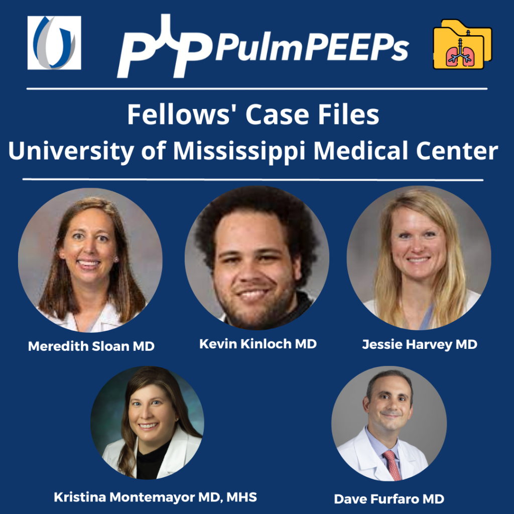
Meredith Sloan is a pulmonary and critical care fellow at the University of Mississippi. She completed her medical school at the Medical University of South Carolina College of Medicine, and her residency at the University of Mississippi.

Kevin Kinloch is a senior fellow at the University of Mississippi Medical Center where he also completed his internal medicine residency. He completed medical school at Meharry Medical College.

Jessie Harvey is an Associate professor of Medicine at the University of Mississippi and is the Pulmonary and Critical Care Program Director. She is also the Director of the MICU, and has been at MMC since medical school. She is a dedicated educator and leads the POCUS curriculum for IM residents and PCCM fellows

Patient Presentation

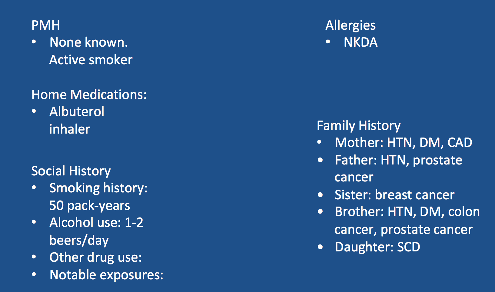
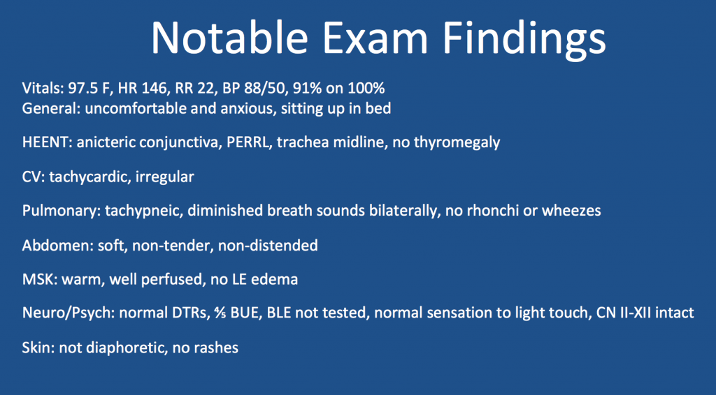
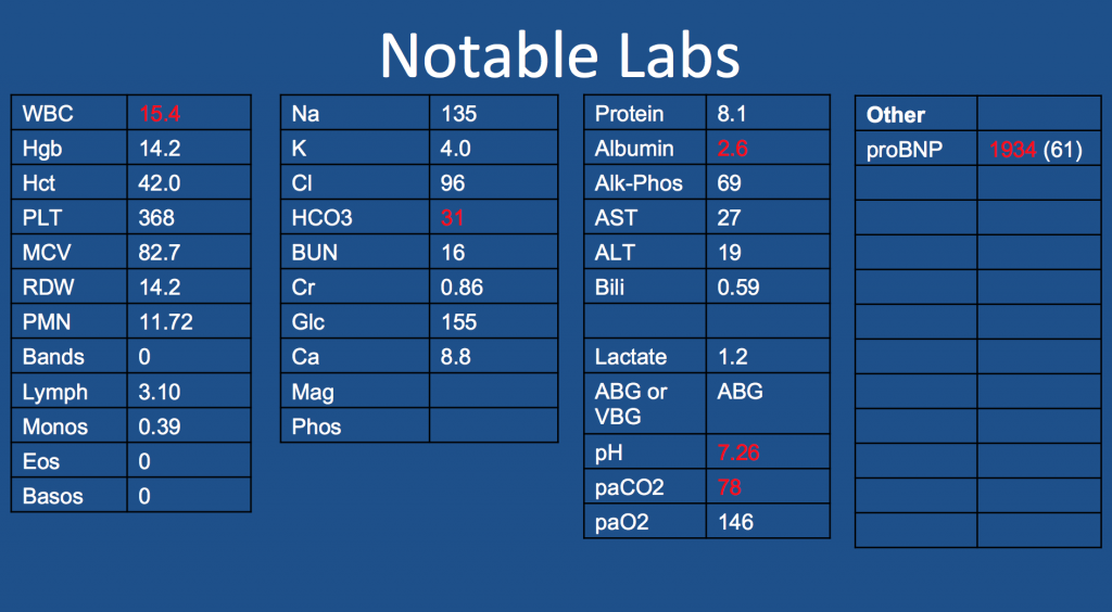
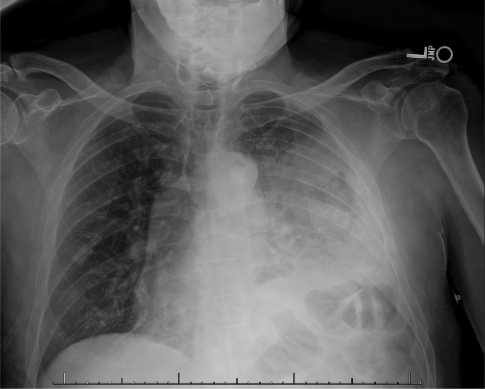
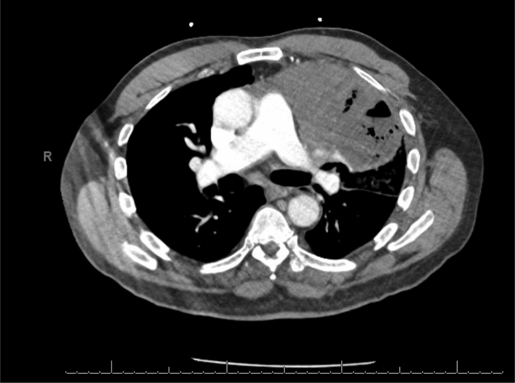
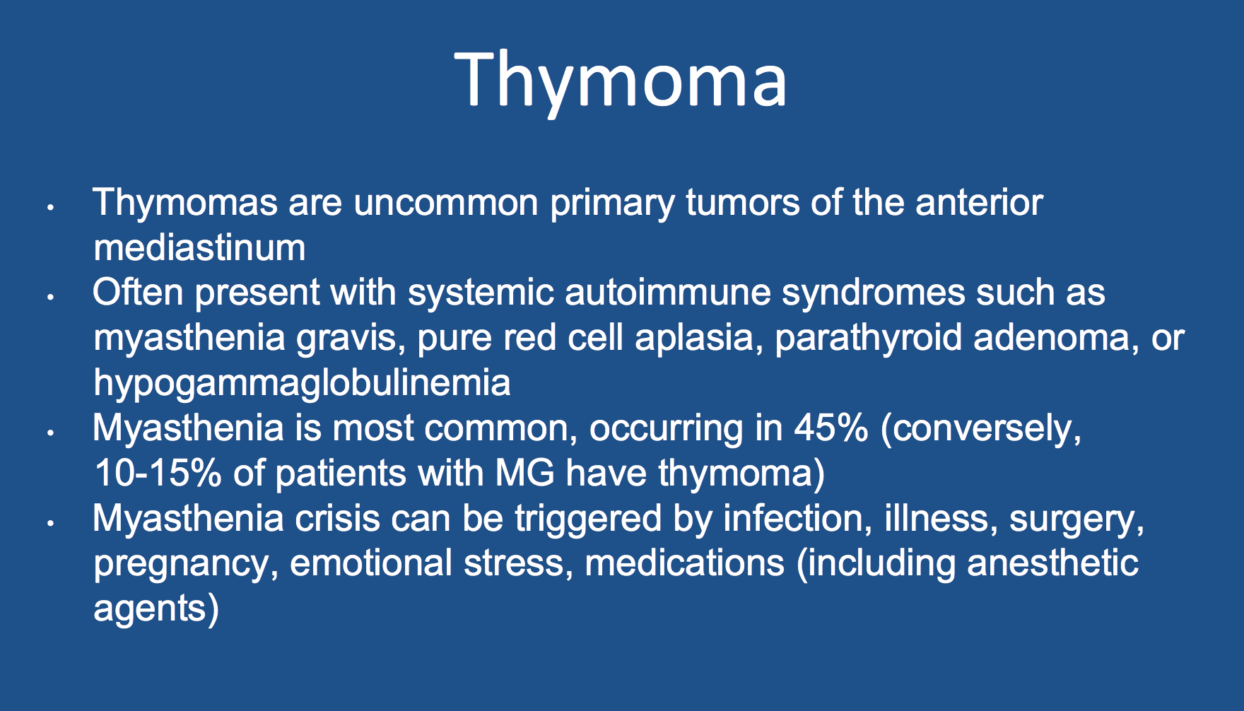
A 65-year-old man presented to the ED with worsening hemoptysis over the last several days after a recent lung biopsy. The patient is an active smoker with at least a 50-pack-year history, and he had been having a cough with small-volume hemoptysis. He ultimately had a chest CT that revealed a large LUL mass (10.3 x 6.4 cm). Given this suspicious mass, three days prior to his ED presentation, he was taken for bronchoscopy with BAL, transbronchial biopsies, endobronchial biopsy, EBUS guided TBNA of 11L, along with TBNA, brushing and radial EBUS TBNA of his left upper lobe mass.

Key Learning Points

**Spoilers Ahead** If you want to think through the case on your own we advise listening to the episode first before looking at these points.

Staging procedures for masses

  • Enough tissue so we can make a diagnosis and do molecular testing
  • Highest staging when getting your biopsy

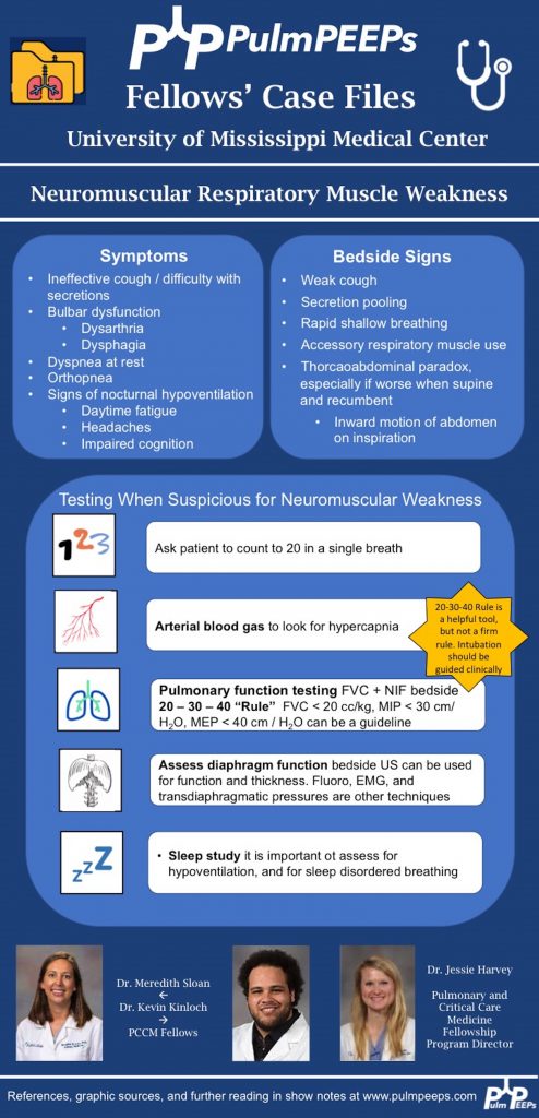
POCUS for respiratory failure

  • Absence of lung slidings
    • Especially post procedure
  • The presence of a new pleural effusion after a procedure could indicate hemothorax
    • Hematocrit sign – an echogenic layering of material in an effusion
  • New B-lines, especially if prior there were only A-lines
    • Cardiogenic or non-cardiogenic pulmonary edema, alveolar hemorrhage, or infection
  • Diaphragmatic function
    • Excursion
    • Diaphragm thickness

References and Further Reading

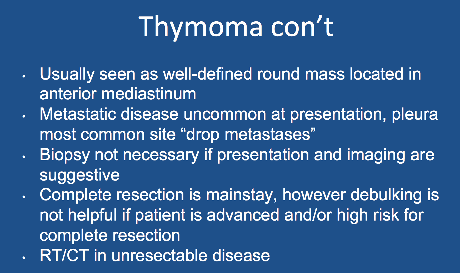
1.Scorsetti M, Leo F, Trama A, D’Angelillo R, Serpico D, Maerelli M, Zucali P, Gatta G, Garassino MC. Thymoma and thymic carcinomas. Critical Reviews in Oncology/Hematology. 2016; 99:332-350.

2. Singh TD, Wijdicks EFM. Neuromuscular respiratory failure. Neurol Clin 2021; 39:333-353.

28. Fellows’ Case Files: Harvard – MGH & BIDMC

Welcome back to our Pulm PEEPs Fellows’ Case Files series! We are joined this week by a fellow and the program director from the Harvard combined PCCM fellowship at Massachusettes General Hospital and Beth Israel Deaconess Medical Center. Listen in for a great learning case and let us know on Twitter, if you have a great case to share!

Meet our Guests

Brian Rosenberg is a third year fellow at the Harvard MGH/BI program. He completed his undergraduate degree at Harvard, received his MD  from Yale where he also got a PhD in cell biology, and then did his internal medicine residency at Columbia University Medical Center in NYC.

Asha is an Assistant Professor Medicine at Beth Israel Deaconess Medical Center and Harvard Medical School, and is the Program Director of the Harvard MGH/BI combined fellowship. She is also the Director of the Pulmonary Consult Service at BIDMC, was a Rabkin Fellow in Medical Education and has received multiple leadership and teaching awards

26. A Case of AMS, Renal Failure, and Hemolysis

This week on Pulm PEEPs, we have another great case episode. We’re switching up the format a bit, and instead of introducing our guests in the beginning, we’ll bring them in consultants as we need to. Luckily, we’re joined by Pulm PEEPs Associated Editor Luke Hedrick to walk us through the case. Let us know your thoughts and if you have any other pearls to add!

Meet Our Guests

Rakhi Naik an Associate Professor of Medicine at Johns Hopkins Hospital and the Associate Director for the Hematology / Oncology Fellowship program. She also has a Masters in Health Sciences from the Johns Hopkins Bloomberg School of Public Health. She has expertise in an array of non-malignant hematology disorders and focuses specifically on sickle cell in her research. She is also an outstanding and dedicated educator and serves as the Chair of the American Society of Hematology Hematology-Focused Training Program Consortium to develop innovative training pathways for non-malignant heme.

Patient Presentation

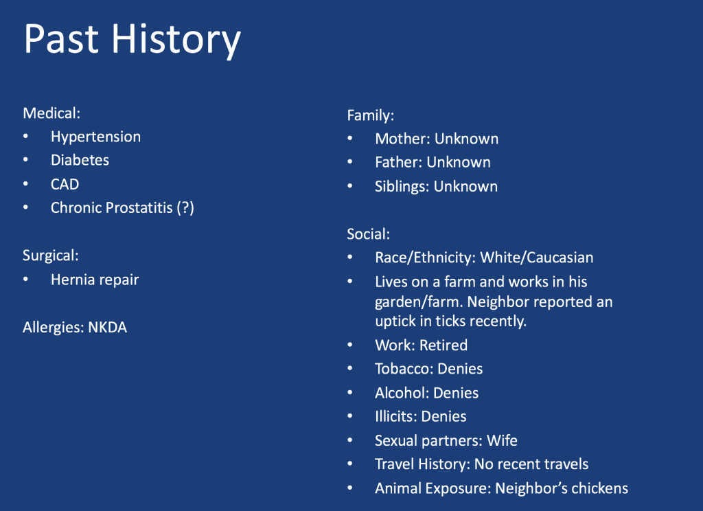
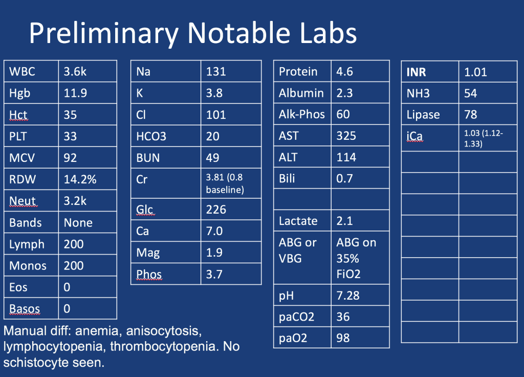
A 60-year-old woman with a past medical history of hypertension, diabetes, stage 4 chronic kidney disease, COPD, HFpEF, chronic pain on methadone, hyperparathyroidism s/p parathyroidectomy that was c/b hypothyroidism now on thyroid hormone replacement, and a recent admission for nonconvulsive status epilepticus is brought to an outside hospital by EMS with encephalopathy and shaking. 

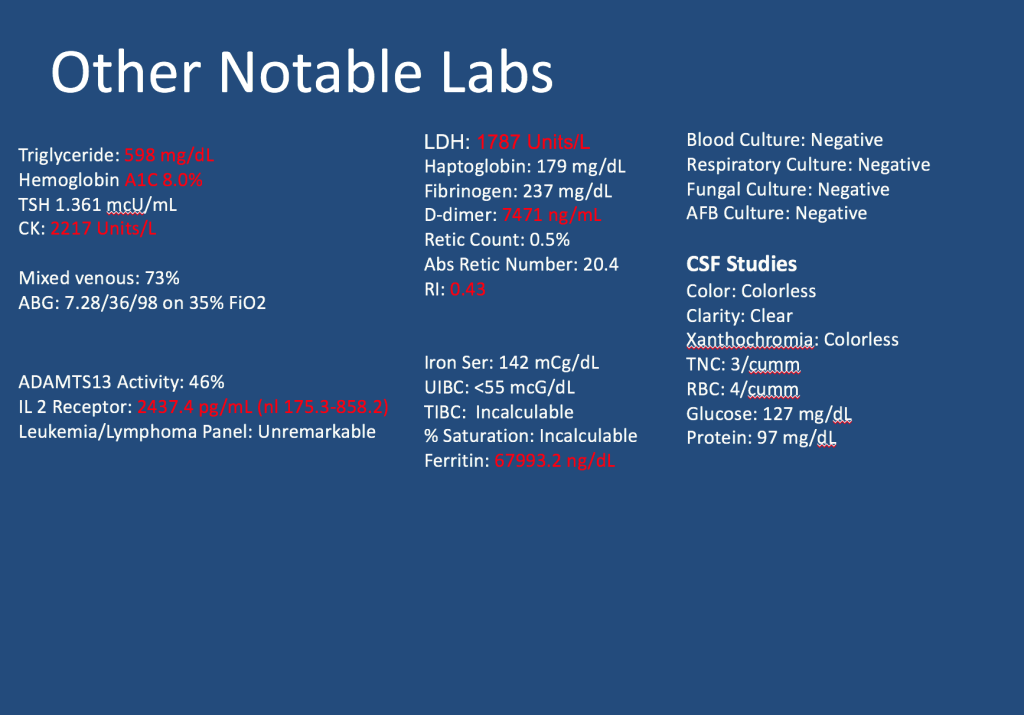
When EMS gets her to the other hospital her GCS was 5, so she was intubated for airway protection and started on fentanyl and midazolam drips. Details of labs and imaging are scarce, but we know that she had a CT head that was normal, a CXR with a report of pulmonary edema, and labs with a Cr of 2.4, serum bicarbonate of 14, and a pH from a VBG of 7.1 with pCO2 of 38.

Key Learning Points

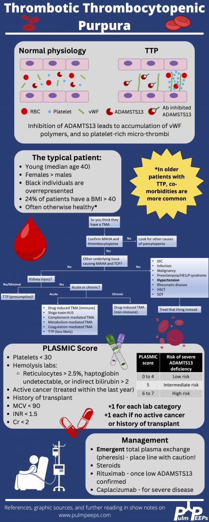
*Spoilers ahead* The infographic below highlighting key points gives away the diagnosis in this case so if you want to work through the case on your own, we recommend listening to the episode first.

References and further reading

  1. George JN. Thrombotic Thrombocytopenic Purpura. New England Journal of Medicine. 2006;354(18):1927-1935. doi:10.1056/NEJMcp053024
  2. Joly BS, Coppo P, Veyradier A. Thrombotic thrombocytopenic purpura. Blood. 2017;129(21):2836-2846. doi:10.1182/blood-2016-10-709857
  3. Kremer Hovinga JA, Coppo P, Lämmle B, Moake JL, Miyata T, Vanhoorelbeke K. Thrombotic thrombocytopenic purpura. Nat Rev Dis Primers. 2017;3(1):1-17. doi:10.1038/nrdp.2017.20
  4. Scully M, Cataland SR, Peyvandi F, et al. Caplacizumab Treatment for Acquired Thrombotic Thrombocytopenic Purpura. New England Journal of Medicine. 2019;380(4):335-346. doi:10.1056/NEJMoa1806311
  5. Sukumar S, Lämmle B, Cataland SR. Thrombotic Thrombocytopenic Purpura: Pathophysiology, Diagnosis, and Management. J Clin Med. 2021;10(3):536. doi:10.3390/jcm10030536

24. Fellows’ Case Files: Baylor College of Medicine

Welcome to another episode in our Pulm PEEPs Fellows’ Case Files series! The purpose of this series is to highlight and amplify the incredible clinical work that is done by pulmonary and critical care fellows, share fascinating cases, and assemble a diverse network of pulmonary and critical care educators. Today we’re headed to Baylor College of Medicine to hear about a fascinating case. Tune in, let us know what you think on Twitter, and let us know if you have a great case to share!

Meet Our Guests

Benjamin Moss completed his internal medicine residency training at Baylor College of Medicine in Houston, Texas, and is currently a senior pulmonary and critical care fellow there.

Philip Alapat is an Assistant Professor of Medicine and the Program Director of the Pulmonary and Critical Care Fellowship at Baylor. He completed his residency and fellowship training all at Baylor.

Patient Presentation

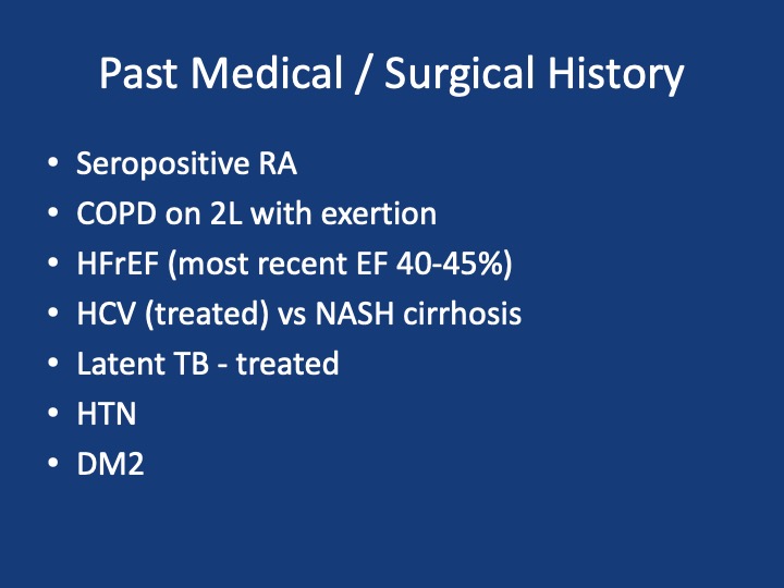
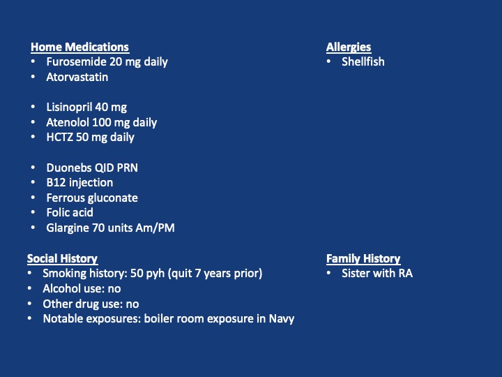
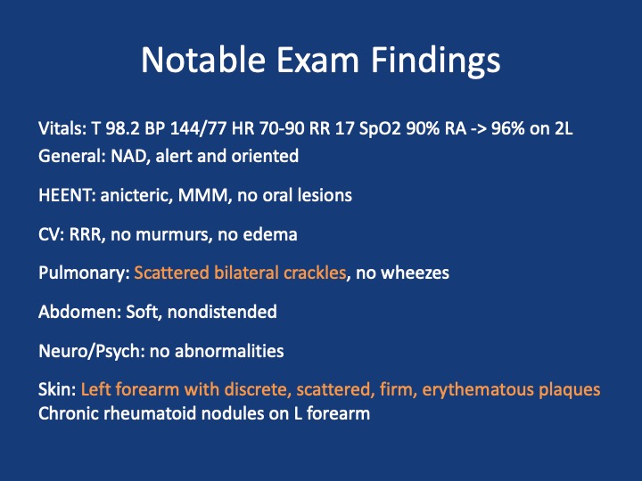
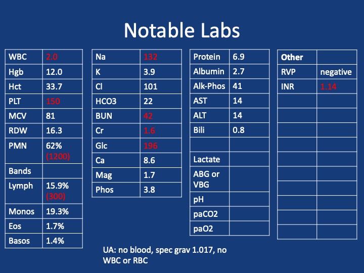
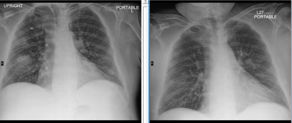
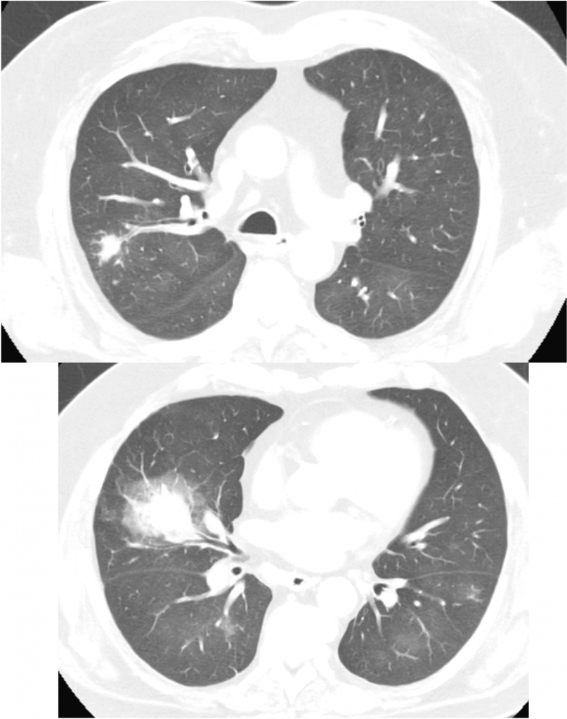
A 60s-year-old man with seropositive RA on Rituximab presents with dyspnea and cough, and overall “not feeling well.”  For the past week, he has had malaise, body aches, and subjective fever. For the past 3 days, he has had acutely worsening dyspnea that is worse with exertion, but present at rest and a cough with scant sputum production. He had been on Methotrexate previously but within the last year developed pancytopenia and MTX was stopped and he was switched to adalimumab/Humira. His pancytopenias did not resolve, and he was ultimately diagnosed with Felty syndrome (a triad of RA, neutropenia, and splenomegaly) and switched to rituximab every 6 months with his last dose being 4 months ago. During the last week, he tried taking prednisone 10 mg a day but his symptoms did not improve.

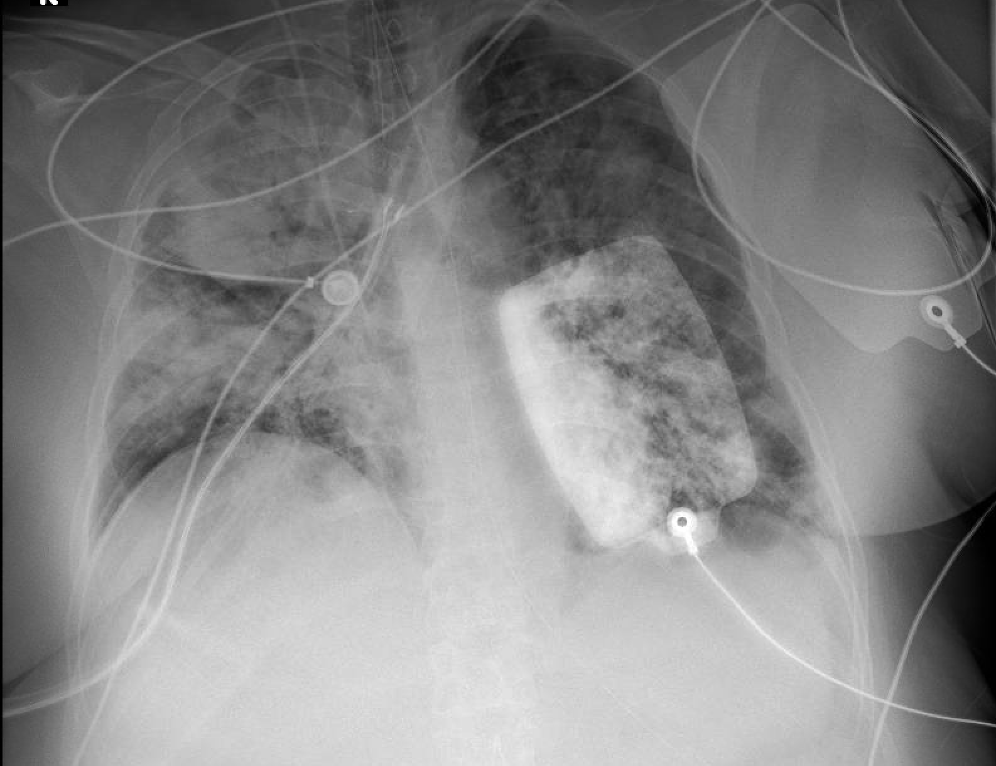
X-ray on presentation (L) X-ray 8 months ago (R)

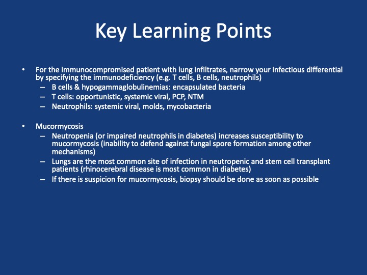
Key Learning Points

**Spoilers Ahead** If you want to think through the case on your own we advise listening to the episode first before looking at these points.

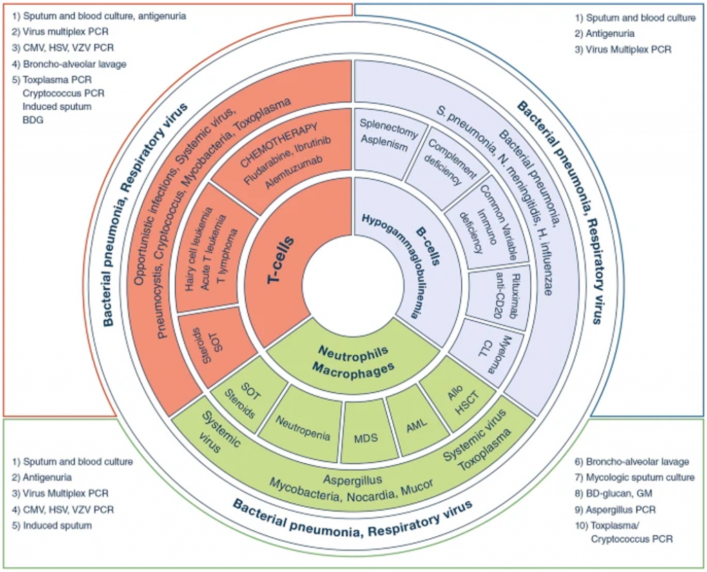
Image Source: Reference 1: https://doi.org/10.1007/s00134-019-05906-5

References and Further Reading

  1. Azoulay E, Russell L, Van de Louw A, et al. Diagnosis of severe respiratory infections in immunocompromised patients. Intensive Care Med. 2020;46(2):298-314. doi:10.1007/s00134-019-05906-5
  2. Cornely OA, Alastruey-Izquierdo A, Arenz D, et al. Global guideline for the diagnosis and management of mucormycosis: an initiative of the European Confederation of Medical Mycology in cooperation with the Mycoses Study Group Education and Research Consortium. Lancet Infect Dis. 2019;19(12):e405-e421. doi:10.1016/S1473-3099(19)30312-3
  3. Ibrahim AS, Spellberg B, Walsh TJ, Kontoyiannis DP. Pathogenesis of mucormycosis. Clin Infect Dis. 2012;54 Suppl 1:S16-22. doi:10.1093/cid/cir865

23. Fellows’ Case Files: University of Washington

We’re very excited for the second episode in our Pulm PEEPs Fellows’ Case Files series! For a reminder, the purpose of this series is to highlight and amplify the incredible clinical work that is done by pulmonary and critical care fellows, share fascinating cases, and assemble a diverse network of pulmonary and critical care educators. This week, we’re visiting the Pacific Northwest and headed to the University of Washington to meet two passionate educators, and hear about an incredible teaching case.

Meet Our Guests

Robin Stiller is a third-year pulmonary and critical care fellow at the University of Washington. Robin completed internal medicine residency training at the University of Washington and her clinical and research interests include procedural education and curriculum development.

Başak Çoruh Associate Professor of Medicine at the University of Washington School of Medicine and is the Program Director for the Pulmonary and Critical Care Fellowship. She completed her fellowship and the Teaching Scholars Program at UW. Başak has received numerous teaching and mentoring awards throughout her career and has leadership roles with ATS, CHEST as well as the APCCMPD.

Patient Presentation

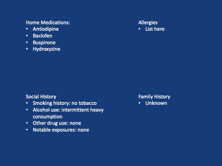
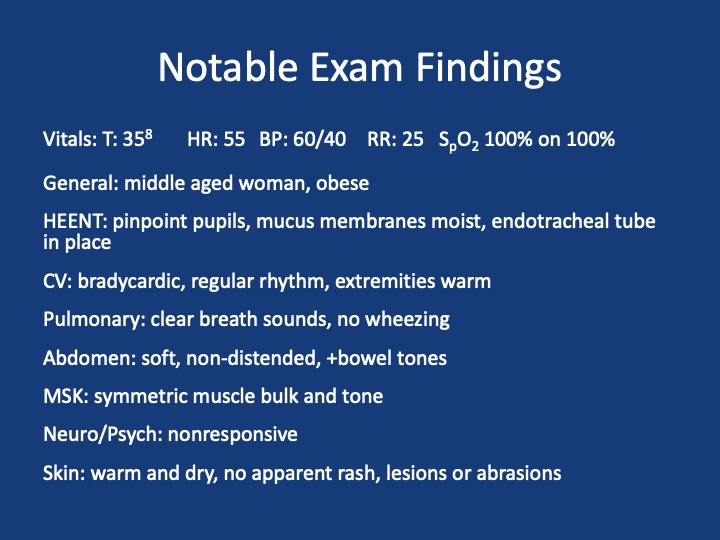
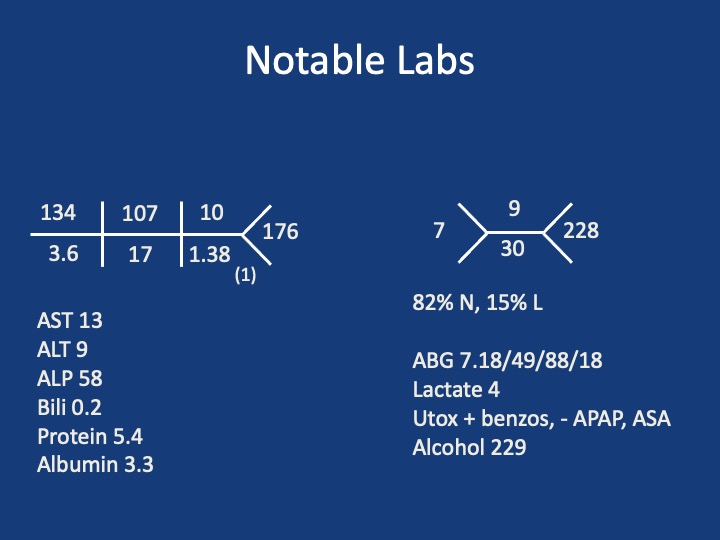
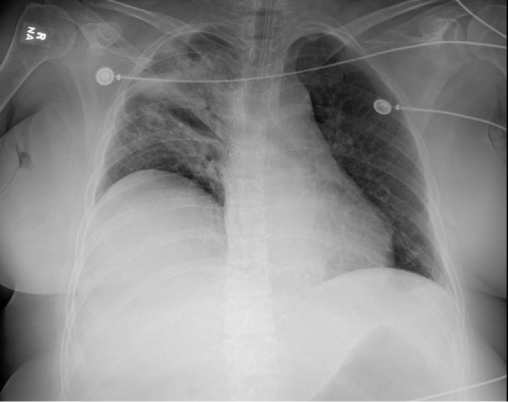
A 56-year-old woman with a history of alcohol use and depression presents after being found down at home by her boyfriend with an unknown downtime. She was found to be unresponsive and in the supine position. Her physical exam did not show any obvious trauma but the paramedics did note vomitus on her face. She received 1 L of crystalloids in the field and was intubated and brought to the ED for further management. A  bag of pill bottles was found and brought with her. Her home medications include amlodipine, baclofen, buspirone, and hydroxyzine.

Key Learning Points

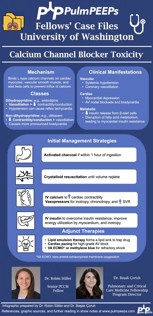
**Spoilers Ahead** If you want to think through the case on your own we advise listening to the episode first before looking at the infographic below

References and Further Reading

  1. Boyer EW, Shannon M. Treatment of calcium-channel-blocker intoxication with insulin infusion. N Engl J Med. 2001;344(22):1721-1722. doi:10.1056/NEJM200105313442215
  2. Cole JB, Arens AM, Laes JR, Klein LR, Bangh SA, Olives TD. High dose insulin for beta-blocker and calcium channel-blocker poisoning. Am J Emerg Med. 2018;36(10):1817-1824. doi:10.1016/j.ajem.2018.02.004
  3. Proano L, Chiang WK, Wang RY. Calcium channel blocker overdose. Am J Emerg Med. 1995;13(4):444-450. doi:10.1016/0735-6757(95)90137-X
  4. St-Onge M, Dubé PA, Gosselin S, et al. Treatment for calcium channel blocker poisoning: a systematic review. Clin Toxicol (Phila). 2014;52(9):926-944. doi:10.3109/15563650.2014.965827