22. Fellows’ Case Files: University of Maryland

This week we are absolutely thrilled to be launching a new series here at Pulm PEEPs. This is the first episode in our new Fellows’ Case Files series. The purpose of this series is to highlight the incredible clinical work that is done by pulmonary and critical care fellows everywhere, share fascinating cases from across the world, and assemble a diverse network of pulmonary and critical care educators. For each episode, we will visit a different institution, and be joined by a current fellow and the Pulmonary and Critical Care Fellowship Program Director. Our aim is to learn from them, amplify some incredible teaching points, and hear about their program. We hope you enjoy it, and if you have a case you want to bring on the series reach out to us on Twitter or at our email pulmpeeps@gmail.com.


Meet Our Guests

Fahid Alghanim is a senior pulmonary and critical care fellow at the University of Maryland. He attended medical school at the Lebanese American University Gilbert and Rose-Marie Chagoury School of Medicine and completed his internal medicine residency at Johns Hopkins Bayview. He has published on topics ranging from lung transplants to patient navigators in the ICU.

Dr. Van Holden is an Associate Professor of  Medicine at the University of Maryland School of Medicine and the Pulmonary and Critical Care Fellowship Program director. Clinically, she specializes in interventional pulmonology. She is also an accomplished educator and is very active with the American Thoracic Society. She helped write the 2021 Critical Care Core Curriculum and helped coordinate the 2022 Resident Boot Camp.

Patient Presentation

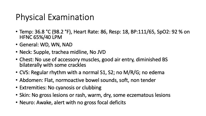
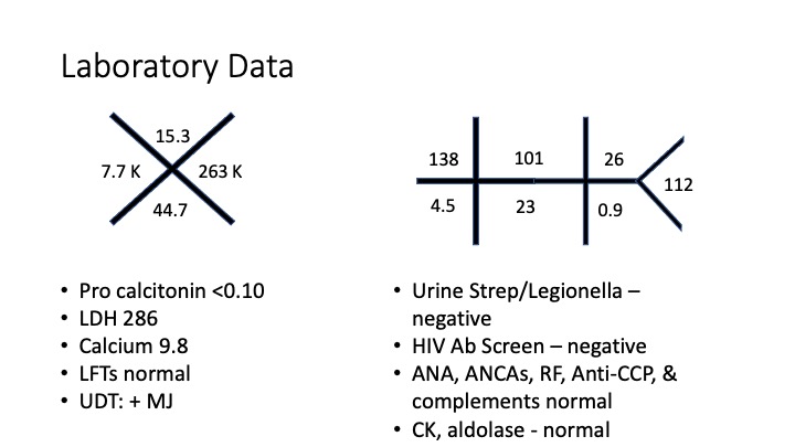
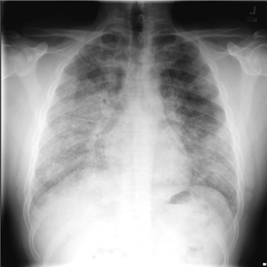
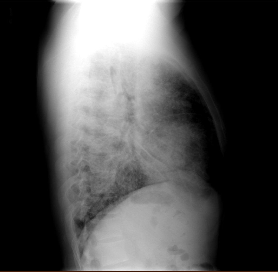
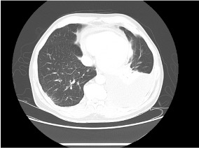
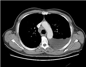
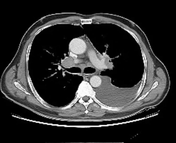
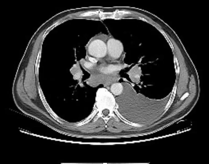
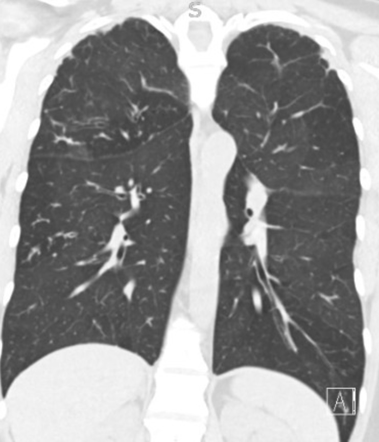
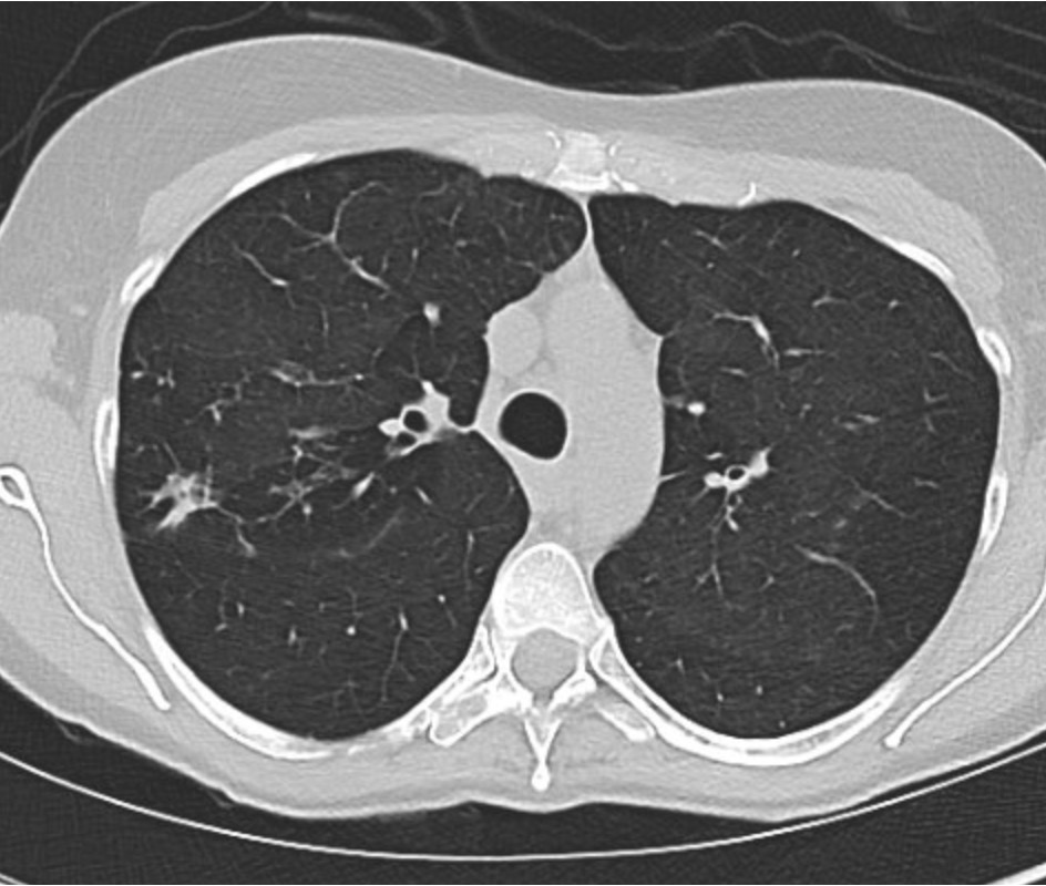
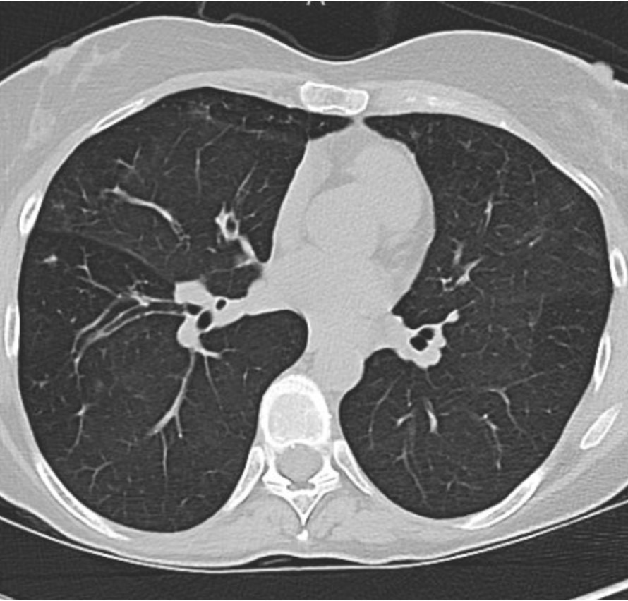
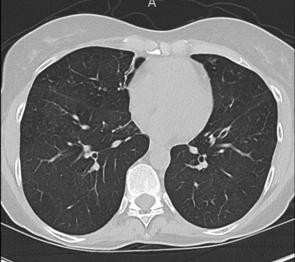
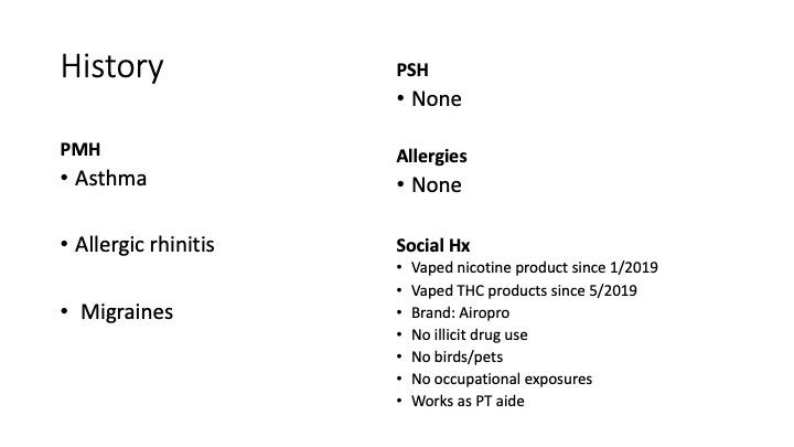
A 26-year-old man presents to his primary care doctor with 1.5 months of intermittent dyspnea, cough, chest tightness, and fatigue. His dyspnea was initially exertional, and he noticed he could do less at the gym. However, in the past 3-4 weeks it has progressed to being even with mild movement. His brother was recently diagnosed and treated for acute bronchitis so he thought this could be similar. In the office, he is noted to be tachypneic with an oxygen saturation of 83% breathing ambient air. A chest X-ray is obtained and he is sent urgently to the emergency department.
Key Learning Points

**Spoilers Ahead** If you want to think through the case on your own we advise listening to the episode first before looking at the infographics below

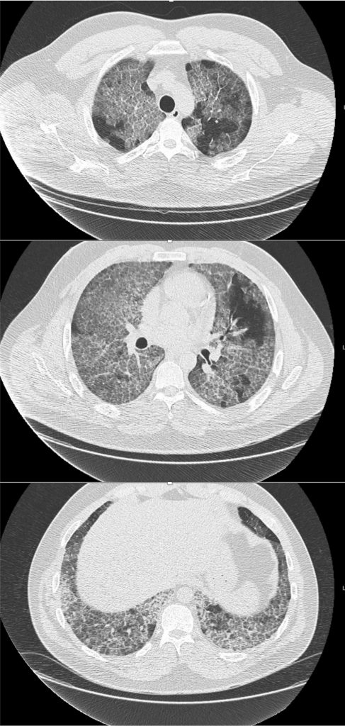
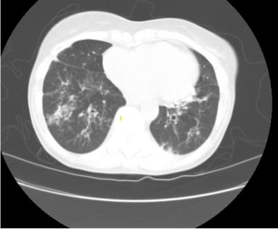
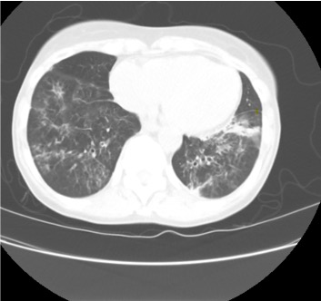
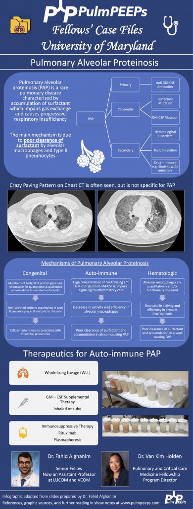
  1. Crazy Paving is a radiological term describing ground glass opacities with superimposed interlobular septal thickening. The differential diagnosis is broad and includes infectious, neoplastic, and autoimmune processes. It is not limited to just Pulmonary alveolar proteinosis (PAP) but is suggestive in an appropriate clinical setting.
  2. PAP is a disorder of surfactant production or clearance and its etiology is divided into three major subgroups. Primary or autoimmune; Secondary such as from toxic inhalations, hematological disorders, or medications; and Congenital
  3. PAP is diagnosed by positive Periodic acid-Schiff (PAS) staining of lipo-proteinaceous material in the distal bronchioles and alveoli on lung biopsy. The diagnosis can be made with PAS-positive BAL staining, but this has limited sensitivity and lung biopsy is necessary for the diagnosis in up to 30 – 35% of cases.
  4. It is important not to anchor on a diagnosis when a patient presents to you for re-evaluation even if seen by a prior expert. This was pivotal in this case!
  5. Please don’t put anything in your lung. Any toxic inhalation exposure could result in significant damage to lung parenchyma and morbidity as a result.
References and Further Reading

  1. Borie R, Danel C, Debray MP, et al. Pulmonary alveolar proteinosis. Eur Respir Rev. 2011;20(120):98-107. doi:10.1183/09059180.00001311
  2. Carey B, Trapnell BC. The molecular basis of pulmonary alveolar proteinosis. Clin Immunol. 2010;135(2):223-235. doi:10.1016/j.clim.2010.02.017
  3. Inoue Y, Trapnell BC, Tazawa R, et al. Characteristics of a large cohort of patients with autoimmune pulmonary alveolar proteinosis in Japan. Am J Respir Crit Care Med. 2008;177(7):752-762. doi:10.1164/rccm.200708-1271OC
  4. Kavuru MS, Malur A, Marshall I, et al. An open-label trial of rituximab therapy in pulmonary alveolar proteinosis. Eur Respir J. 2011;38(6):1361-1367. doi:10.1183/09031936.00197710
  5. Michaud G, Reddy C, Ernst A. Whole-lung lavage for pulmonary alveolar proteinosis. Chest. 2009;136(6):1678-1681. doi:10.1378/chest.09-2295
  6. Smith BB, Torres NE, Hyder JA, et al. Whole-lung Lavage and Pulmonary Alveolar Proteinosis: Review of Clinical and Patient-centered Outcomes. J Cardiothorac Vasc Anesth. 2019;33(9):2453-2461. doi:10.1053/j.jvca.2019.03.047
  7. Tazawa R, Ueda T, Abe M, et al. Inhaled GM-CSF for Pulmonary Alveolar Proteinosis. New England Journal of Medicine. 2019;381(10):923-932. doi:10.1056/NEJMoa1816216
  8. Tung AH, Grace J, O’Kane GM, Kumar K. Transbronchial lung biopsy (TBLB) in diagnosing pulmonary alveolar proteinosis (PAP): forgotten role in Australia? Respirology Case Reports. 2015;3(4):145-147. doi:10.1002/rcr2.129
  9. Werner AK, Koumans EH, Chatham-Stephens K, et al. Hospitalizations and Deaths Associated with EVALI. New England Journal of Medicine. 2020;382(17):1589-1598. doi:10.1056/NEJMoa1915314

18. A Case of Severe Weakness in the ICU

We are thrilled here @PulmPEEPS to have our first episode with one of our new Associate Editors Luke Hedrick, and our first nephrology consultant Jeff William. Luke will walk us through an interesting case presentation, and we will discuss an approach to severe weakness in our patient in the ICU.
Meet Our Guests

Jeff William is an Assistant Professor of Medicine at Harvard Medical School and Beth Israel Deaconess Medical Center, where he is also the Associate Director of the Nephrology Fellowship Program. He completed a Medical Education Research Fellowship at Harvard Medical School, and is very involved in residency, fellowship and medical student education.

Patient Presentation

We have a man in his 40s with a past medical history of asthma, hypertension, and acid reflux who was brought in by EMS with back pain and profound proximal lower extremity weakness. He reports mild weakness in his legs which started 2 days ago, but this morning his weakness acutely worsened to the point that he can’t lift his legs out of the bed. He also has some cramping pain in his thighs. He additionally has had mild shortness of breath and yesterday went to an urgent care where he was given steroids and swabbed for COVID (which was negative).

Key Learning Points

**Spoilers Ahead** If you want to think through the case on your own we advise listening to the episode first before looking at the infographics below

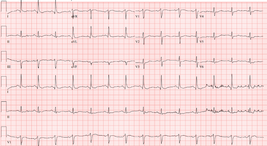
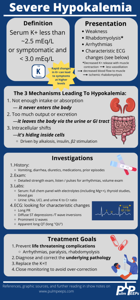
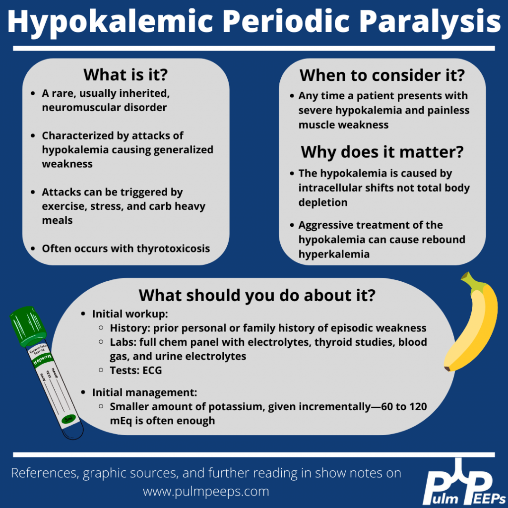
Although our patient’s etiology of severe hypokalemia was thought to be secondary to thiazide diuretic use, it is important to be familiar with hypokalemic periodic paralysis.

References

  1. Knochel JP, Schlein EM. On the mechanism of rhabdomyolysis in potassium depletion. J Clin Invest. 1972 Jul;51(7):1750-8. doi: 10.1172/JCI106976.
  2. Wang X, Han D, Li G. Electrocardiographic manifestations in severe hypokalemia. J Int Med Res. 2020 Jan;48(1):300060518811058. doi: 10.1177/0300060518811058.
  3. Venance SL, Cannon SC, Fialho D, Fontaine B, Hanna MG, Ptacek LJ, Tristani-Firouzi M, Tawil R, Griggs RC; CINCH investigators. The primary periodic paralyses: diagnosis, pathogenesis and treatment. Brain. 2006 Jan;129(Pt 1):8-17. doi: 10.1093/brain/awh639.
  4. Lin SH, Lin YF, Halperin ML. Hypokalaemia and paralysis. QJM. 2001 Mar;94(3):133-9. doi: 10.1093/qjmed/94.3.133. 
  5. Lin SH, Lin YF, Chen DT, Chu P, Hsu CW, Halperin ML. Laboratory tests to determine the cause of hypokalemia and paralysis. Arch Intern Med. 2004 Jul 26;164(14):1561-6. doi: 10.1001/archinte.164.14.1561.

16. A Case of Hemoptysis and Hypertension

We are thrilled here at Pulm PEEPs to have our first episode with our new Associate Editor Tess Litchman. Tess will walk us through an interesting case presentation of hemoptysis and we’ll use the approach from our Top Consults episode on hemoptysis to come to a key pulmonary and critical care diagnosis.

Meet Our Guests

Tess Litchman is a second-year internal medicine resident at Beth Israel Deaconess Medical Center. She received her undergraduate degree from Wesleyan University in Middletown, CT where she studied neuroscience and internal relations. She attended medical school at the Yale School of Medicine in New Haven, CT. She is currently completing her internal medicine residency at BIDMC. She is interested in medical education and pulmonary and critical care medicine.

Patient Presentation

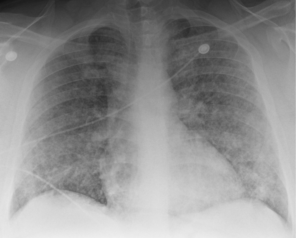
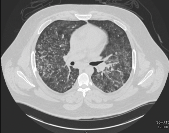
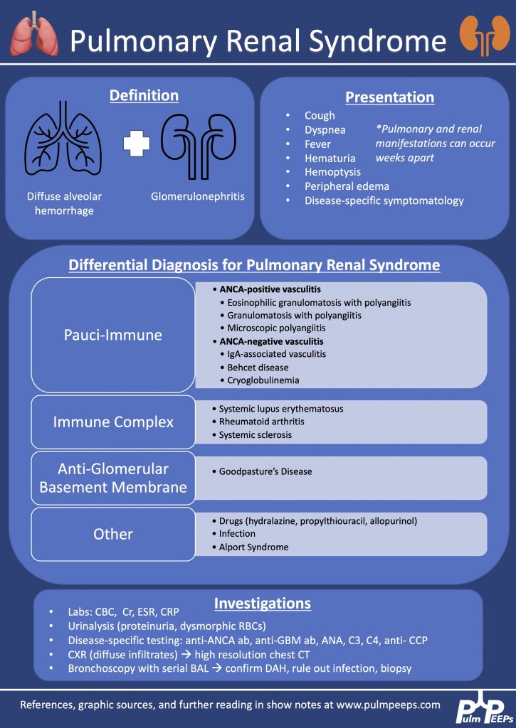
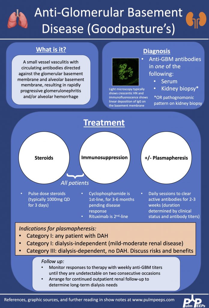
A young man in his 20s presented to the emergency department with one week of cough and small volume hemoptysis. He has been experiencing several episodes of hemoptysis per day during this time. He says he coughs up about 1/4 cup of blood with each episode. He also adds that for the past 2 weeks he also has noticed worsening nausea, vomiting, headaches, and fatigue. He saw his primary care doctor and he was diagnosed with new hypertension and started on clonidine 0.1 mg three times a day, and provided cough medication. However, his symptoms continued. Given the increasing frequency of the hemoptysis and worsening nausea, he presented to the emergency department.

Key Learning Points

**Spoilers Ahead** If you want to think through the case on your own we advise listening to the episode first before looking at the infographics below

References and links for further reading

  1. Radchenko C, Alraiyes AH, Shojaee S. A systematic approach to the management of massive hemoptysis. J Thorac Dis. 2017;9(Suppl 10):S1069-S1086. doi:10.21037/jtd.2017.06.41
  2. Lara AR, Schwarz MI. Diffuse Alveolar Hemorrhage. CHEST. 2010;137(5):1164-1171. doi:10.1378/chest.08-2084
  3. Gallagher H, Kwan JTC, Jayne DRW. Pulmonary renal syndrome: A 4-year, single-center experience. American Journal of Kidney Diseases. 2002;39(1):42-47. doi:10.1053/ajkd.2002.29876
  4. Sanders JSF, Rutgers A, Stegeman CA, Kallenberg CGM. Pulmonary-Renal Syndrome with a Focus on Anti-GBM Disease. Semin Respir Crit Care Med. 2011;32(3):328-334. doi:10.1055/s-0031-1279829
  5. Hudson BG, Tryggvason K, Sundaramoorthy M, Neilson EG. Alport’s syndrome, Goodpasture’s syndrome, and type IV collagen. N Engl J Med. 2003;348(25):2543-2556. doi:10.1056/NEJMra022296
  6. McAdoo SP, Pusey CD. Anti-Glomerular Basement Membrane Disease. Clin J Am Soc Nephrol. 2017;12(7):1162-1172. doi:10.2215/CJN.01380217
  7. Maxwell AP, Nelson WE, Hill CM. Reversal of renal failure in nephritis associated with antibody to glomerular basement membrane. BMJ. 1988;297(6644):333-334. doi:10.1136/bmj.297.6644.333

10. A Case of Fevers, Night Sweats, and Dyspnea

This week, we have another great case episode on Pulm PEEPs! We are joined by Emily Fridenmaker who helps us think through a fascinating case presenation.

Meet Our Guests

Emily Fridenmaker is a Pulmonary, Critical Care, and Sleep Medicine fellow at the University of Kentucky College of Medicine. She went to medical school at West Virginia school of medicine and did her internal medicine residency at Charleston Area Medical Center

Patient Presentation

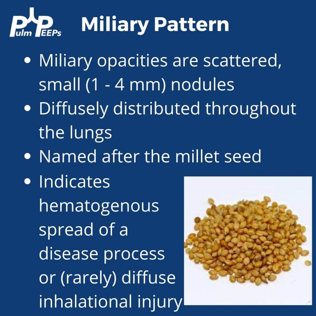
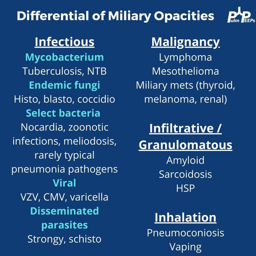
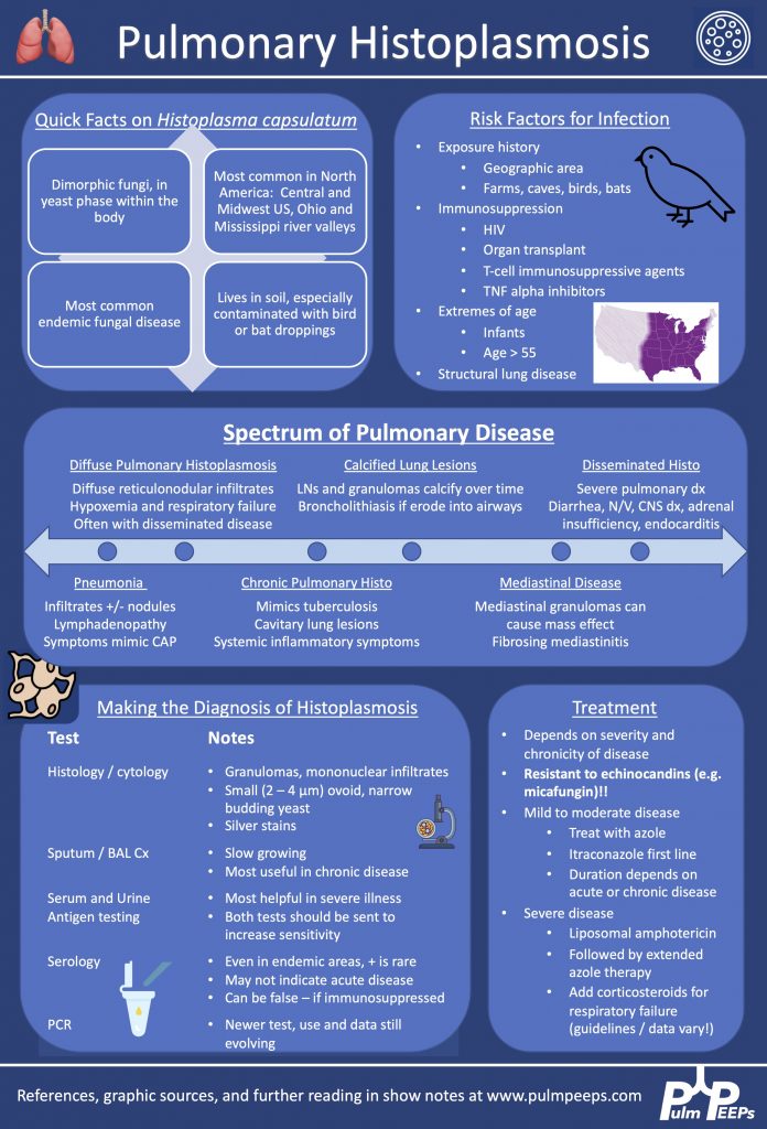
A middle-aged woman presents with 2-3 weeks of mild but progressive shortness of breath with exertion associated with low-grade fevers, worsening night sweats, and fatigue. Further history reveals a progressive non-productive cough and weight loss. She has a past medical history of neurologic dysfunction over two years and a working diagnosis of chronic inflammatory demyelinating polyneuropathy and is pursuing treatment in Mexico due to cost limitations. She has been receiving prednisone 10 mg daily and azathioprine. Aside from travel history to Mexico, her social history is notable for prior employment in a candle factory, and for hiking with some cave exploration. She is a former 20 pack-year smoker and has rare alcohol use.

Key Learning Points

References and links for further reading

  1. McKinsey DS, McKinsey JP. Pulmonary histoplasmosis. Semin Respir Crit Care Med. 2011;32(6):735-744. doi:10.1055/s-0031-1295721
  2. Cuellar-Rodriguez J, Avery RK, Lard M, et al. Histoplasmosis in solid organ transplant recipients: 10 years of experience at a large transplant center in an endemic area. Clin Infect Dis. 2009;49(5):710-716. doi:10.1086/604712
  3. Wheat LJ, Slama TG, Norton JA, et al. Risk Factors for Disseminated or Fatal Histoplasmosis. Ann Intern Med. 1982;96(2):159-163. doi:10.7326/0003-4819-96-2-159
  4. Smith JA, Kauffman CA. Endemic fungal infections in patients receiving tumour necrosis factor-alpha inhibitor therapy. Drugs. 2009;69(11):1403-1415. doi:10.2165/00003495-200969110-00002
  5. Poplin V, Smith C, Milsap D, Zabel L, Bahr NC. Diagnosis of Pulmonary Infections Due to Endemic Fungi. Diagnostics. 2021;11(5):856. doi:10.3390/diagnostics11050856
  6. Wheat LJ, Freifeld AG, Kleiman MB, et al. Clinical Practice Guidelines for the Management of Patients with Histoplasmosis: 2007 Update by the Infectious Diseases Society of America. Clinical Infectious Diseases. 2007;45(7):807-825.
  7. Azar MM, Hage CA. Clinical Perspectives in the Diagnosis and Management of Histoplasmosis. Clinics in Chest Medicine. 2017;38(3):403-415. doi:10.1016/j.ccm.2017.04.004
  8. Threadcraft MA, Case R. Vape-Associated Pulmonary Injury (VAPI) Presenting With a “Miliary” Pattern on Imaging. Cureus. 13(2):e13385. doi:10.7759/cureus.13385
  9. Raoof S, Amchentsev A, Vlahos I, Goud A, Naidich DP. Pictorial essay: multinodular disease: a high-resolution CT scan diagnostic algorithm. Chest. 2006;129(3):805-815. doi:10.1378/chest.129.3.805
  10. Sharma BB. Miliary nodules on chest radiographs: A diagnostic dilemma. Lung India. 2015;32(5):518-520.
  11. Purek L, Laroumagne S, Dutau H, Maldonado F, Astoul P. Miliary mesothelioma: a new clinical and radiological presentation in mesothelioma patients with prolonged survival after trimodality therapy. J Thorac Oncol. 2011;6(10):1753-1756. doi:10.1097/JTO.0b013e31822e295a

8. A Case of Dyspnea and Lymphadenopathy

Dave Furfaro, Kristina Montemayor, and Ansa Razzaq are back to tackle another pulmonary case! Listen in and solve the case yourself, and we’ll share some diagnostic pearls along the way. Let us know any additional thoughts on Twitter.

Patient Presentation

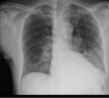
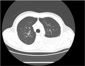
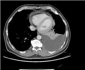
The is patient is 57-year-old man with hypertension and asthma who presents with dyspnea and left-sided pleuritic chest pain for 3 weeks. He was in his usual state of health until 3 weeks prior to admission, when he developed dyspnea and sharp left-sided chest pain that worsens with deep breathing. His symptoms are accompanied by unintentional 30-pound weight loss over the past several months as well as an intermittent cough that is nonproductive.

On physical exam, he is mildly tachypneic and saturating well on room air with otherwise normal vital signs. He has decreased breath sounds at the right lung base.

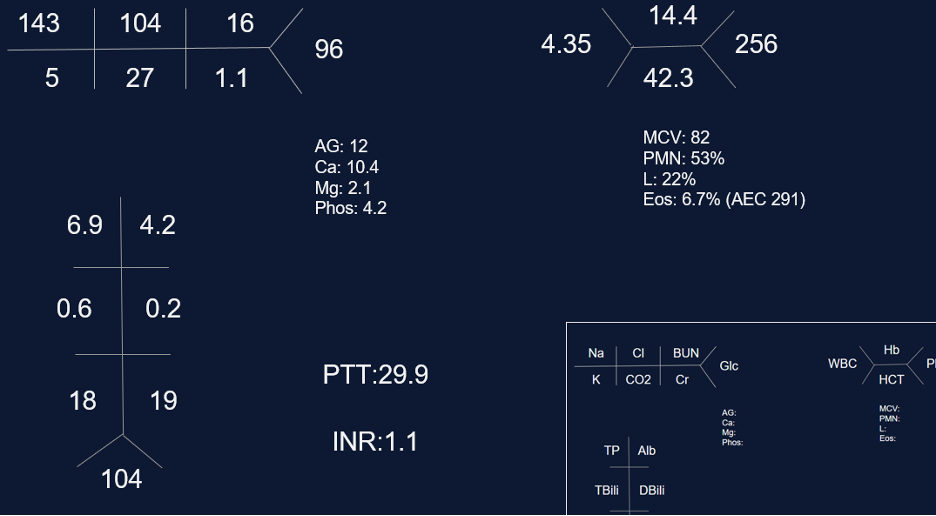
Initial labs

Key Learning Points

**Spoilers Ahead** If you want to think through the case on your own we advise listening to the episode first before looking at the infographics below

Physical Exam Pearls

Reasons for decreased breath sounds on physical exam

1. Increased thickness of chest wall

2. Reduced airflow to part of the lung

3. Overinflation to part of the lung

4. Something between the lung and chest wall — air or fluid

Determine bradypnea and tachypnea quickly by matching your breahting rate to the patient’s respiratory rate

Pleural Effusions

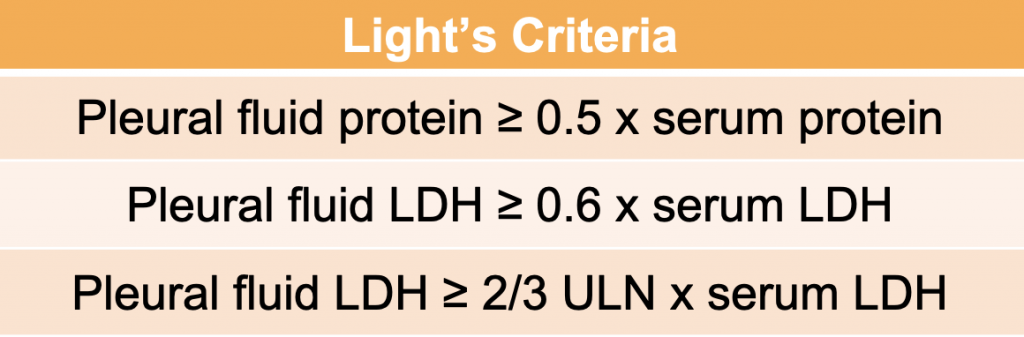
Transudative effusion partial differential: heart failure, nephrotic syndrome, hepatohydrothorax, fluid overload, hypoalbuminemia, urinothorax, amyloidosis*, chlothorax*, hypothyrodism*, malignancy*, sarcoidosis*

*these effusions often present as exudates but can be transudates

Exudative effusion partial differential: Infection, hemothorax, malignancy, connective tissue disease, chylothorax, pancreatitis, esophageal perforation.

Sarcoidosis

Key Elements of Diagnosis:

1. Is there a compatible presentation (imaging, physical exam)

2. Detection of non-necrotizing granulomatous inflammation in one or more tissue samples

3. Exclusion of other disease that may present similarily

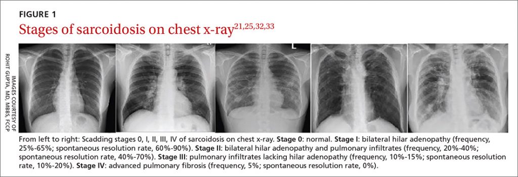
Pulmonary stages of sarcoidosis:

Key to remember that patients don’t always progress through these stages. The system is useful for prognosticating and determining treatment based on the risk for disease progression.

Image source: Gupta Et Al. J Fam Pract. 2021 April;70(3):E4-E15 | 10.12788/jfp.0177

Treatment for pulmonary sarcoidosis

Patients with stage 1, and even many with stage 2, often don’t require treatment

The first-line agent is oral glucocorticoids and the typical starting dose is prednisone 20 – 40 mg by mouth daily. The patient should be evaluated closely, and ideally, this dose can be tapered starting at about 4 – 6 weeks. Following this, the prednisone dose is tapered slowly over 6 months – 1 year while monitoring for symptom recurrence.

Second-line steroid-sparing agents are methotrexate, azathioprine, or mycophenolate. These are often used if the patient relapses, or is on more then 10mg daily for 3 months after the initial taper with intolerance of steroids

Third-line agents: tumor necrosis factor (TNF)- alpha antagonists (small molecule or monoclonal antibody therapy)

References and links for further reading

  1. Light RW. Pleural Effusion. New England Journal of Medicine. 2002;346(25):1971-1977. doi:10.1056/NEJMcp010731
  2. Feller-Kopman D, Light R. Pleural Disease. New England Journal of Medicine. 2018;378(8):740-751. doi:10.1056/NEJMra1403503
  3. Sarcoidosis: An FP’s primer on an enigmatic disease. MDedge Family Medicine. 2021;70(3). doi:10.12788/jfp.0177
  4. Iannuzzi MC, Rybicki BA, Teirstein AS. Sarcoidosis. New England Journal of Medicine. 2007;357(21):2153-2165. doi:10.1056/NEJMra071714
  5. Drent M, Crouser ED, Grunewald J. Challenges of Sarcoidosis and Its Management. New England Journal of Medicine. 2021;385(11):1018-1032. doi:10.1056/NEJMra2101555

5. A Case of Chronic, Productive Cough

The Pulm PEEPs are joined again by Natalie West to discuss a patient who presented with a chronic, productive cough. Listen in today as we work through our differential diagnosis, interpret basic pulmonary testing, and share our clinical reasoning along the way. We have some fantastic diagnostic and treatment teaching points, so once you’ve solved the case check out the takeaways and infographics below. Please let us know any additional insights you have on Twitter!

Patient Presentation

A 50-year-old woman, who is a never smoker, with a past medical history of recurrent pancreatitis presents to the pulmonary clinic with a chronic, productive cough. Her cough has been present for 3 years and has increased in frequency to now being present daily. In the last three months, the cough has also worsened and is productive of small amounts of yellow to green sputum. She has a history of chronic post-nasal drip and sinus infections, and uses intranasal steroids, but has not noted changes in these symptoms. There is no significant family history of pulmonary disease, and an exposure history review of symptoms is negative.

On physical exam, she was a thin woman who appeared her stated age and was breathing comfortably on room air. Her exam was notable for mild expiratory wheezing, primarily on auscultation of the right posterior lung field. She had no cyanosis, clubbing, evidence of volume overload, or abdominal tenderness.

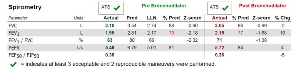
Basic Spirometry Values
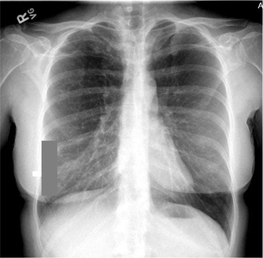
Chest X-ray

Key Learning Points

**Spoilers Ahead** If you want to think through the case on your own we advise listening to the episode first before looking at the infographics below.

Differential Diagnosis of Chronic Cough

Three most common causes: upper airway cough syndrome, GERD, cough variant asthma

Additional etiologies to consider: chronic bronchitis, post-infectious after a respiratory tract infection, bronchiectasis, ACE inhibitors, lung cancer, eosinophilic bronchitis, interstitial lung disease


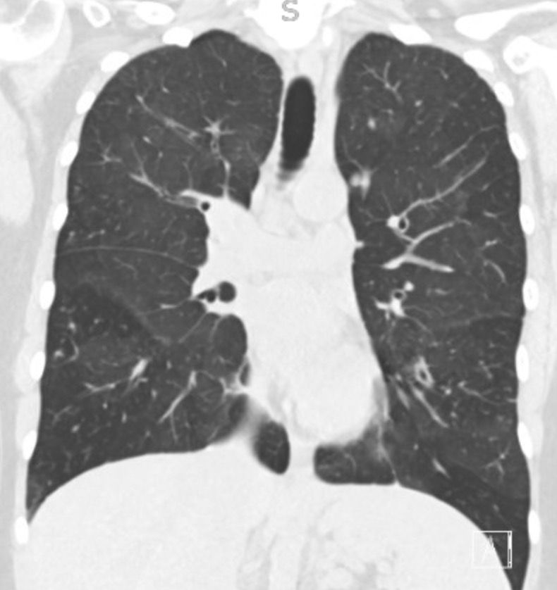
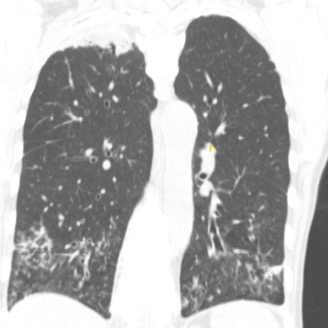
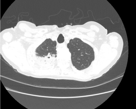
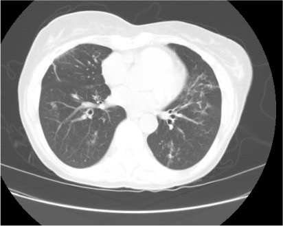
Imaging Pearl


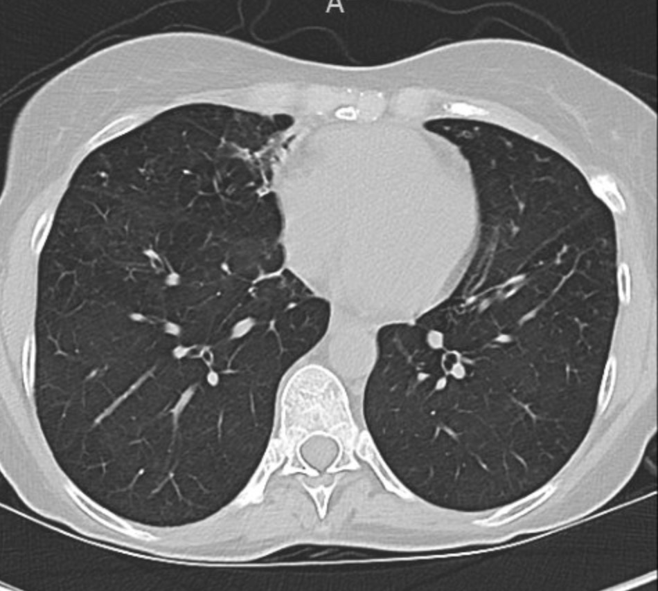
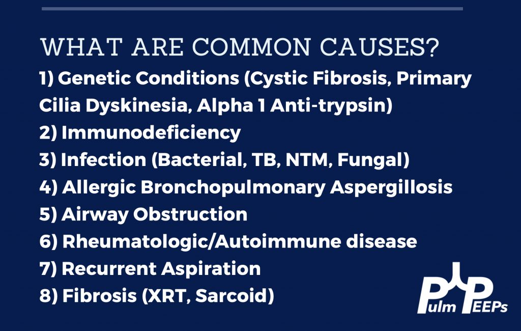
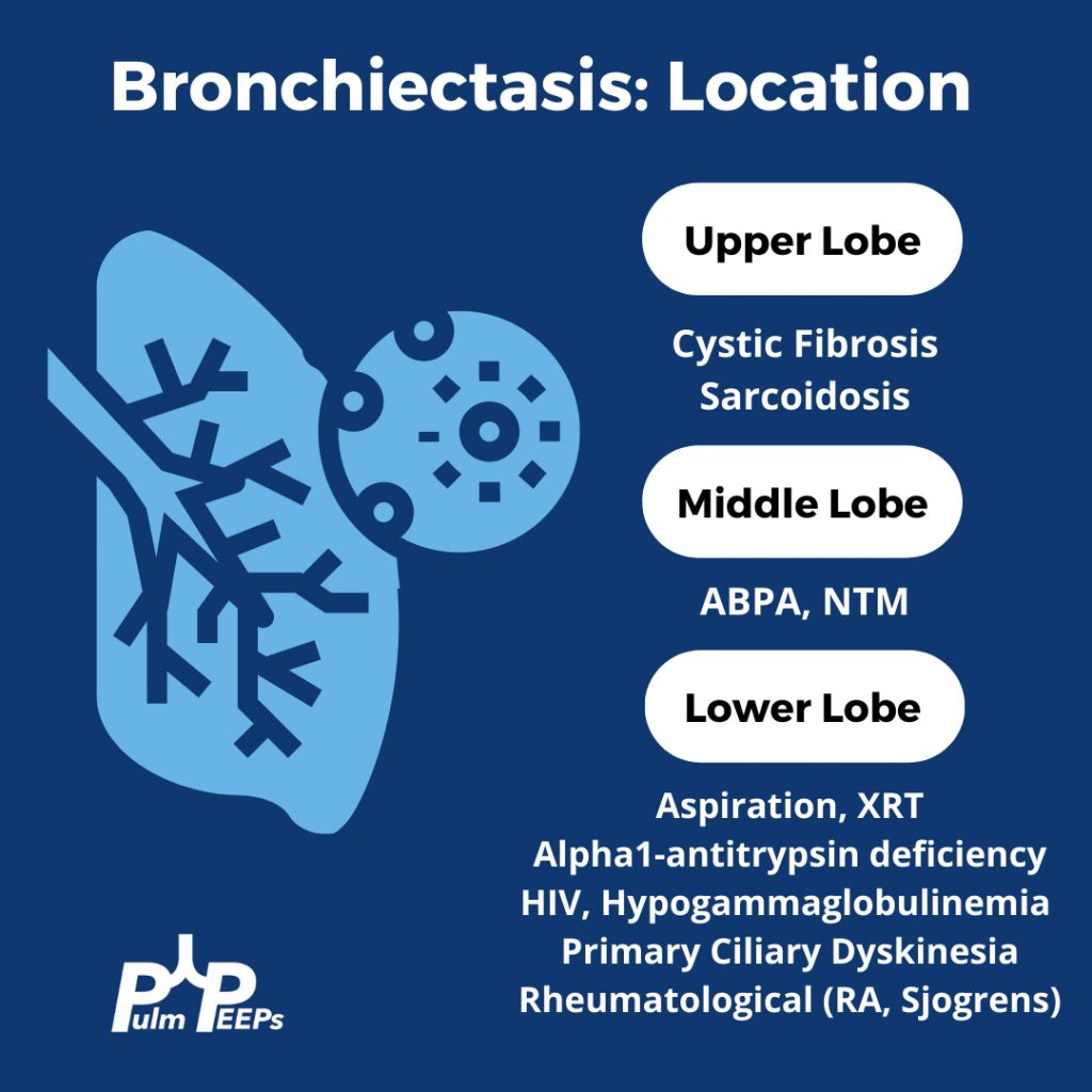
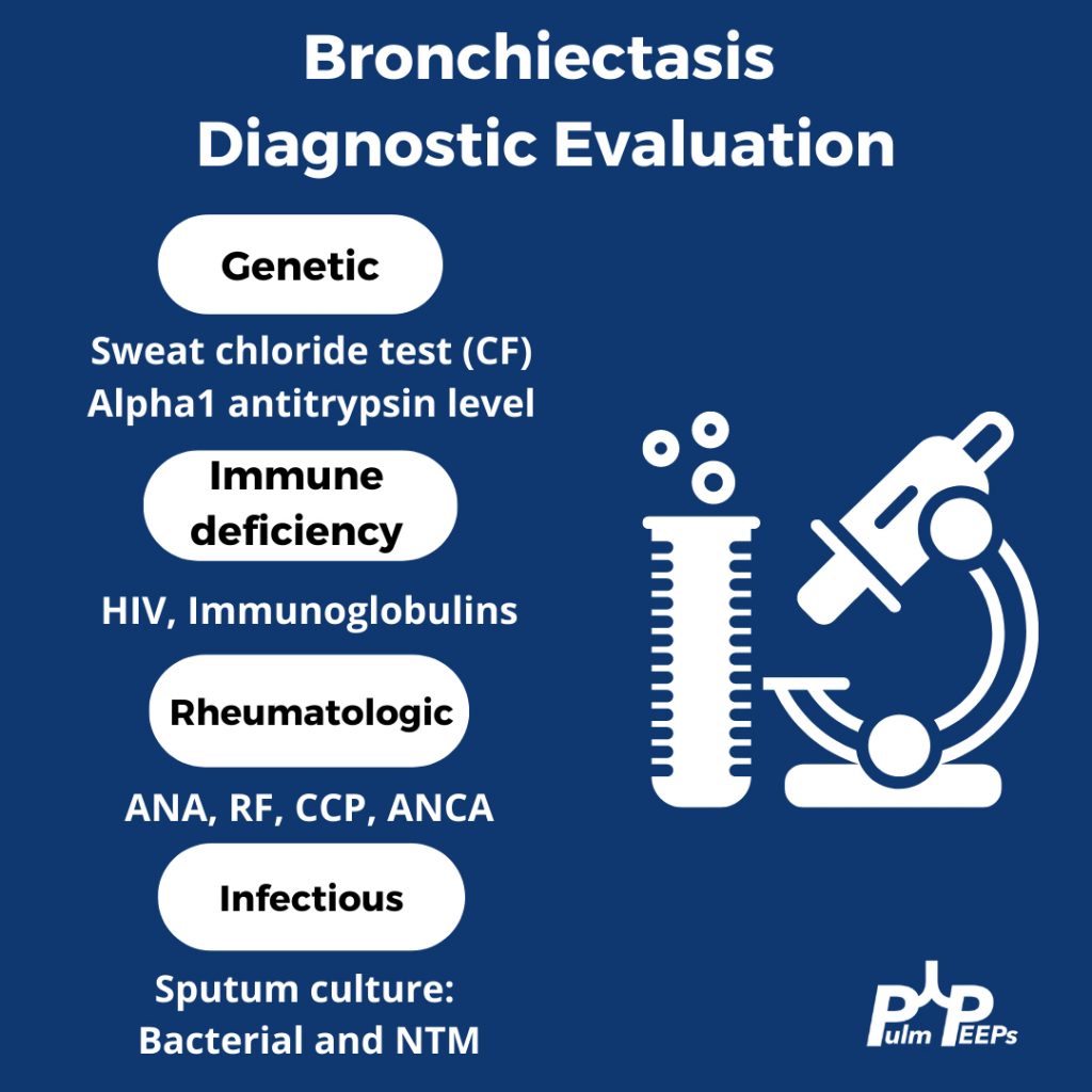
Evaluating Bronchiectasis


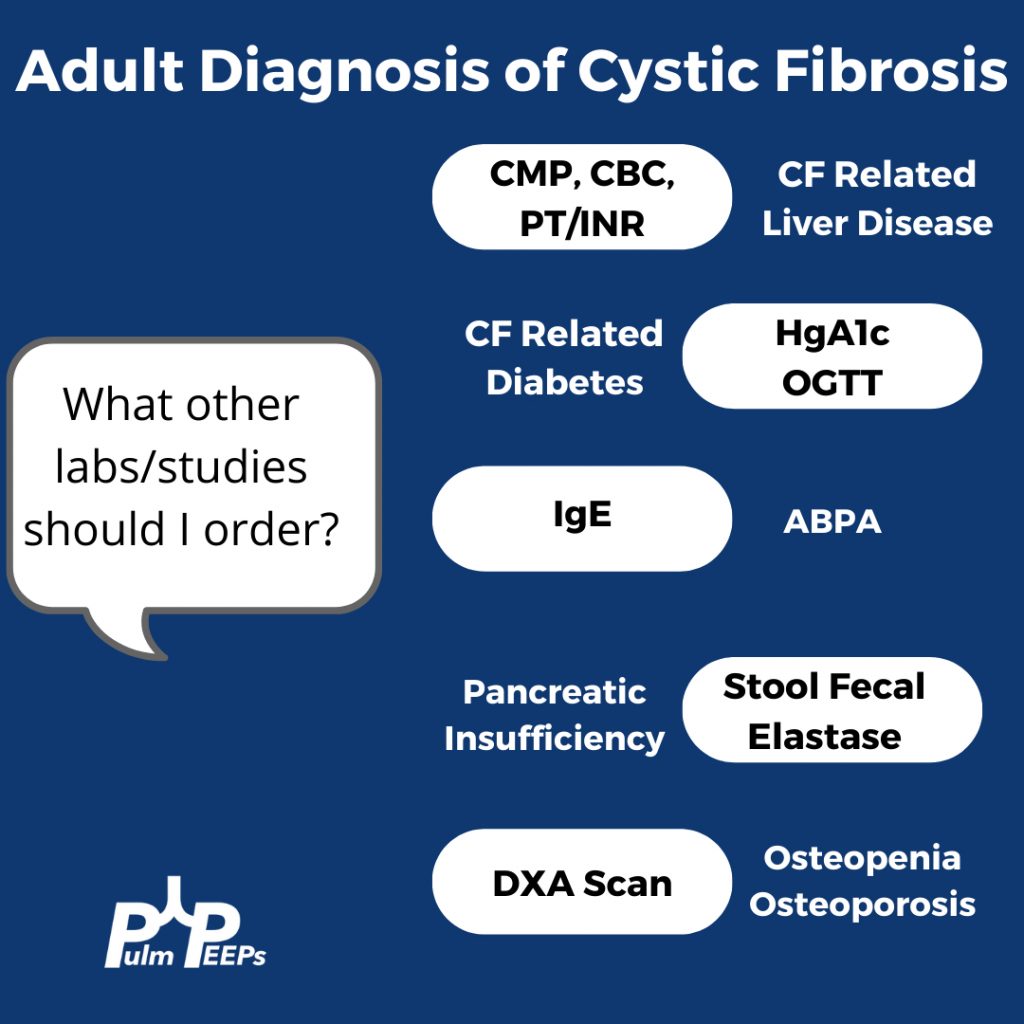
Making a New Diagnosis of Cystic Fibrosis in an Adult

Sweat testing

Sweat testing should be done in CF accredited center. Inform patients that there are no needles involved. Pilocarpine and electrical stimulation are applied to the arm or leg to stimulate the sweat gland, and then sweat is collected on filter paper, a gauze, or a plastic coil. From there, the amount of chloride in the sweat is calculated

Results

< 30 normal

31 – 60 indeterminate

> 60 is positive and Cystic Fibrosis is likely

What do you do with an Indeterminate test?

Patients with milder phenotypes of Cystic Fibrosis can have a normal or indeterminate sweat chloride level, and 10% of adults diagnosed with CF have a normal sweat chloride. If the sweat chloride test is indeterminate or normal, but suspicion is high for CF, then genetic testing for the whole array of mutations should be performed

References and links for further reading

  1. Morice AH, Millqvist E, Bieksiene K, et al. ERS guidelines on the diagnosis and treatment of chronic cough in adults and children. European Respiratory Journal. 2020;55(1). doi:10.1183/13993003.01136-2019
  2. Barker AF. Bronchiectasis. New England Journal of Medicine. 2002;346(18):1383-1393. doi:10.1056/NEJMra012519
  3. Bronchiectasis: a case-based approach to investigation and management | European Respiratory Society. Accessed November 23, 2021. https://err.ersjournals.com/content/27/149/180016
  4. Rowe SM, Miller S, Sorscher EJ. Cystic Fibrosis. New England Journal of Medicine. 2005;352(19):1992-2001. doi:10.1056/NEJMra043184
  5. Shteinberg M, Haq IJ, Polineni D, Davies JC. Cystic fibrosis. The Lancet. 2021;397(10290):2195-2211. doi:10.1016/S0140-6736(20)32542-3
  6. Jain R. Diagnosing Cystic Fibrosis in Adults: Better Late Than Never. Annals ATS. 2018;15(10):1140-1141. doi:10.1513/AnnalsATS.201806-432ED

3. A Case of Worsening Episodic Dyspnea

The Pulm PEEPs are excited to bring our first mystery case! Kristina Montemayor and Dave Furfaro hear a fascinating case presentation from Pulm PEEPs senior editor Ansa Razzaq. Join us as we work through this case together to come to a diagnosis, and share our thought process along the way. Come back to these show notes afterward, or once you’ve solved the case yourself, for some key teaching pearls and representative images.

Patient Presentation

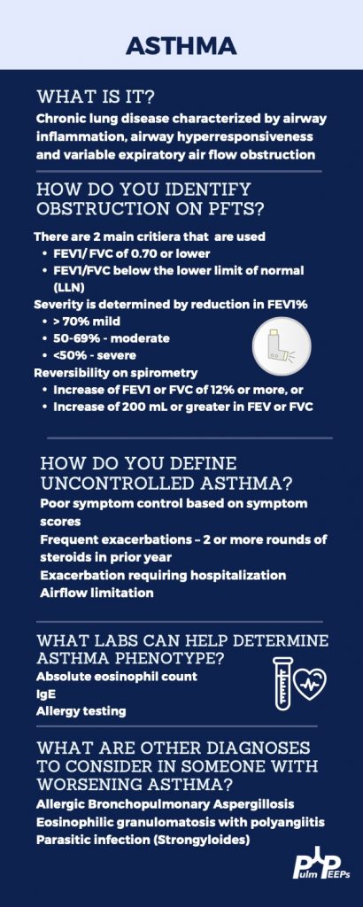
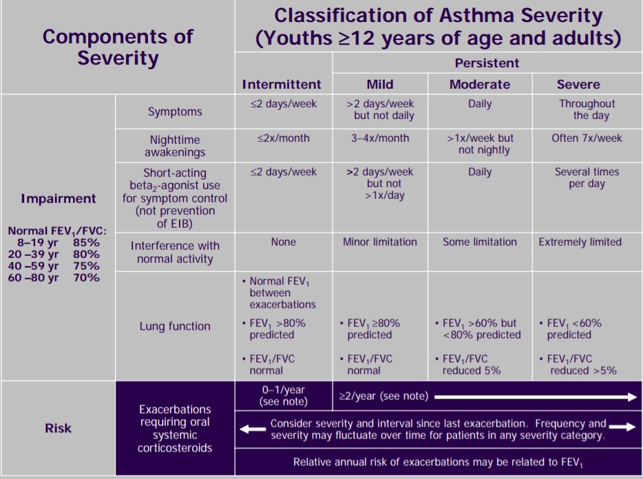
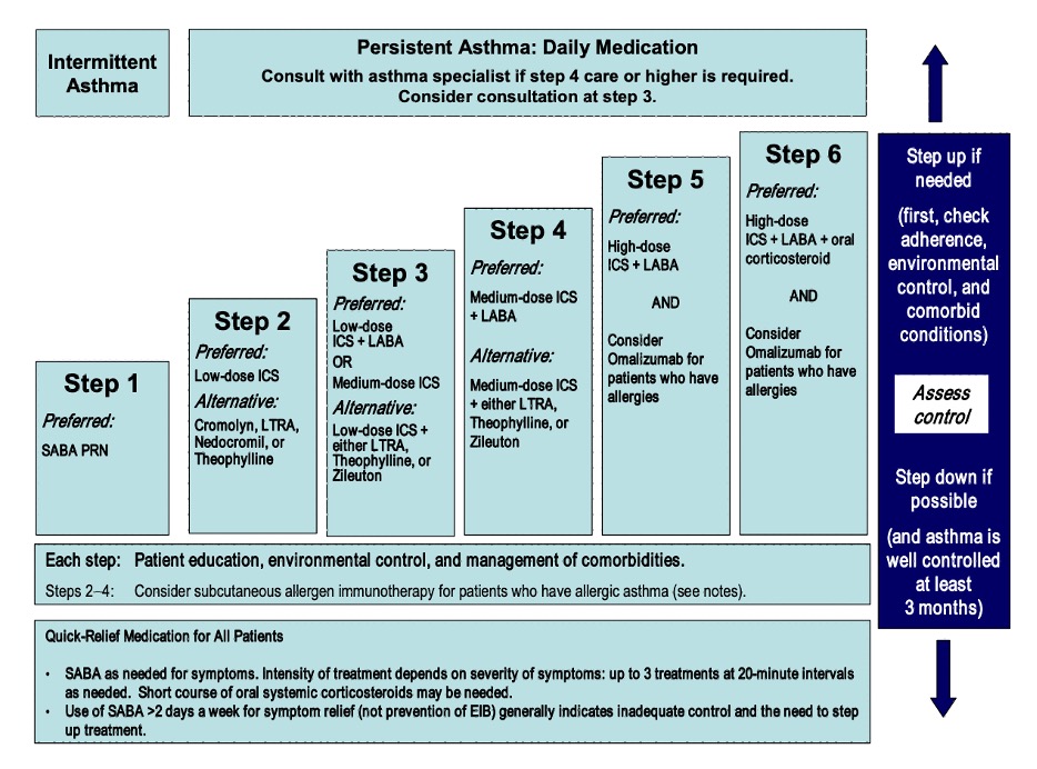
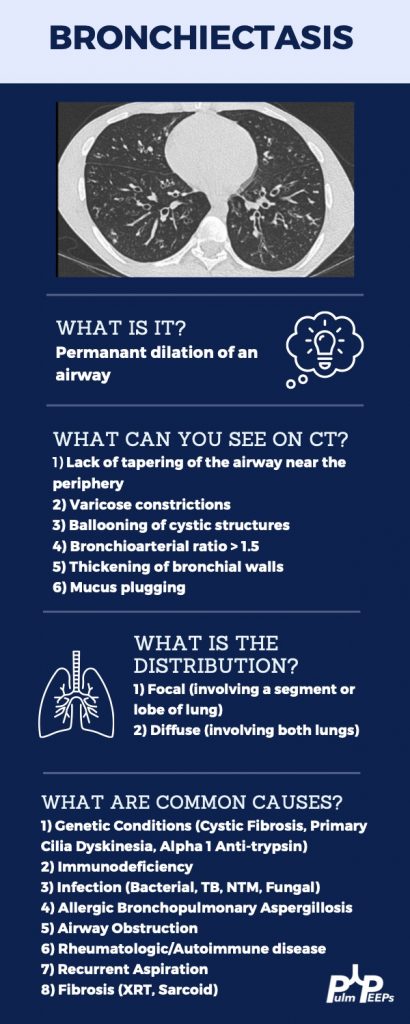
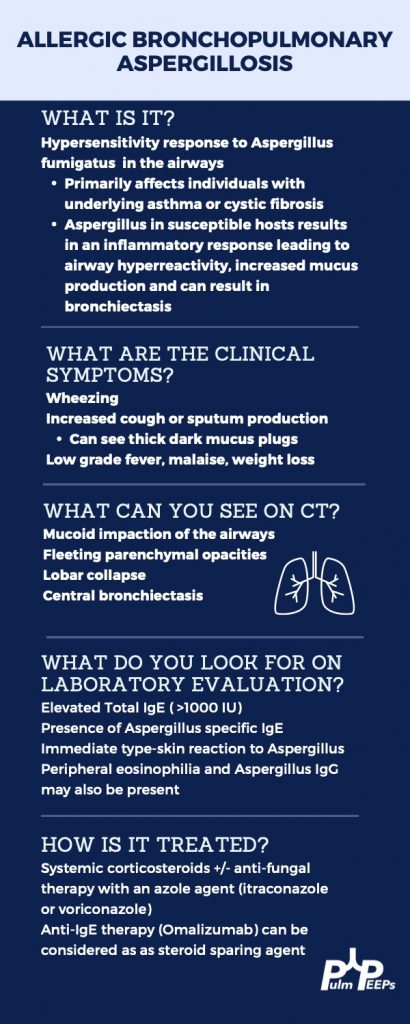
A 66-year-old woman with no smoking history and past medical history of previously well-controlled asthma is referred to pulmonary clinic after multiple recent episodes of dyspnea, wheezing, and coughing. The episodes have features consistent with asthma exacerbations; however, they are also associated with migratory infiltrates. She has been treated with multiple courses of antibiotics and steroids, and despite escalating therapy, the episodes are occurring more frequently and she was worsening overall exercise tolerance. Listen in to hear more and try to solve the case!

Key Learning Points

**Spoilers Ahead** If you want to think through the case on your own we advise listening to the episode first before looking at the infographics below.

References and links for further reading

  1. Israel E, Reddel HK. Severe and Difficult-to-Treat Asthma in Adults. New England Journal of Medicine. 2017;377(10):965-976. doi:10.1056/NEJMra1608969
  2. Asthma NAE and PP Third Expert Panel on the Diagnosis and Management of. Expert Panel Report 3: Guidelines for the Diagnosis and Management of Asthma. National Heart, Lung, and Blood Institute (US); 2007.
  3. Chung KF, Wenzel SE, Brozek JL, et al. International ERS/ATS guidelines on definition, evaluation and treatment of severe asthma. European Respiratory Journal. 2014;43(2):343-373. doi:10.1183/09031936.00202013
  4. Barker AF. Bronchiectasis. New England Journal of Medicine. 2002;346(18):1383-1393. doi:10.1056/NEJMra012519
  5. Chen T hsu, Hollingsworth H. Allergic Bronchopulmonary Aspergillosis. New England Journal of Medicine. 2008;359(6):e7. doi:10.1056/NEJMicm055764
  6. Agarwal R, Dhooria S, Singh Sehgal I, et al. A Randomized Trial of Itraconazole vs Prednisolone in Acute-Stage Allergic Bronchopulmonary Aspergillosis Complicating Asthma. Chest. 2018;153(3):656-664. doi:10.1016/j.chest.2018.01.005