107. Fellows’ Case Files: University of Kansas Medical Center KUMC

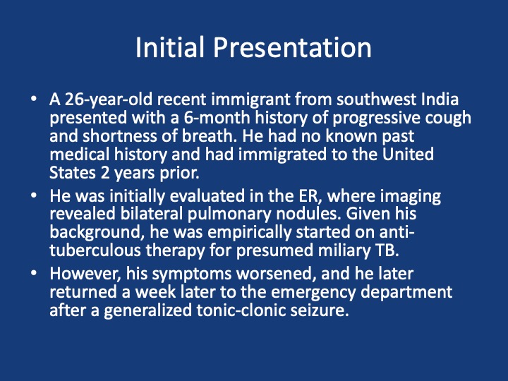
After a brief hiatus, we are excited to be back today with another Fellows’ Case Files! Today we’re virtually visiting the University of Kansas Medical Center (KUMC) to hear about a fascinating pulmonary presentation. There are some fantastic case images and key learning points. Take a listen and see if you can make the diagnosis along with us. As always, let us know your thoughts and definitely reach out if you have an interesting case you’d like to share.
Dr. Vishwajit Hegde completed his internal medicine residency at University of Kansas Medical Center where he stayed for fellowship and is currently a second year Pulmonary and Critical Care medicine fellow. 

Dr. Sahil Pandya is an Associate Professor of Medicine and Program Director of the PCCM Fellowship at KUMC.

1) Initial frame & diagnostic mindset

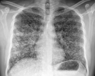
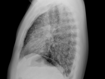
  • Young (26), subacute → chronic dyspnea/cough with diffuse pulmonary nodules; avoid premature closure on TB.
  • Use a Bayesian approach: combine pre-test probability (epidemiology, exposures, tempo) with targeted tests to decide next steps.
  • Always confirm TB when possible (micro/path + resistance testing); empiric RIPE may be reasonable but shouldn’t replace tissue when stakes are high.

2) Imaging pearls—nodular pattern recognition

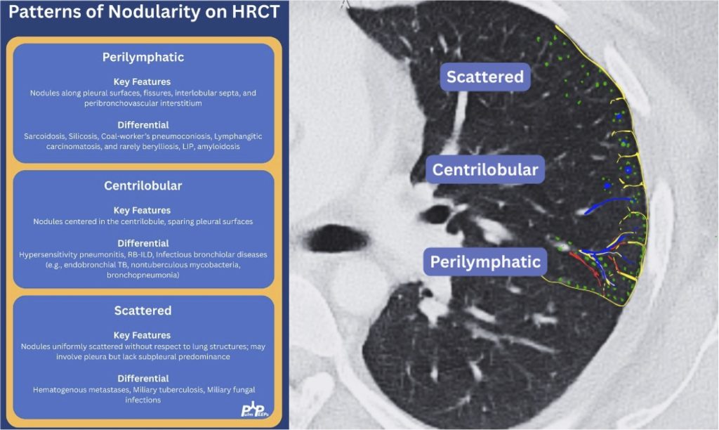
  • Ask three things: craniocaudal distribution, symmetry, central vs peripheral.
  • Centrilobular (spares pleura/fissures): airway-centered (e.g., NTM, bronchiolitis, tree-in-bud).
  • Perilymphatic (tracks fissures/pleura & septa): sarcoid, lymphangitic spread.
  • Random/diffuse (involves pleural surfaces): hematogenous spread → think miliary TB, disseminated fungal, septic emboli, metastatic disease.
  • Interval change matters: new cavitation and confluence can upweight infection or aggressive malignancy.

3) Neuro findings—ring-enhancing lesions

  • Differential: septic emboli/abscess, nocardia, fungal, TB, parasites, metastases, vasculitis, sarcoid.
  • Partner with neuroradiology for pattern nuances; treat seizures but keep searching for the unifying diagnosis.

4) Lab/serology strategy

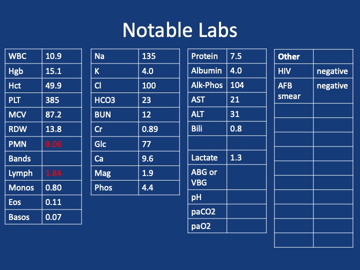
  • Broad infectious workup (AFB × multiple, fungal serologies), HIV and basic immune screen.
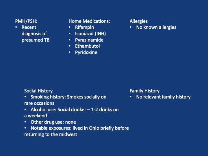
  • Negative/indeterminate tests don’t end the search—revisit history (e.g., Ohio travel → histo/blasto risk).

5) “Tissue is the issue”—choosing the procedure

  • For diffuse nodules with mediastinal adenopathy and stable patient: EBUS-TBNA + BAL, consider transbronchial or cryobiopsy.
  • Cryobiopsy pros: larger, less crush artifact, better for molecular testing; cons: ↑ bleeding/pneumothorax vs forceps.
  • VATS still best for certain ILD questions or if less invasive routes are non-diagnostic—but weigh patient preference and stage/likelihood of yield.
6) ROSE (rapid on-site evaluation) in bronchoscopy

  • Confirms adequacy in real time, steers you away from necrotic zones, helps decide when you’ve got enough for molecular studies, and when to pivot sites—reduces anesthesia time and repeat procedures.

7) Final diagnosis & management

  • Path: TTF-1+, CK7+, napsin A → pulmonary adenocarcinoma with a fusion driver.
  • Therapy: Targeted TKI (crizotinib) → dramatic radiographic response of miliary lung disease and CNS lesions.
  • Teaching point: even “miliary TB-like” lungs + CNS lesions in a 20-something can be driver-positive lung cancer—don’t let age or pattern blind you.

  1. Desai, S., Devaraj, A., Lynch, D., & Sverzellati, N. (2020). Webb, Müller and Naidich’s high-resolution CT of the lung (6th ed.). Lippincott Williams & Wilkins.
  2. Rajeswaran, G., Becker, J. L., Michailidis, C., Pozniak, A. L., & Padley, S. P. G. (2006). The radiology of IRIS (immune reconstitution inflammatory syndrome) in patients with mycobacterial tuberculosis and HIV co-infection: appearances in 11 patients. Clinical radiology, 61(10), 833-843
  3. Poletti, V., Ravaglia, C., & Tomassetti, S. (2016). Transbronchial  cryobiopsy in diffuse parenchymal lung diseases. Current opinion in pulmonary medicine, 22(3), 289-296.
  4. Norman, G. R., Monteiro, S. D., Sherbino, J., Ilgen, J. S., Schmidt, H. G., & Mamede, S. (2017). The causes of errors in clinical reasoning: cognitive biases, knowledge deficits, and dual process thinking. Academic Medicine, 92(1), 23-30.

70. Bronchoscopy Emergencies with Critical Care Time

We’re super excited to have a joint episode this week with Dr. Cyrus Askin and Dr. Nick Mark from Critical Care Time! We discuss all the ways that bronchoscopy can be your best friend in the ICU and how to be prepared for the unexpected scary situations that arise in the ICU. This ranges from airway bleeds, difficult intubations, lobar collapse, and trach emergencies. Don’t miss this great discussion!

Utility of bronchoscopy in people with critical illness

  • Bronchoscopy can be both diagnostic and therapeutic; both are potentially lifesaving. 
  • General situations where bronchoscopy is useful in the ICU:
    • Placing (or confirming placement of) an endotracheal tube or tracheostomy tube
    • Removing a foreign body or mucous plugs from the lungs
    • Localizing the source of pulmonary hemorrhage or performing interventions to stop/contain the bleed
    • Diagnosing certain rare conditions, particularly those where the diagnosis can substantially change management (e.g. DAH, AEP, rare infections, etc).
  • Proficiency with bronchoscopy is important to realize the benefits. Simply “having the equipment” is insufficient, regular practice/simulation is essential
    • Anesthesiologists, emergency physicians, and other specialists may have limited experience with bronchoscopy in training. Even experienced pulmonologists, who may be good at diagnostic bronchoscopy often have limited experience deploying bronchial blockers, using retrieval baskets, etc.
    • Remember: “People don’t rise to the occasion, they sink to the level of their training.”
    • If you haven’t regularly practiced with a bronchoscope, you are not going to be able to use it effectively under stress when performing high acuity low occurrence (HALO) procedures such as in emergent airways, deploying bronchial blockers, retrieving foreign bodies, etc.

Practice practice practice: High fidelity bronchoscopy simulators are available. Low cost bronchoscopy simulators (e.g. 3D printed DIY) are available.

Difficult Airways

  • Two broad situations where a bronchoscope is generally used:
    • Awake intubation in the anticipated difficult airway (e.g. someone with abnormal anatomy, airway tumor, etc)
    • Rescue method in the unanticipated difficult airway (e.g. very anterior cords, difficulty with Bougie, etc)
  • Nasal vs Oral approach:
    • Oral approach is usually used in an unanticipated difficult airway
    • Nasal approach: More common if performing an awake intubation. Nasal is often better tolerated however epistaxis can make a difficult airway almost impossible.
  • Sedation strategy:
    • Full topicalization: lidocaine vs cocaine (equally effective and lidocaine is normally preferred, however the vasoconstriction action of cocaine may be helpful in preventing epistaxis).
      • Which types of topicalization work best?
        • Spray as you go w/ or w/o and atomizer 
        • Nebulization (maybe better? maybe)
        • Gurgling (Nick: from personal experience lidocaine is super gross)
      • Remember total dose of lidocaine: < 8 mg/kg
    • Ketamine
      • Ideal because it’s dissociative and analgesic, maintains respiratory drive and (maybe) airway reflexes
      • Consider scopolamine patch to reduce oral secretions
    • Dexmedetomidine
      • Great adjunct
  • One vs two operator
    • Especially in unanticipated difficult airways; the second operator can use VL/DL to facilitate visualization of the vocal cords.
    • Second operator can also be preparing for a surgical airway.
  • Equipment considerations:
    • Preload the endotracheal tube onto the bronchoscope. Use the bronchoscope as a bougie to guide the ETT through the vocal cords.
    • Suction! You want two – one connected to the bronch and one connected to a yankuer.
    • Disposable vs “good” scope
    • Remember to load the tube first!
    • Also remember to lube the tube!


Tracheostomy troubleshooting 

  • Similarly to intubation, bronchoscopy can be very useful to confirm placement
  • Mechanics are similar to above
  • Goal is to avoid inadvertent placement of the tracheostomy tube into the soft tissues of the neck and to avoid putting air into those tissues (false lumen).
  • Advanced trick for exchanging tubes: You can use a disposable bronchoscope to exchange tubes: you can get it in, confirm placement, then cut it with trauma shears! Now you can slide the old tube out and put a new one in. (Don’t try this on a $40,000 fiberoptic bronchoscope!)
  • Ideally you should load the ETT onto the bronchoscope in advance (red arrow). If necessary however, you can cut the ETT and turn the disposable bronchoscope into a improvised exchange catheter. This technique is very useful for exchanging tracheostomy tubes.


Foreign Body Removal from airways

  • Bronchoscopy is invaluable for both diagnosis and treatment of foreign body aspirations. 
  • Most commonly these aspirations are food (nuts, seeds, etc), teeth, pills, etc
  • Great overview of the procedure.
  • Intubated vs awake
    • Intubated is harder in many cases: no cough to help, hard to get foreign body out of the ETT.
  • Flexible vs rigid
    • Most objects can be retrieved using flexible bronchoscope; however 15-20% require rigid bronchoscopy 
    • Flexible can reach smaller foreign bodies that are lodged more distally.
    • Rigid bronchoscopy is usually done if flexible bronchoscopy fails; an interventional pulmonologist wielding a rigid is superior but more invasive (requires GA)
  • Many different retrieval devices; technique depends on what equipment is available.
    • Forceps
      • Many types: shark tooth, rat tooth, alligator are most common
    • Basket
    • Grasper
    • Snare
    • Net (GI device repurposed)
    • Cryoprobe can be especially useful for frangible materials (e.g. food)


Mucous Plugs & Lobar collapse

  • Presentation can be subtle or dramatic.
  • Bronchoscopy can remove mucous plugs and help re-expand collapsed lung areas, which is potentially life saving.
  • Additionally, bronchoscopy can permit diagnosis of tracheal bronchus (bronchus sui)
    • Pig bronchus – 1-3% of people – have a RUL bronchus that comes off the trachea. 
    • Often presents with RUL collapse in an intubated person.
  • Suction considerations and bronchoscope size
    • Remember that suctioning force is highly dependent (i.e. radius raised to the fourth power!) upon the working channel size. Use the largest size bronchoscopy possible when suctioning.
  • Remember that other interventions: regular inline suctioning, chest PT, adequate hydration, mucolytics are also important to prevent recurrent mucous plugging.


Localization & Isolation of Pulmonary Hemorrhage

  • Pre-bronch interventions
    • Stabilization
    • Nebulized TXA
    • Bad side down → counter-intuitive because shifting blood flow, but also the goal is to protect the non-bleeding lung.
    • etc
  • Bronch can localize the bleeding site. Bronch can also perform interventions such as:
    • Cold saline
    • Epinephrine 1:100,000
    • Bronchial blockers – comparison of types
      • CRE balloon
      • Fogarty
    • Cryo probe – great for removing clots
    • Delivering ETT to contralateral side → single lung ventilation


Making “bronchoscopy only” diagnoses

  • Diffuse Alveolar Hemorrhage (DAH)
    • Finding: Increasingly bloody returns on serial lavages
  • Infections not covered by empiric therapies:
    • Invasive fungal infection (e.g. mucor), azole resistant fungi (C glabrata)
    • Rare/unusual infections (PJP, histoplasmosis, etc)
  • Infection mimics:
    • Acute eosinophilic pneumonia (AEP) and chronic eosinophilic pneumonia (CEP)
      • Finding: eosinophils > 20%
    • E-Cigarette Vaping Associated Lung Injury (EVALI)
      • Foamy lymphocytes
    • Organizing Pneumonia
    • Others
  • Remember to always send a cell count on a BAL! And cytology!
  • How often does bronchoscopy change management? Surprisingly often!
    • A study of how often bronchoscopy changes management in an oncology population. 500+ patients with AML or high grade myeloid neoplasms who underwent bronchoscopy at one center over 5+ years.
    • 1) an unexpected diagnosis was made and followed by a management change (as the most rigorous estimate of utility)
      • 13% of the time a diagnosis was only made because of bronchoscopy which changed management 
    •  2) the post-bronchoscopy diagnosis was discordant from the leading diagnosis considered before this procedure and was followed by a management change
      • 48% of the time pre and post procedure leading diagnoses were different
      • 26% of the time the change in leading diagnosis led to a change in therapy
    • 3) a change in management was made following bronchoscopy regardless of whether the diagnosis was expected or considered.
      • 32% escalation of antibiotics
      • 30% de-escalation of antibiotics
      • 9% addition of steroids
      • 2% mold → surgery
  • Remember that in critically ill patients whose symptoms are unexplained or failing to resolve with therapy, diagnostic flexible bronchscopy can provide useful insights.


Radiology Rounds – 3/12/24

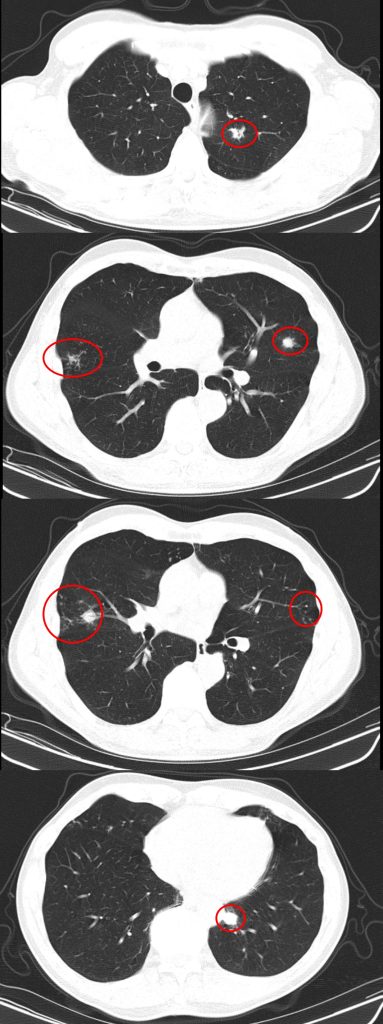
A #RadiologyRounds case with 3 different imaging modalities! A 65+ year old man never smoker, former marathoner has had 2-3 years of progressive non-productive, incessant cough with decreasing exercise tolerance. Some select CT scan slices are below

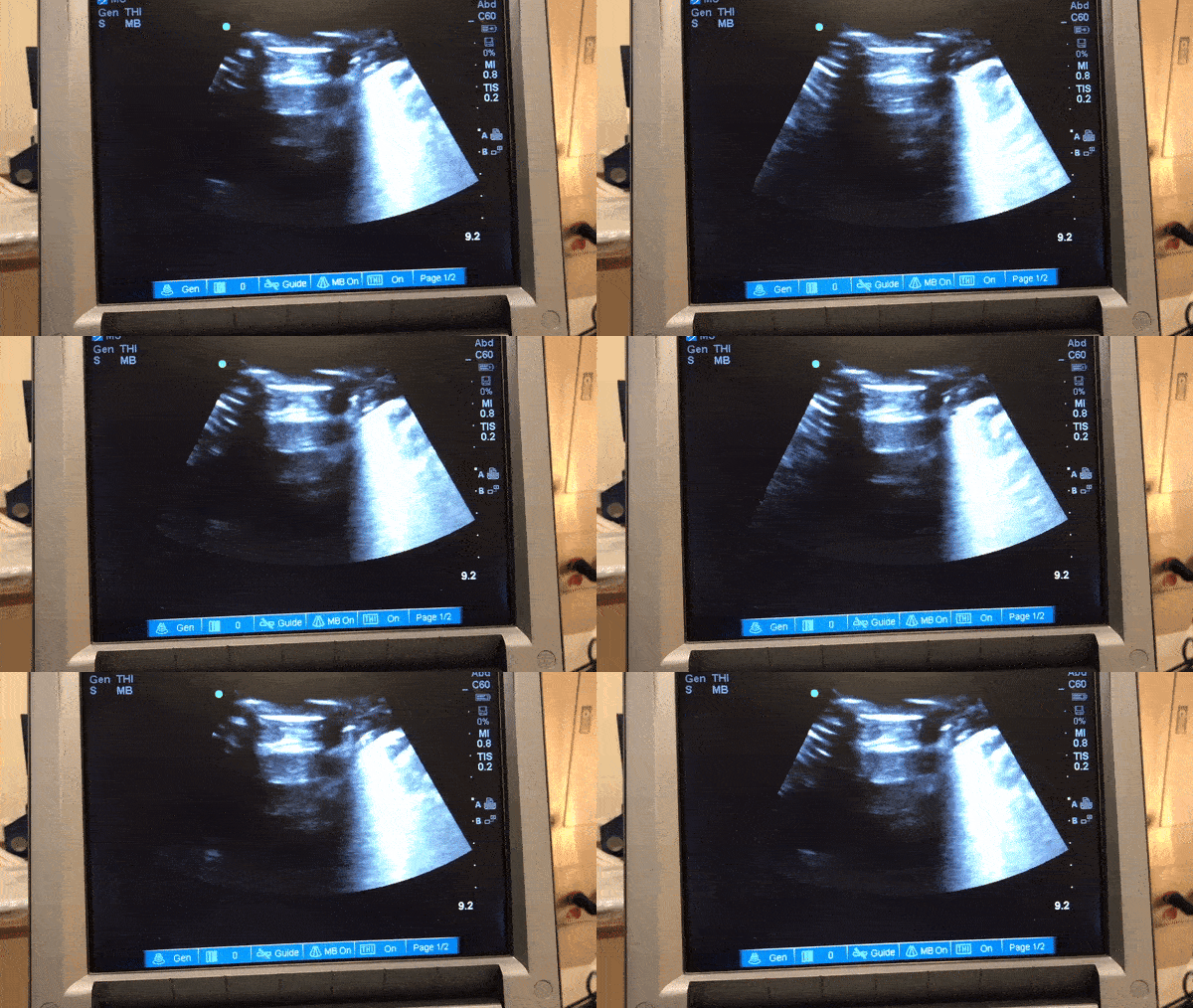
You are considering multiple etiologies including airway bleeding, pneumothorax, and hemothorax. You grab an ultrasound and perform a lung / pleural POCUS. Here is what you see

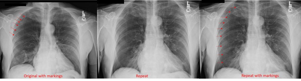
The long POCUS shows an area of lung sliding and an area without any lung sliding. This is called lung point and is diagnostic of a pneumothorax. To get a better look at this, you can use M-mode.

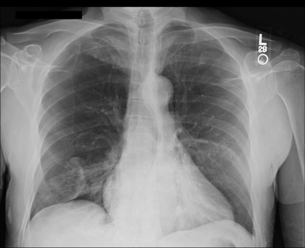
A CXR confirmed the finding of a pneumo. He was trialed on 100% oxygen but repeat CXR showed the pneumothorax was expanding. He had a chest tube placed with re-expansion of his lung and no air leak. It was able to be removed the next morning without incident

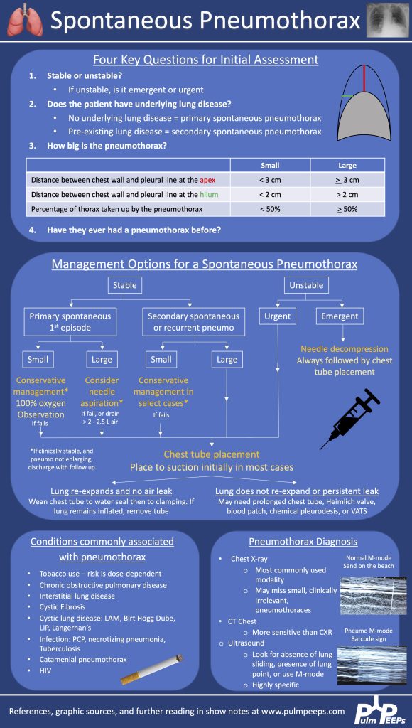
Here is our algorithm for pneumothorax!

Both BAL and tbbx returned positive for MAI complex. He was HIV neagative. Given his persistent and bothersome symptoms, he was started on treatment for pulmonary MAC with a macrolide, ethambutol and a rifamycin with plan for 6 months of therapy. He improved with this

30. Fellows’ Case Files: University of Mississippi Medical Center

We’re excited to be back with another episode in our Pulm PEEPs Fellows’ Case Files series! This is a particularly exciting case since it is our first episode where some intrepid fellows reached out to us with an interesting case they had encountered. If you have a great case, please let us know and you can follow in their footsteps! Pack your bags, and let’s head to Mississippi to learn about another great pulmonary and critical care case.

Meet our Guests

Meredith Sloan is a pulmonary and critical care fellow at the University of Mississippi. She completed her medical school at the Medical University of South Carolina College of Medicine, and her residency at the University of Mississippi.

Kevin Kinloch is a senior fellow at the University of Mississippi Medical Center where he also completed his internal medicine residency. He completed medical school at Meharry Medical College.

Jessie Harvey is an Associate professor of Medicine at the University of Mississippi and is the Pulmonary and Critical Care Program Director. She is also the Director of the MICU, and has been at MMC since medical school. She is a dedicated educator and leads the POCUS curriculum for IM residents and PCCM fellows

Patient Presentation

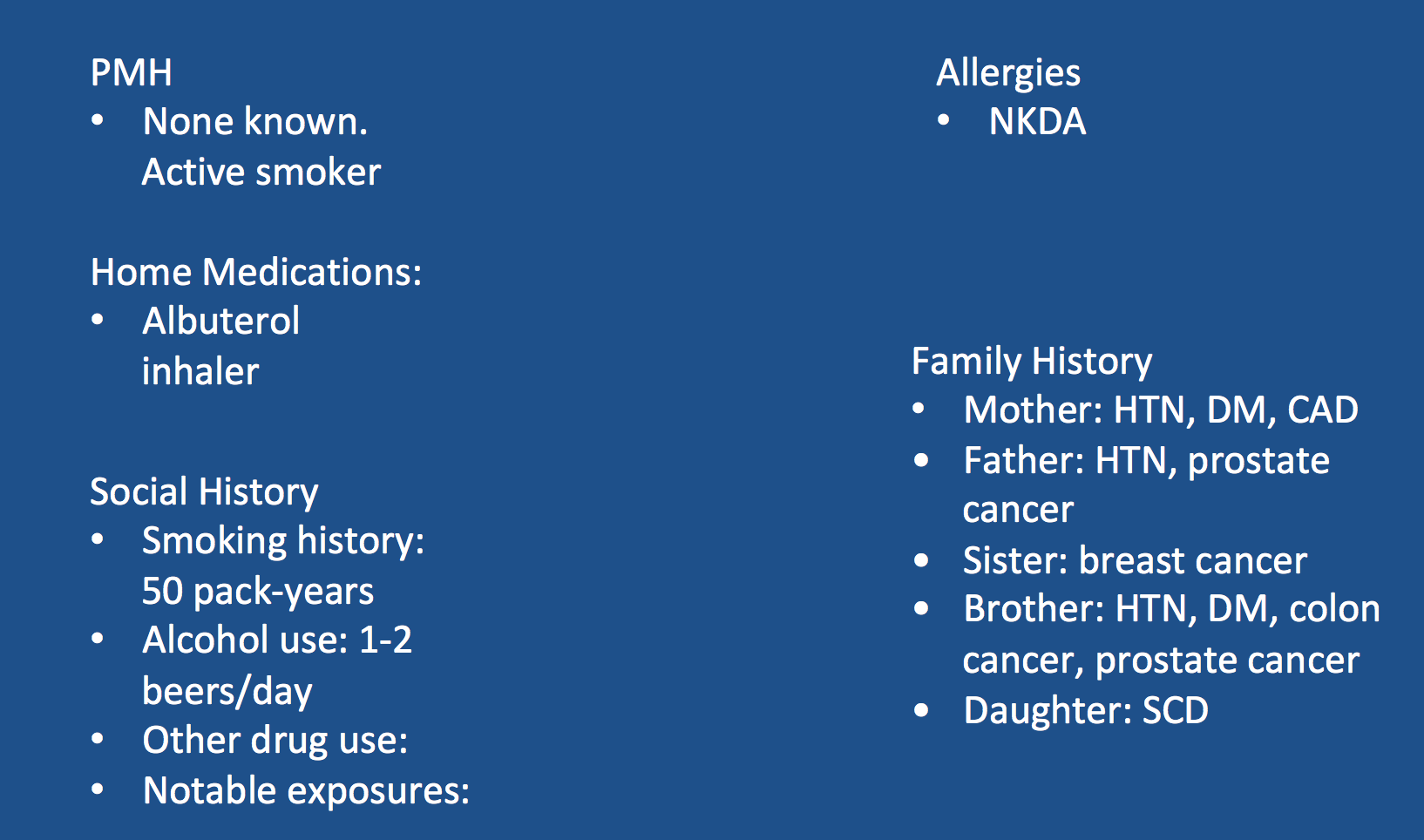
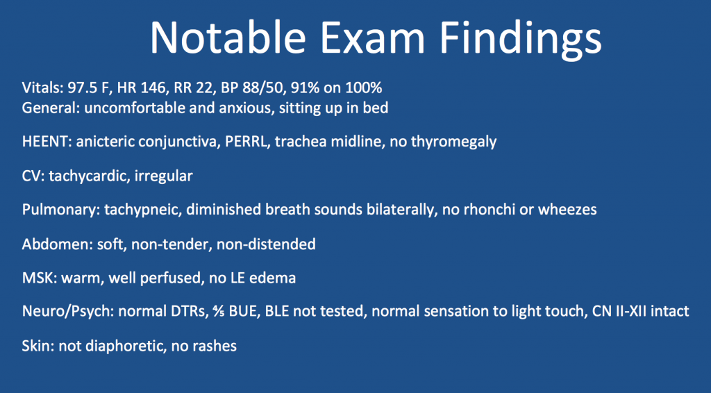
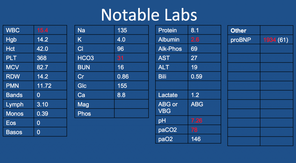
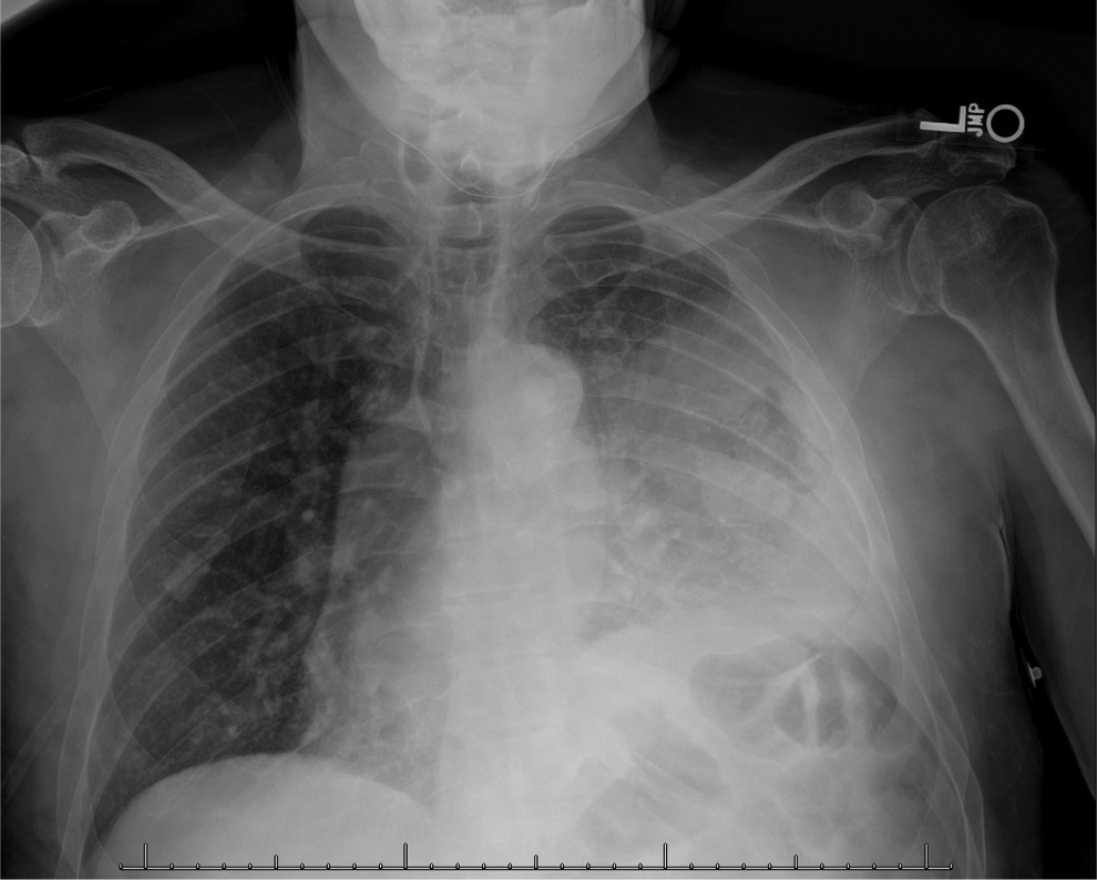
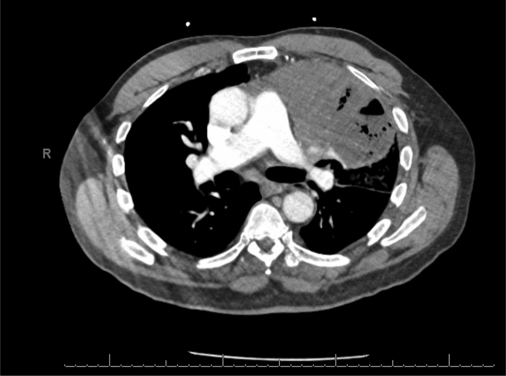
A 65-year-old man presented to the ED with worsening hemoptysis over the last several days after a recent lung biopsy. The patient is an active smoker with at least a 50-pack-year history, and he had been having a cough with small-volume hemoptysis. He ultimately had a chest CT that revealed a large LUL mass (10.3 x 6.4 cm). Given this suspicious mass, three days prior to his ED presentation, he was taken for bronchoscopy with BAL, transbronchial biopsies, endobronchial biopsy, EBUS guided TBNA of 11L, along with TBNA, brushing and radial EBUS TBNA of his left upper lobe mass.

Key Learning Points

**Spoilers Ahead** If you want to think through the case on your own we advise listening to the episode first before looking at these points.

Staging procedures for masses

  • Enough tissue so we can make a diagnosis and do molecular testing
  • Highest staging when getting your biopsy

POCUS for respiratory failure

  • Absence of lung slidings
    • Especially post procedure
  • The presence of a new pleural effusion after a procedure could indicate hemothorax
    • Hematocrit sign – an echogenic layering of material in an effusion
  • New B-lines, especially if prior there were only A-lines
    • Cardiogenic or non-cardiogenic pulmonary edema, alveolar hemorrhage, or infection
  • Diaphragmatic function
    • Excursion
    • Diaphragm thickness

References and Further Reading

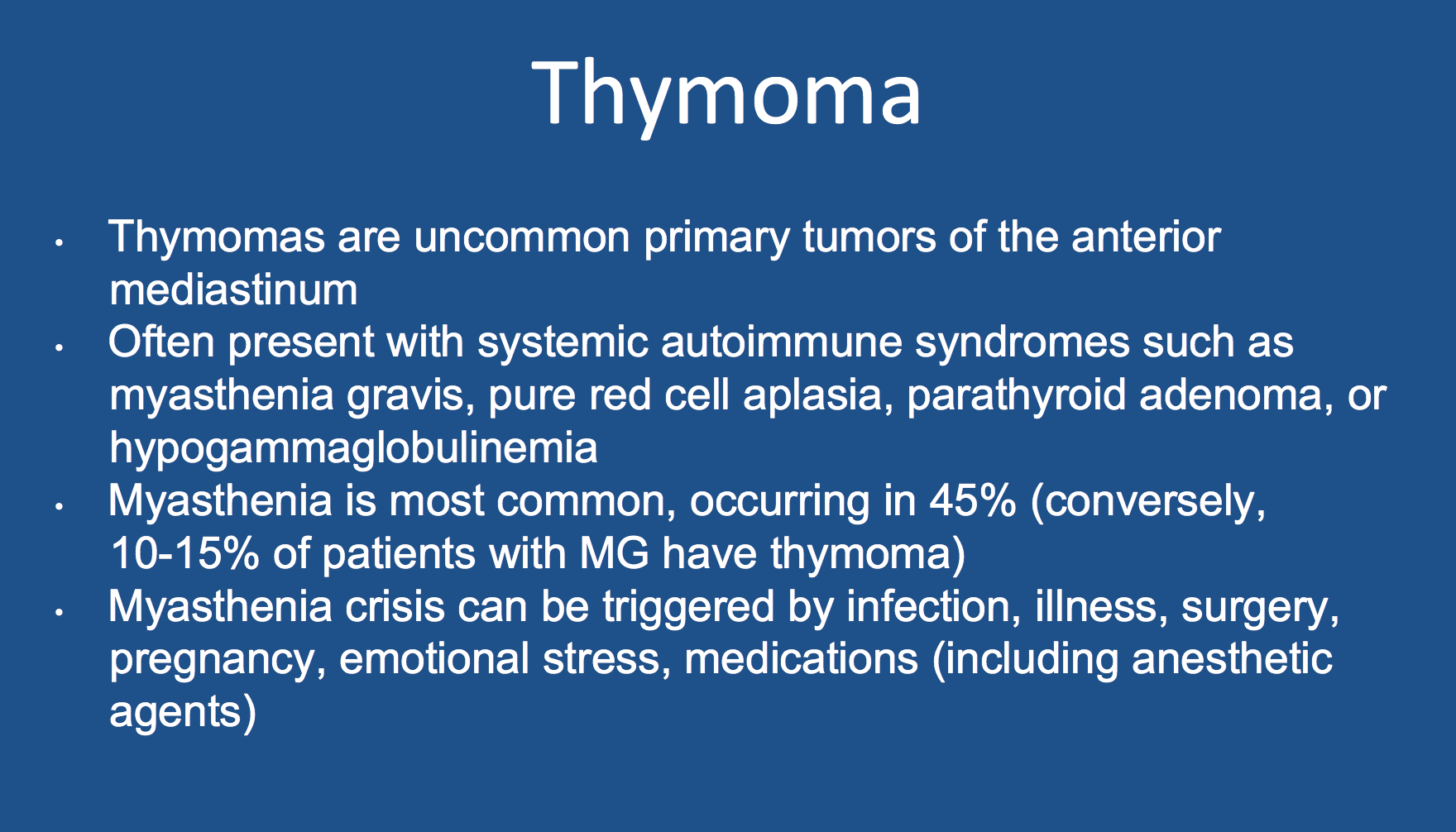
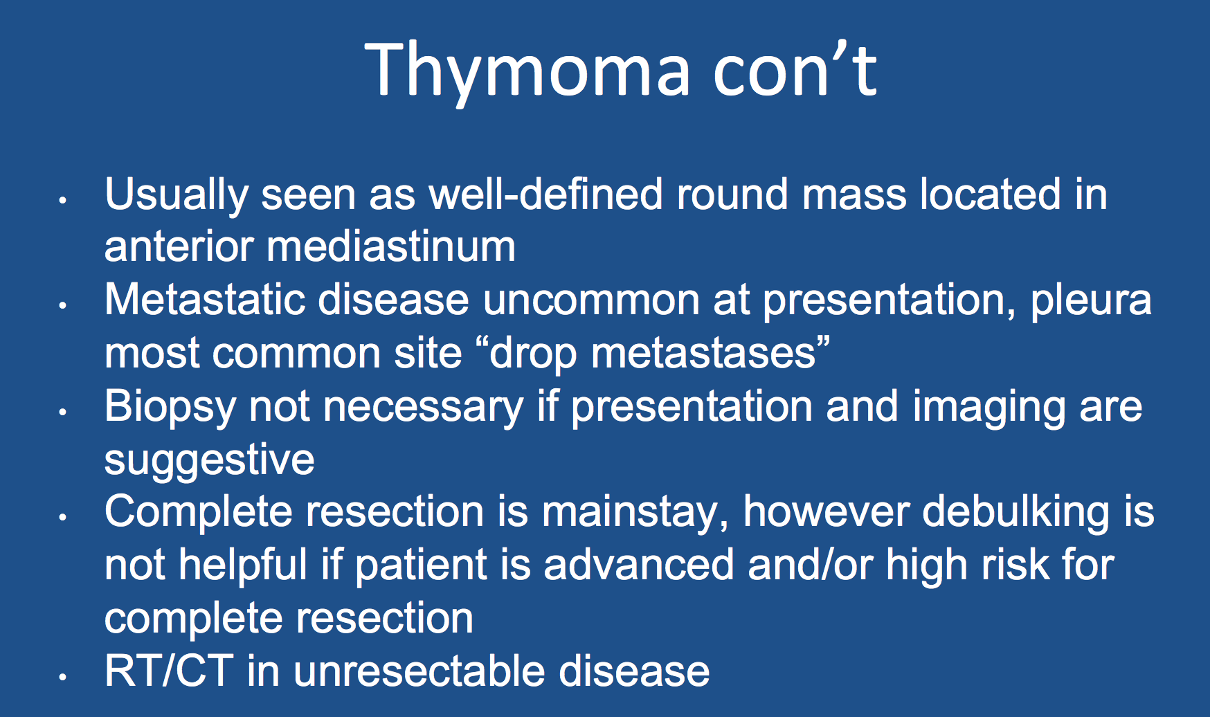
1.Scorsetti M, Leo F, Trama A, D’Angelillo R, Serpico D, Maerelli M, Zucali P, Gatta G, Garassino MC. Thymoma and thymic carcinomas. Critical Reviews in Oncology/Hematology. 2016; 99:332-350.

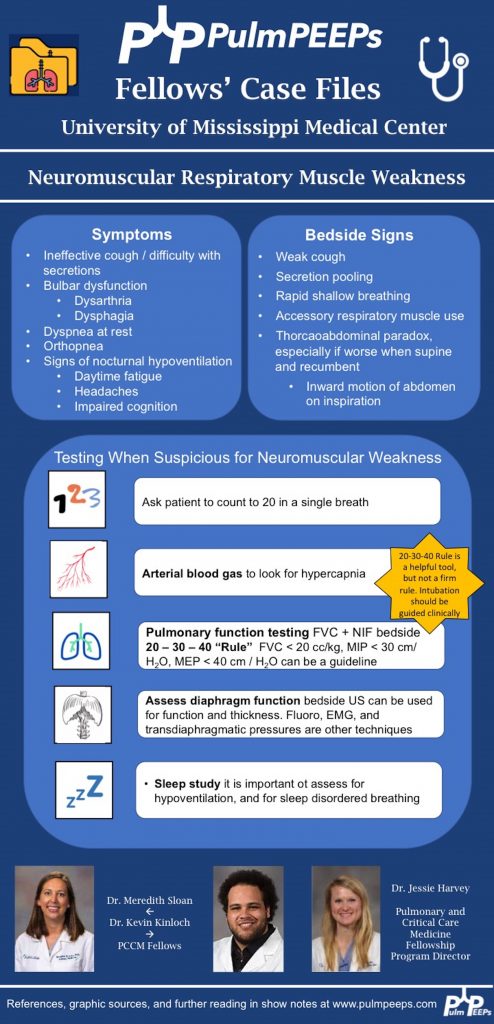
2. Singh TD, Wijdicks EFM. Neuromuscular respiratory failure. Neurol Clin 2021; 39:333-353.

14. Radiology Rounds Revisited: Right Heart Catheterization

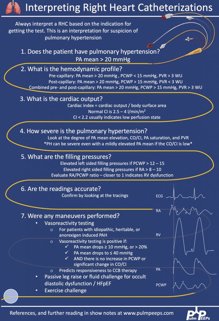
Today we have a special edition of Pulm PEEPs! We are revisiting our Radiology Rounds from 4 weeks ago to dive further into Right Heart Catheterizations and how to interpret them. We are joined by two experts in the field, Allison Tsao and Stephen Mathai.

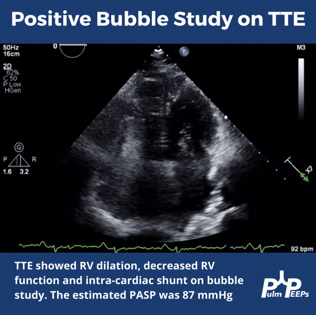
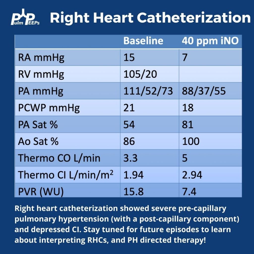
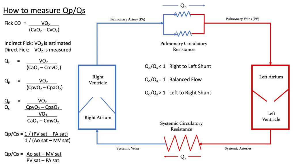
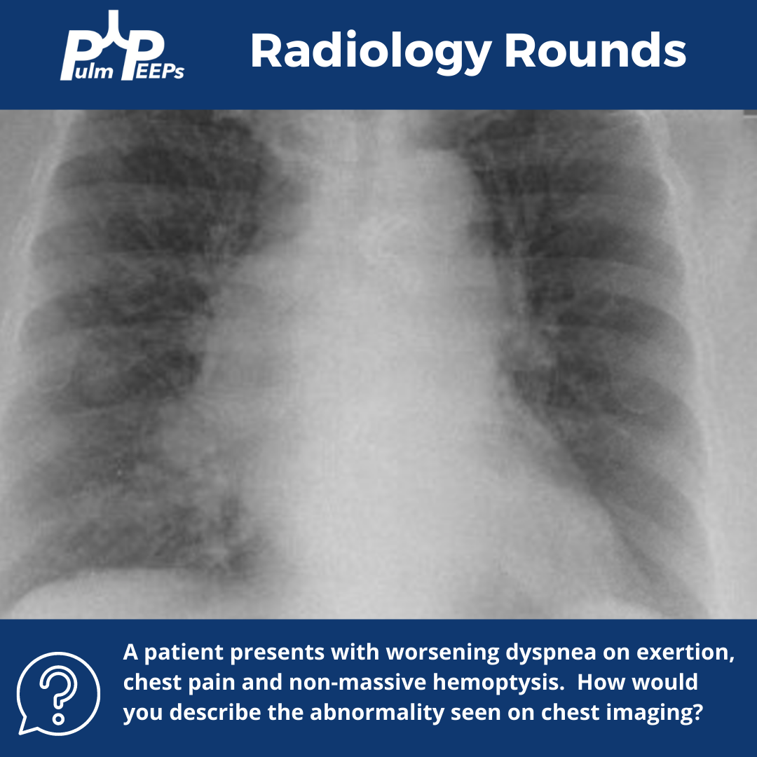
For a reminder, in that Radiology Rounds, we met a woman in her 50s with GERD, Raynaud’s, and multiple positive auto-antibodies (+ ANA 1:2560, + RNA pol III, + SSA, + anti-centromere) who presented with progressive dyspnea and was found to be hypoxemic. Her workup revealed severe pulmonary hypertension, and RV dysfunction on TTE with right to left shunting.

Meet Our Guests

Dr. Steve Mathai is an Associate Professor of Medicine at Johns Hopkins Hospital and the Director of the Inpatient Pulmonary Service. He specializes in Pulmonary Hypertension and his research focus is on scleroderma-associated PAH.

Dr. Allison Tsao is an Instructor in Medicine at Harvard Medical School and is an interventional cardiologist working at the Boston VA and Brigham and Women’s Hospital. She specializes in adult congenital heart disease and is the assistant director of the Translational Discovery Lab at BWH.

Key Learning Points

References and links for further reading

  1. Bonno EL, Viray MC, Jackson GR, Houston BA, Tedford RJ. Modern Right Heart Catheterization: Beyond Simple Hemodynamics. Advances in Pulmonary Hypertension. 2020;19(1):6-15. doi:10.21693/1933-088X-19.1.6
  2. Callan P, Clark AL. Right heart catheterisation: indications and interpretation. Heart. 2016;102(2):147-157. doi:10.1136/heartjnl-2015-307786
  3. Chokkalingam Mani B, Chaudhari SS. Right Heart Cardiac Catheterization. In: StatPearls. StatPearls Publishing; 2022. Accessed April 18, 2022. http://www.ncbi.nlm.nih.gov/books/NBK557404/
  4. D’Alto M, Dimopoulos K, Coghlan JG, Kovacs G, Rosenkranz S, Naeije R. Right Heart Catheterization for the Diagnosis of Pulmonary Hypertension: Controversies and Practical Issues. Heart Failure Clinics. 2018;14(3):467-477. doi:10.1016/j.hfc.2018.03.011
  5. Galiè N, McLaughlin VV, Rubin LJ, Simonneau G. An overview of the 6th World Symposium on Pulmonary Hypertension. European Respiratory Journal. 2019;53(1). doi:10.1183/13993003.02148-2018
  6. Rosenkranz S, Preston IR. Right heart catheterisation: best practice and pitfalls in pulmonary hypertension. European Respiratory Review. 2015;24(138):642-652. doi:10.1183/16000617.0062-2015

Radiology Rounds – 3/8/22

We’re excited to bring you another Radiology Rounds today that combines pulmonary and critical care.

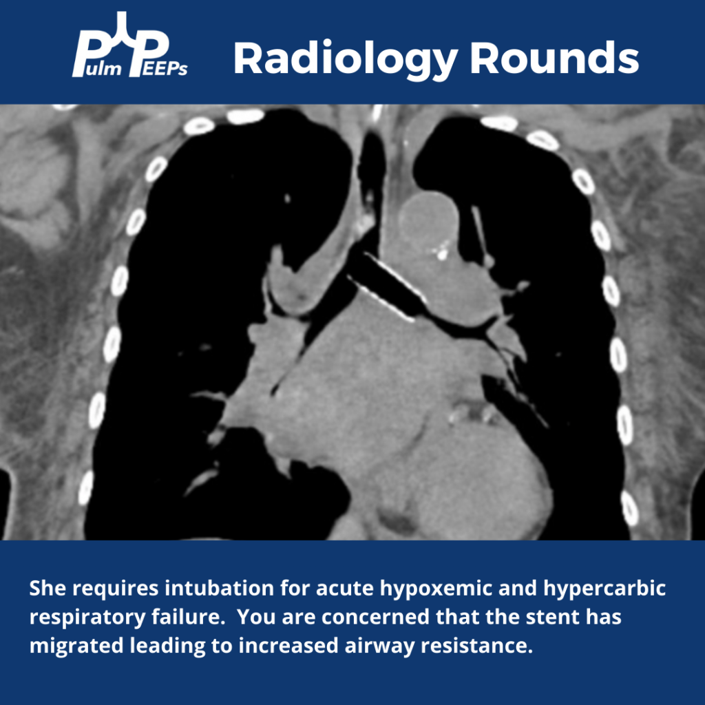
The patient is diagnosed with small cell lung cancer and requires a left bronchial stent. She develops acute hypoxemic and hypercapnic respiratory failure requiring intubation.

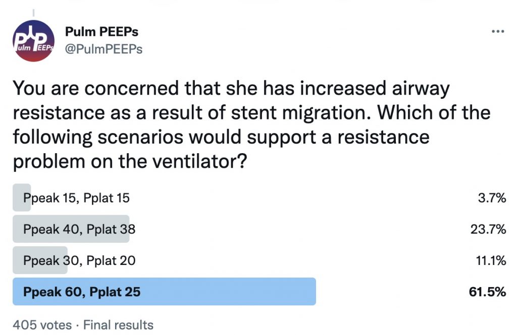
You are concerned that she has increased airway resistance as a result of stent migration. What would you expect to see on the ventilator if this is the case?

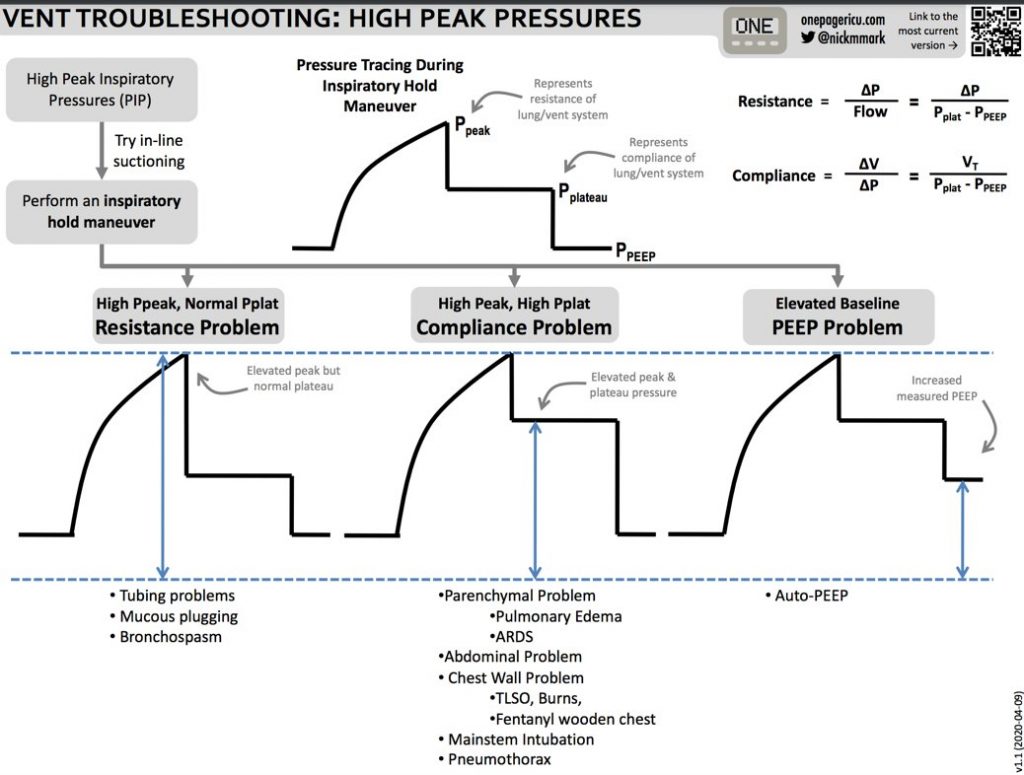
Here are some tips from ICU OnePager on interpreting high peak pressures on the ventilator