
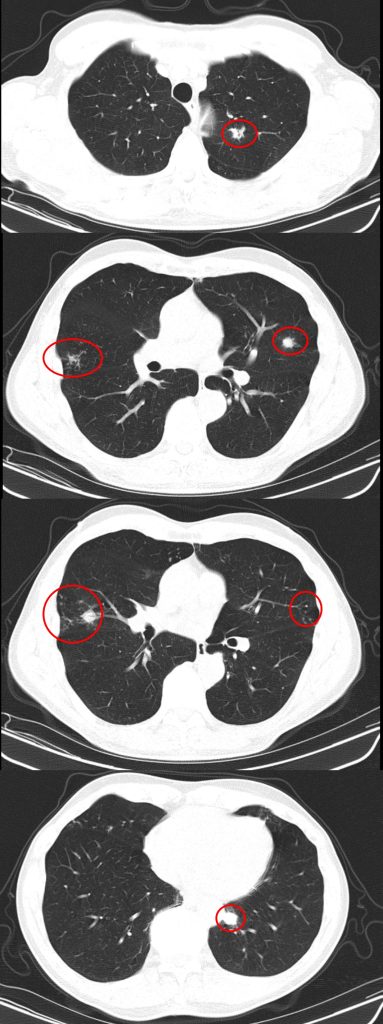
A #RadiologyRounds case with 3 different imaging modalities! A 65+ year old man never smoker, former marathoner has had 2-3 years of progressive non-productive, incessant cough with decreasing exercise tolerance. Some select CT scan slices are below


You are considering multiple etiologies including airway bleeding, pneumothorax, and hemothorax. You grab an ultrasound and perform a lung / pleural POCUS. Here is what you see
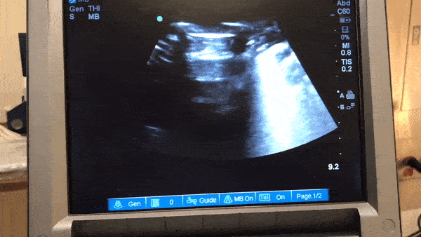
The long POCUS shows an area of lung sliding and an area without any lung sliding. This is called lung point and is diagnostic of a pneumothorax. To get a better look at this, you can use M-mode.

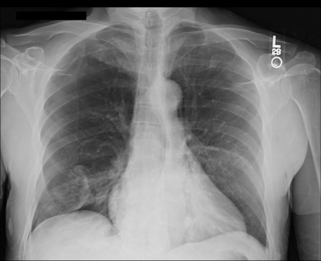
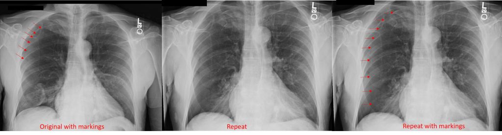
A CXR confirmed the finding of a pneumo. He was trialed on 100% oxygen but repeat CXR showed the pneumothorax was expanding. He had a chest tube placed with re-expansion of his lung and no air leak. It was able to be removed the next morning without incident
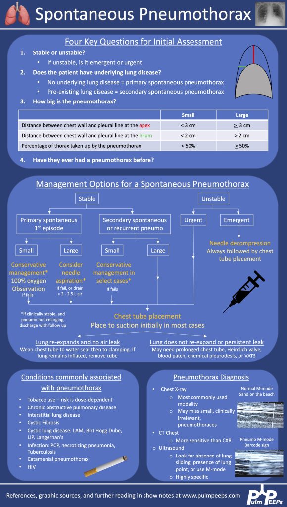
Here is our algorithm for pneumothorax!
Both BAL and tbbx returned positive for MAI complex. He was HIV neagative. Given his persistent and bothersome symptoms, he was started on treatment for pulmonary MAC with a macrolide, ethambutol and a rifamycin with plan for 6 months of therapy. He improved with this