48. Fellows’ Case Files: Boston University

Today we’re back with another stop on our Fellows’ Case Files journey. We’re at Boston University and Boston Medical Center with an outstanding fellow, the program director, and an expert guest to dive into a new case. Tune in to hear about this fascinating case and learn some key pulmonary points along the way.
Meet Our Guests

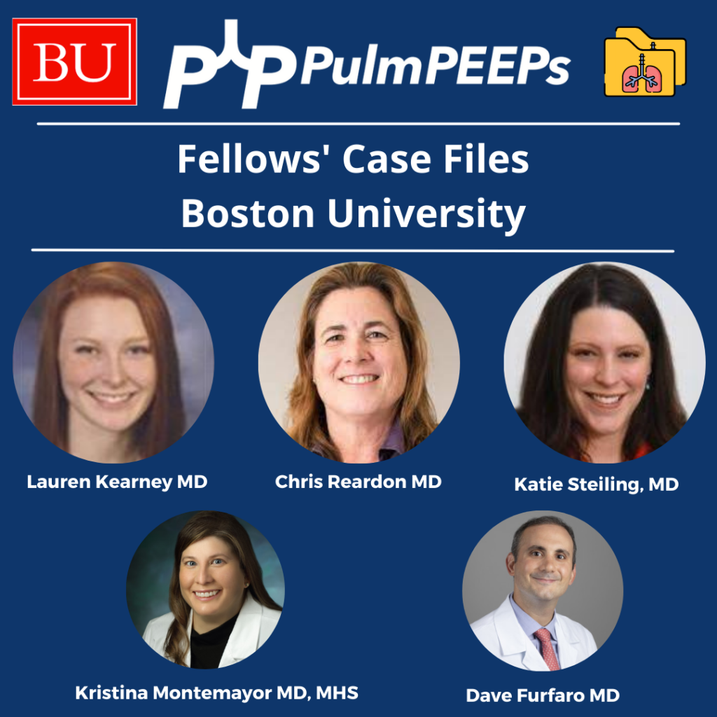
Lauren Kearney is a research fellow at Boston University. She completed her internal medicine residency and chief residency at BU. She is pursuing a research career in health equity and community-based participatory research to improve outcomes for patients with lung cancer and other pulmonary conditions.

Chris Reardon is a Clinical Professor of Medicine at Boston University, where she is also the Fellowship Training Program Director for Pulmonary and Critical Care Medicine. She is additionally the Director of Respiratory Care Services at Pappas Rehab Hospital for Children.

Katie Steiling is an Assistant Professor of Medicine at Boston University and a Member of the Bioinformatics Graduate Program. She founded the Lung Nodule Clinic at Boston Medical Center and co-chairs the Lung Cancer Screening Steering Committee. She is dedicated to improving the equitable treatment of patients with and at risk for lung cancer.

Case Presentation

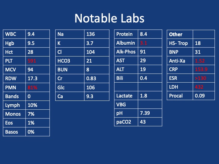
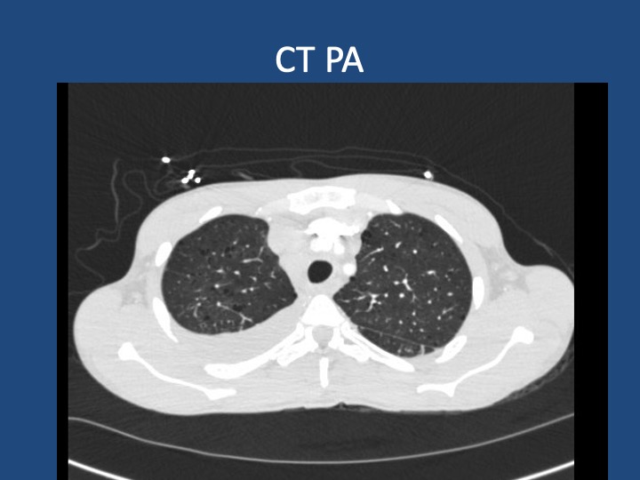
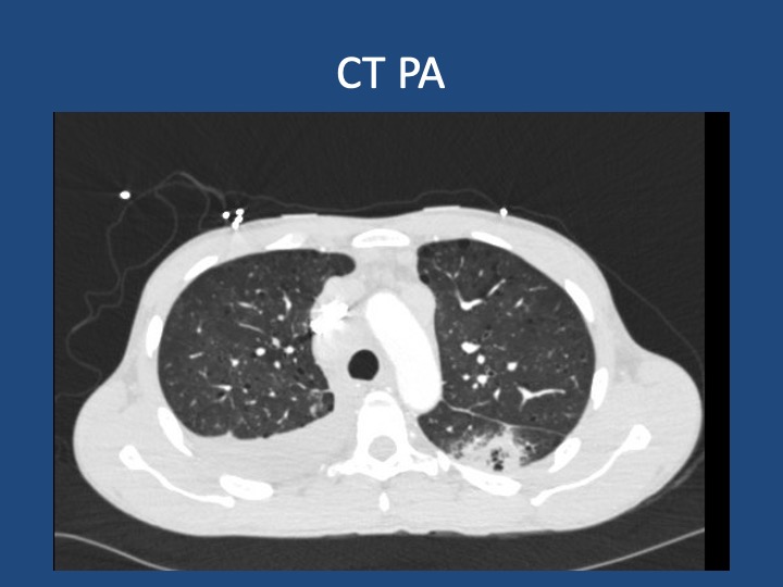
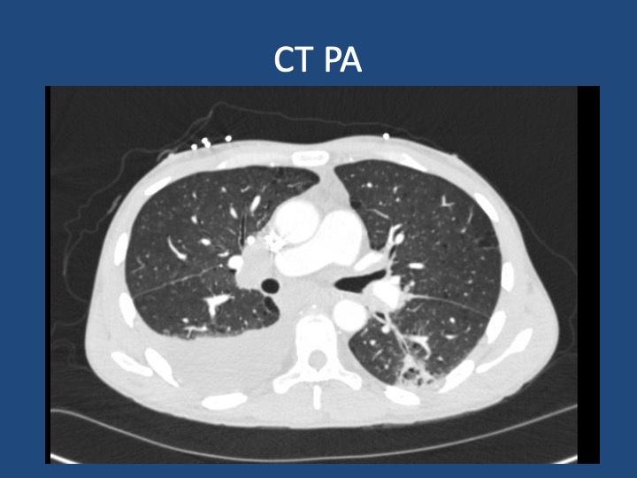
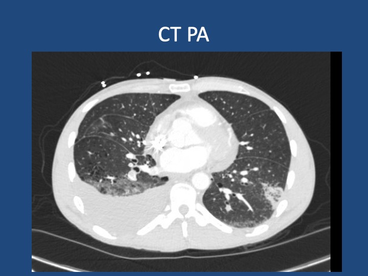
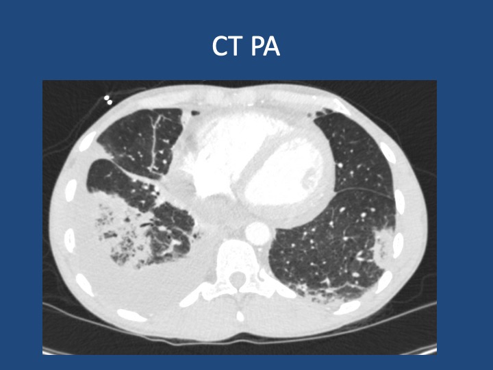
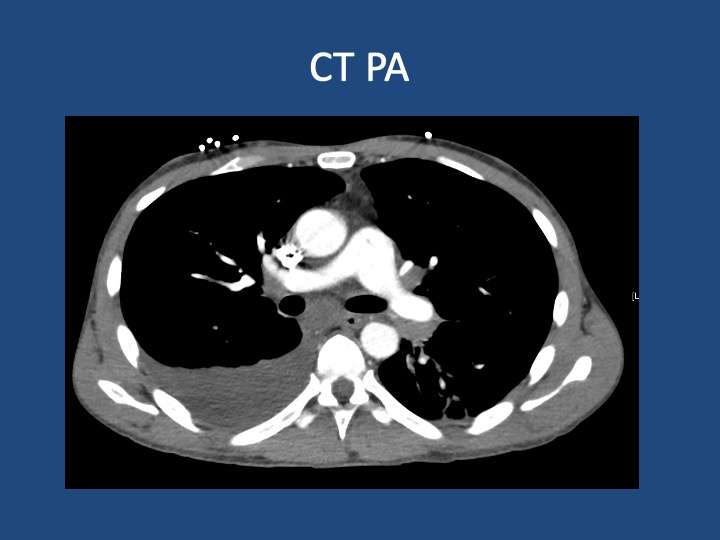
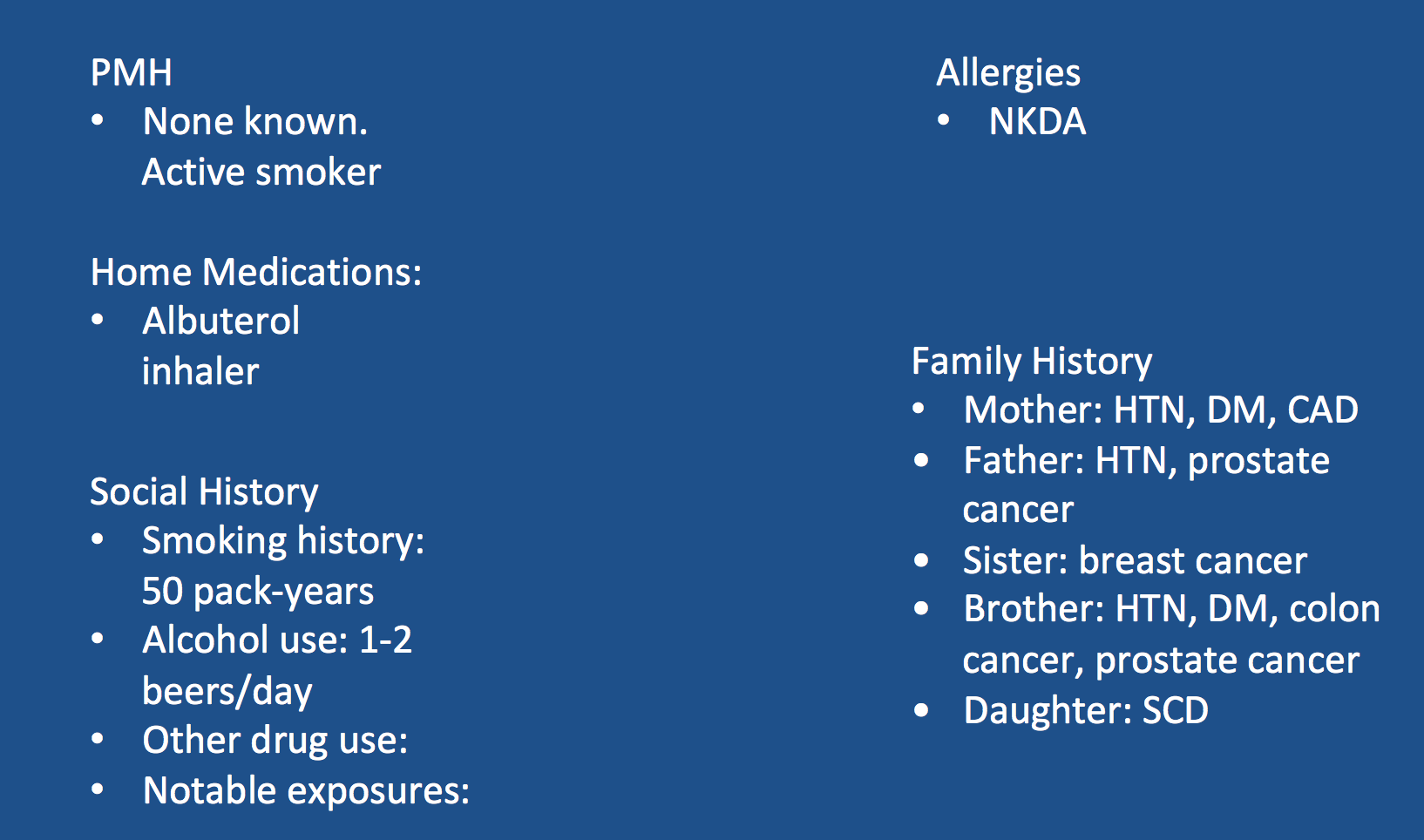
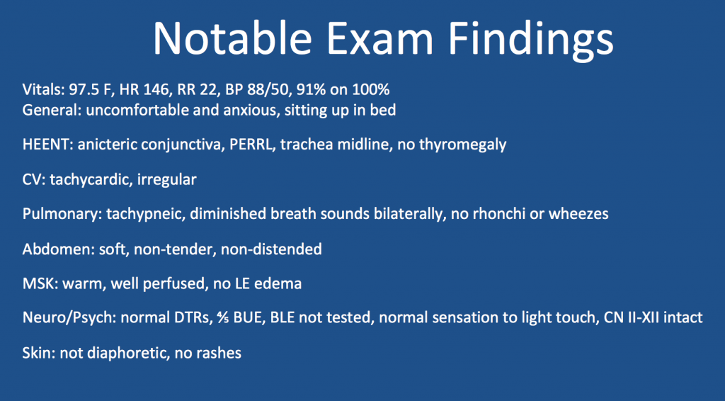
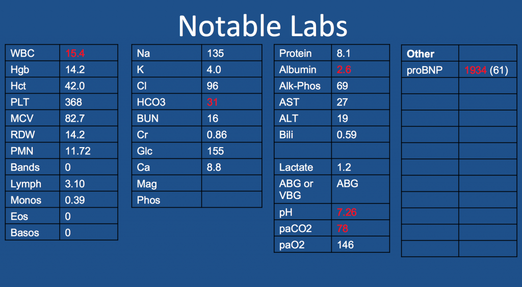
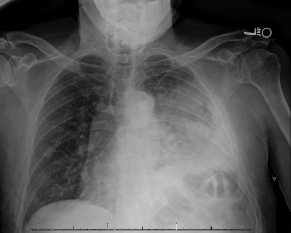
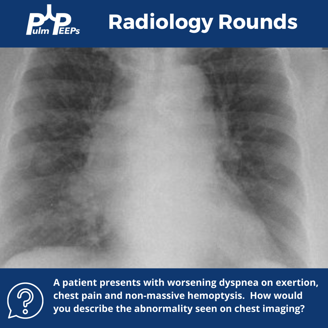
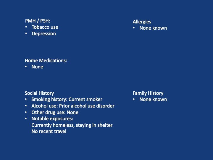
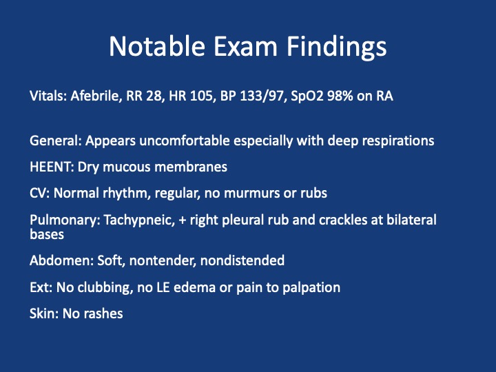
A 44-year-old man who is undomiciled and a current smoker presents with three days of intermittent, progressively worsening pleuritic chest pain with a cough productive of blood-tinged sputum. This is in the setting of 1-2 months of fatigue and anorexia, and 2 weeks of bilateral calf swelling.
Key Learning Points

  1. Factors that may lead to initial hypercoagulability testing in first unprovoked PE: young age, thrombosis in multiple or unusual vascular beds, history of warfarin-induced skin necrosis, arterial thrombosis.
  2. When lung cancer is suspected, identification of metastases to identify the highest radiologic stage and optimal biopsy site is essential.
  3. Given that pleural fluid sensitivity is only ~46%, negative cytology should not assuage further malignancy work-up in a high-risk patient.
  4. Black individuals in the US are at higher risk of developing and dying of lung cancer- the reasons for which are complex & multifactorial. Yet, gaps remain in the targeted identification of these patients, adequate lung cancer screening, and connection to diagnosis & treatment.
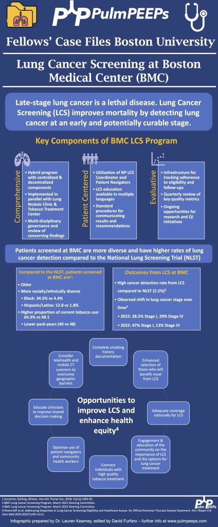
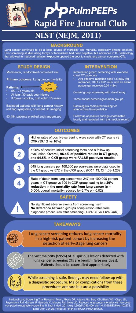
47. Rapid Fire Journal Club 1 – National Lung Screening Trial

We are introducing a new feature today on Pulm PEEPs! In our Rapid Fire Journal Club series, we will be reviewing landmark trials in pulmonary and critical care medicine in 10 minutes or less, and sharing a high-yield summary graphic. We are focusing on pulmonary trials to start since these are often overlooked and under-discussed.

Article and Reference

Our first trial is the National Lung Screening Trial published in the New England Journal of Medicine in 2018. We also suggest you read the accompanying editorial if you are looking for even more discussion after listening to this episode.

Reference: National Lung Screening Trial Research Team; Aberle DR, Adams AM, Berg CD, Black WC, Clapp JD, Fagerstrom RM, Gareen IF, Gatsonis C, Marcus PM, Sicks JD. Reduced lung-cancer mortality with low-dose computed tomographic screening. N Engl J Med. 2011 Aug 4;365(5):395-409. doi: 10.1056/NEJMoa1102873. Epub 2011 Jun 29. PMID: 21714641; PMCID: PMC4356534.

Infographic

This can be downloaded on our website and will be shared on Twitter and Instagram.

Radiology Rounds – 5/30/2023

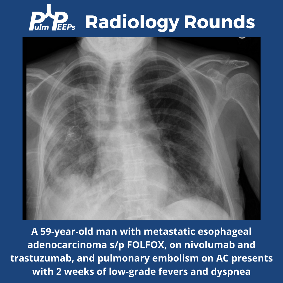
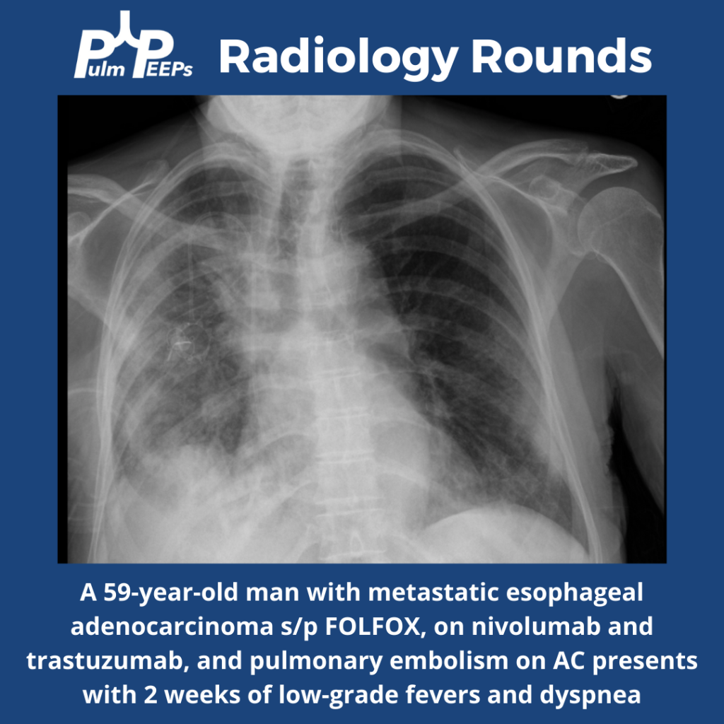
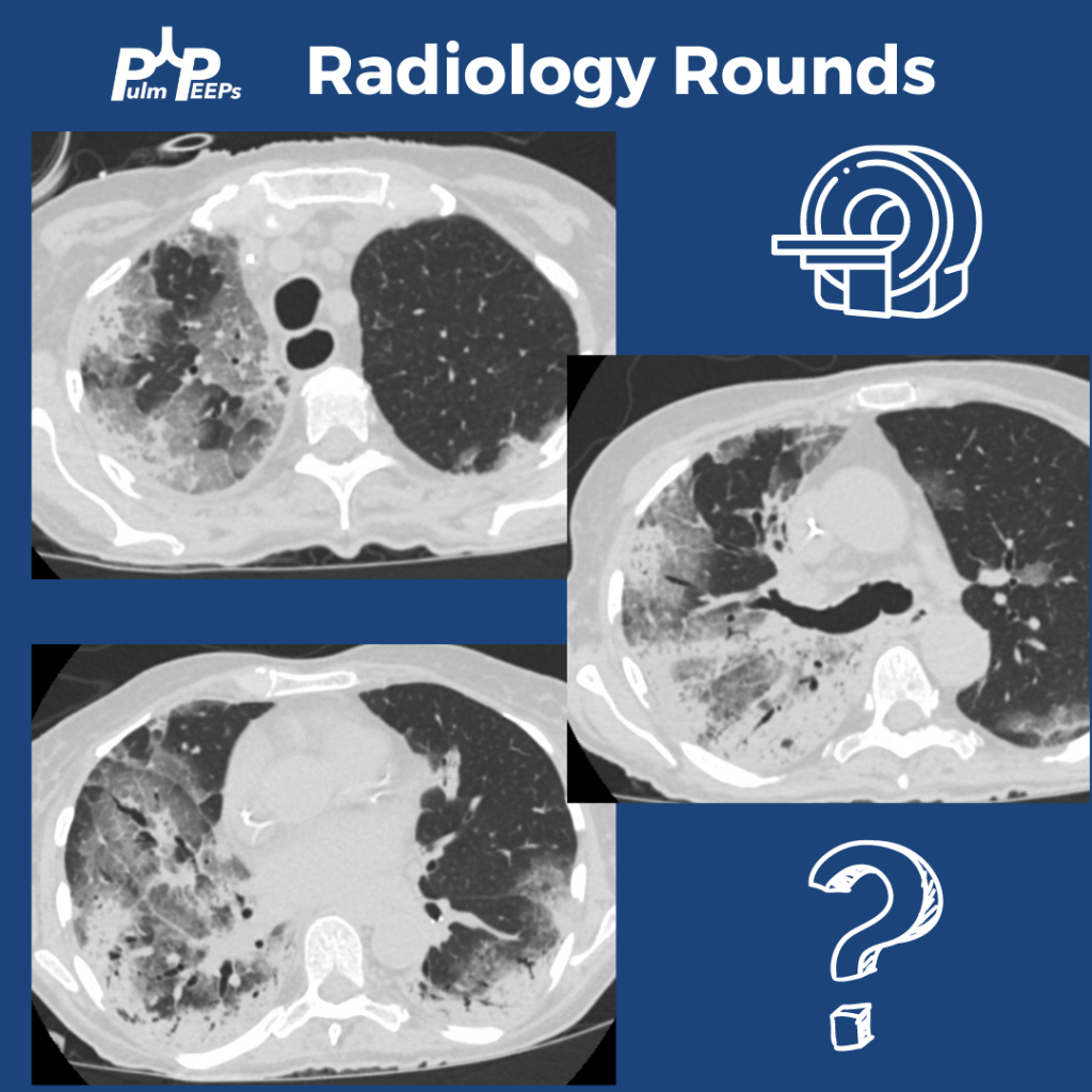
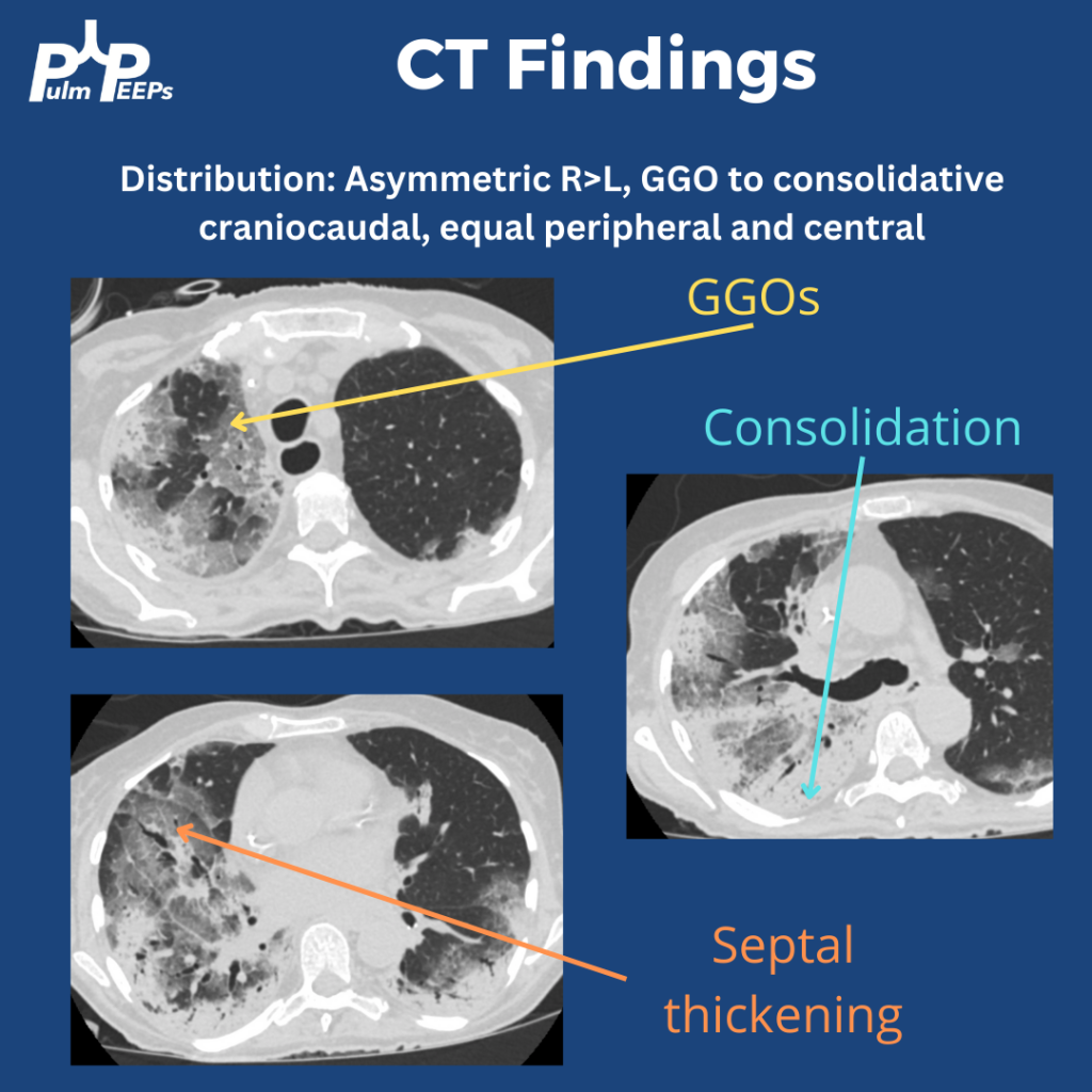
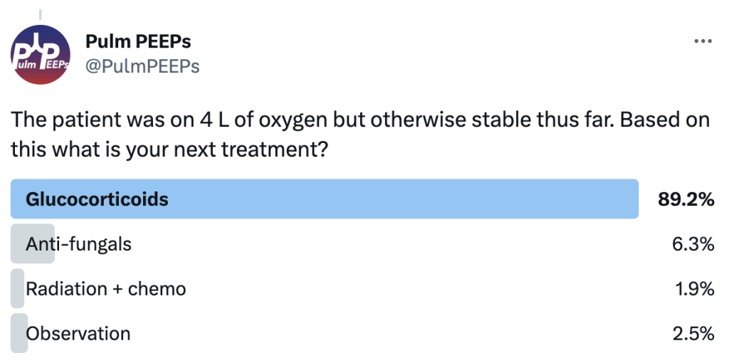
A middle-aged man with metastatic esophageal cancer presents with 2 weeks of low-grade fevers and more recently progressive dyspnea. His exam is notable for late inspiratory crackles throughout the right lung.

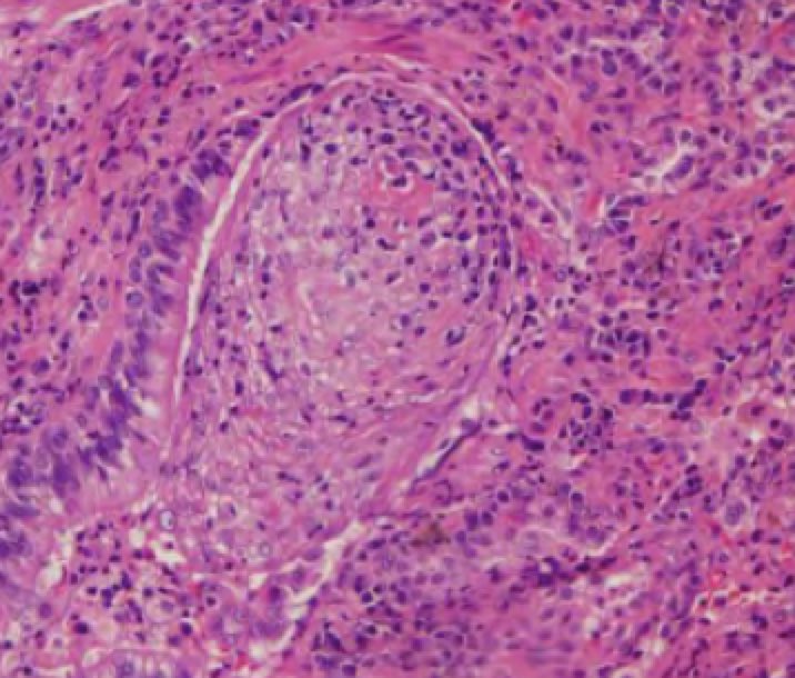
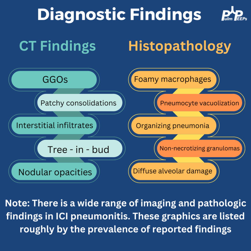
A bronchoscopy was performed and an expanded infectious panel was sent and all were negative, as were serum infectious studies (e.g. histoplasmosis ag, urine legionella etc). A trans bronchial biopsy showed plugs of granulation tissue filling the alveolar spaces

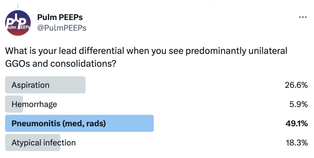
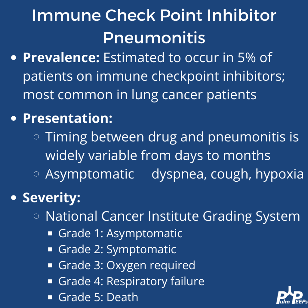
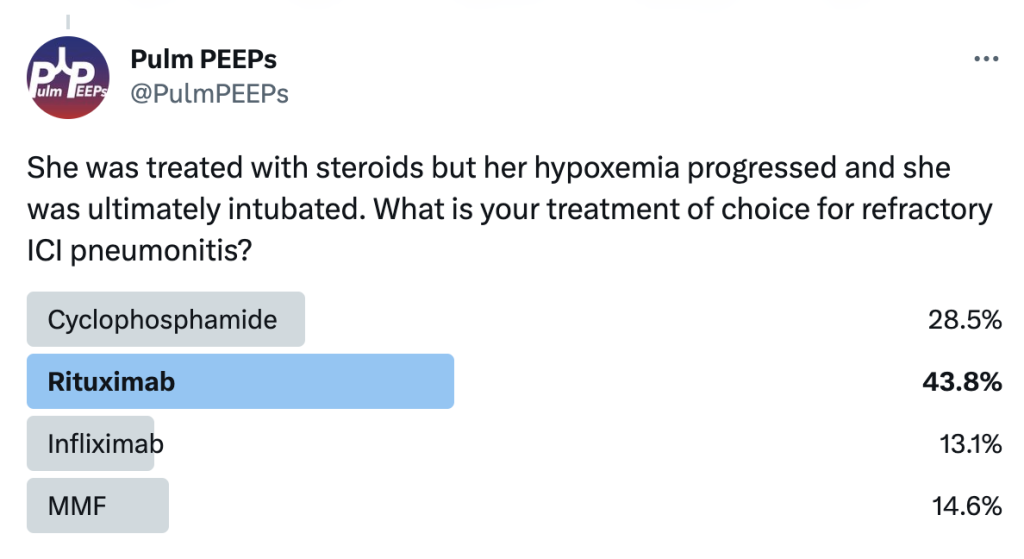
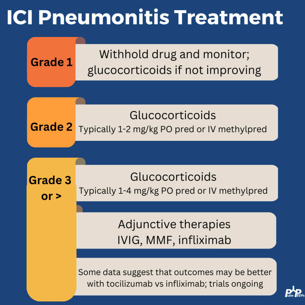
He was treated with steroids but her hypoxemia progressed and she was ultimately intubated. What is your treatment of choice for refractory ICI pneumonitis?

Severe ICI is rare and the treatment for steroid refractory disease is still based on small series and case reports This patient was treated with high-dose steroids (2mg/kg), IVIG, MMF, and tocilizumab. With that, he improved after 10 days intubated and was weaned to room air.

Radiology Rounds – 3/21/2023

We are back with another #RadiologyRounds! This week’s case comes from our Associate Editor @luke_hedrick

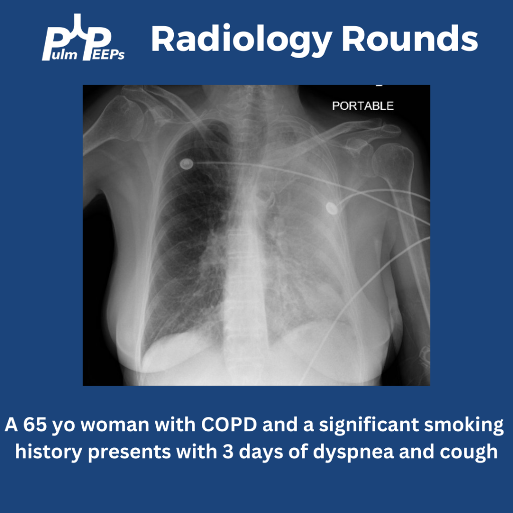
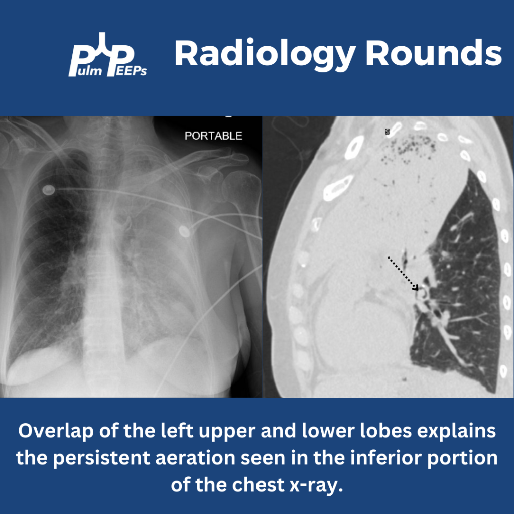
A portable film is obtained on a 65 yo woman with COPD presenting with progressive dyspnea and cough.

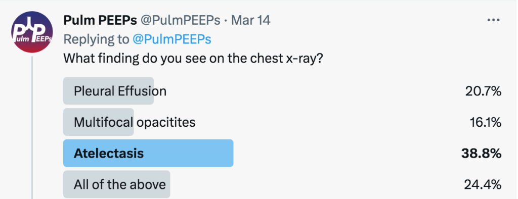
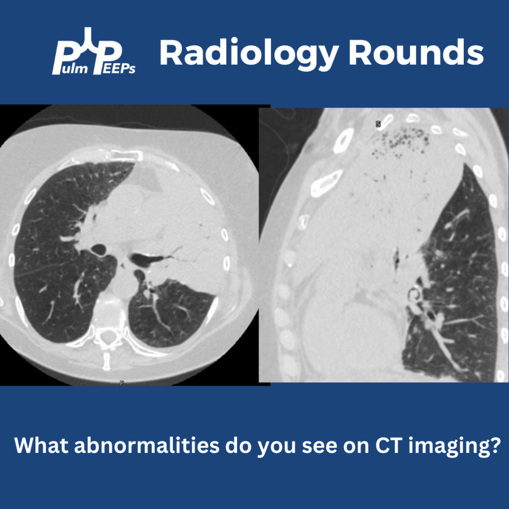
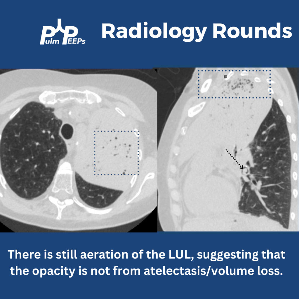
The image shows near complete opacification of the left lung. There is no ipsilateral or contralateral tracheal deviation which you would expect with atelectasis or a large pleural effusion, respectively. A CT chest is obtained to better visualize the parenchyma.

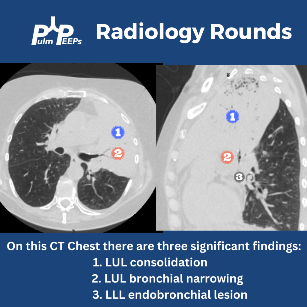
The CT chest shows all three findings as noted below.

Yes! There is still aeration in some of the upper lobe, which would not be the case if this were caused by profound atelectasis. Also, atelectasis of such a large territory of the lung would usually cause traction on surrounding structures.

Having seen the CT, can you explain why the x-ray showed aeration in the inferior, left hemithorax? This is from the overlap of the left upper and lower lobes when viewed anteriorly. The sagittal view of the CT demonstrates this nicely.

30. Fellows’ Case Files: University of Mississippi Medical Center

We’re excited to be back with another episode in our Pulm PEEPs Fellows’ Case Files series! This is a particularly exciting case since it is our first episode where some intrepid fellows reached out to us with an interesting case they had encountered. If you have a great case, please let us know and you can follow in their footsteps! Pack your bags, and let’s head to Mississippi to learn about another great pulmonary and critical care case.

Meet our Guests

Meredith Sloan is a pulmonary and critical care fellow at the University of Mississippi. She completed her medical school at the Medical University of South Carolina College of Medicine, and her residency at the University of Mississippi.

Kevin Kinloch is a senior fellow at the University of Mississippi Medical Center where he also completed his internal medicine residency. He completed medical school at Meharry Medical College.

Jessie Harvey is an Associate professor of Medicine at the University of Mississippi and is the Pulmonary and Critical Care Program Director. She is also the Director of the MICU, and has been at MMC since medical school. She is a dedicated educator and leads the POCUS curriculum for IM residents and PCCM fellows
Patient Presentation

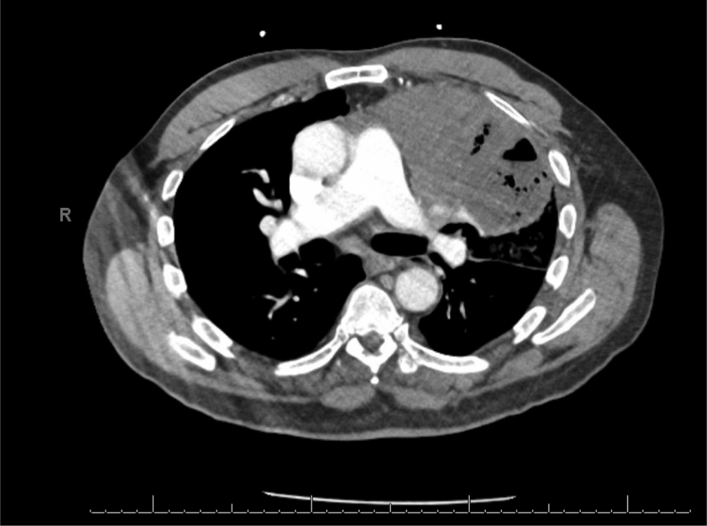
A 65-year-old man presented to the ED with worsening hemoptysis over the last several days after a recent lung biopsy. The patient is an active smoker with at least a 50-pack-year history, and he had been having a cough with small-volume hemoptysis. He ultimately had a chest CT that revealed a large LUL mass (10.3 x 6.4 cm). Given this suspicious mass, three days prior to his ED presentation, he was taken for bronchoscopy with BAL, transbronchial biopsies, endobronchial biopsy, EBUS guided TBNA of 11L, along with TBNA, brushing and radial EBUS TBNA of his left upper lobe mass.

Key Learning Points

**Spoilers Ahead** If you want to think through the case on your own we advise listening to the episode first before looking at these points.

Staging procedures for masses

  • Enough tissue so we can make a diagnosis and do molecular testing
  • Highest staging when getting your biopsy

POCUS for respiratory failure

  • Absence of lung slidings
    • Especially post procedure
  • The presence of a new pleural effusion after a procedure could indicate hemothorax
    • Hematocrit sign – an echogenic layering of material in an effusion
  • New B-lines, especially if prior there were only A-lines
    • Cardiogenic or non-cardiogenic pulmonary edema, alveolar hemorrhage, or infection
  • Diaphragmatic function
    • Excursion
    • Diaphragm thickness

References and Further Reading

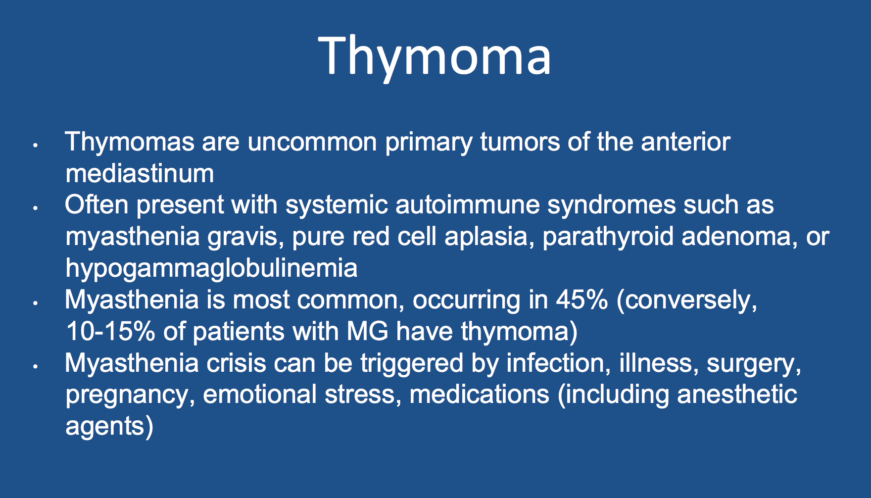
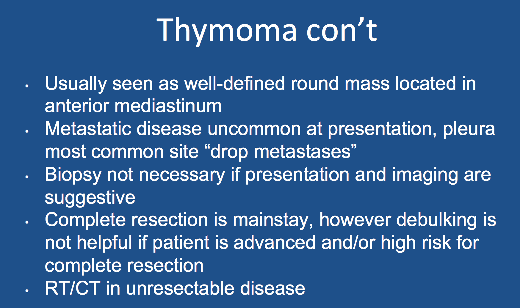
1.Scorsetti M, Leo F, Trama A, D’Angelillo R, Serpico D, Maerelli M, Zucali P, Gatta G, Garassino MC. Thymoma and thymic carcinomas. Critical Reviews in Oncology/Hematology. 2016; 99:332-350.

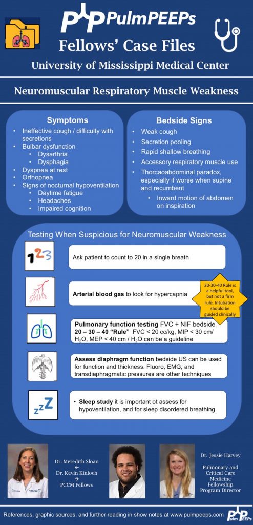
2. Singh TD, Wijdicks EFM. Neuromuscular respiratory failure. Neurol Clin 2021; 39:333-353.

Radiology Rounds – 11/8/22

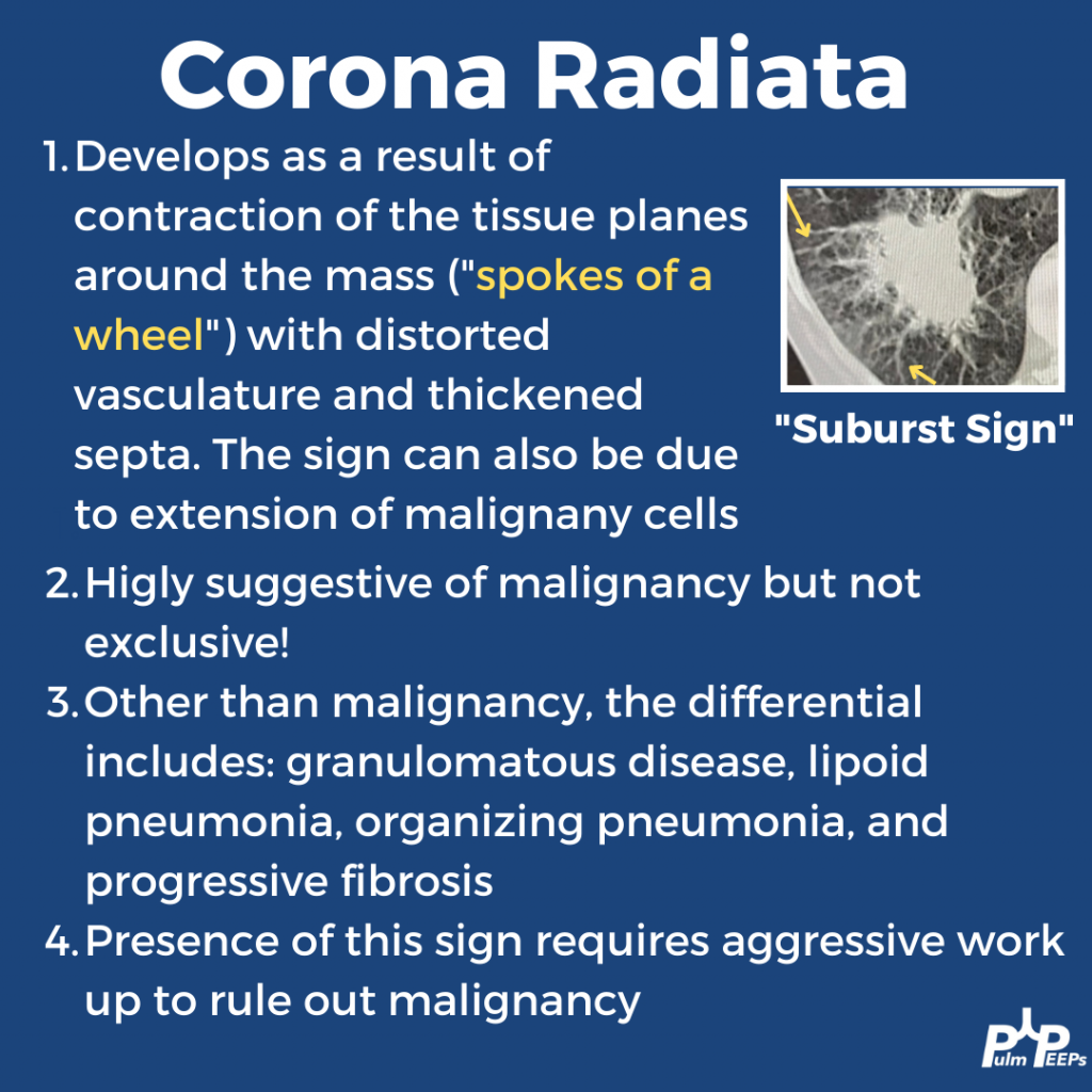
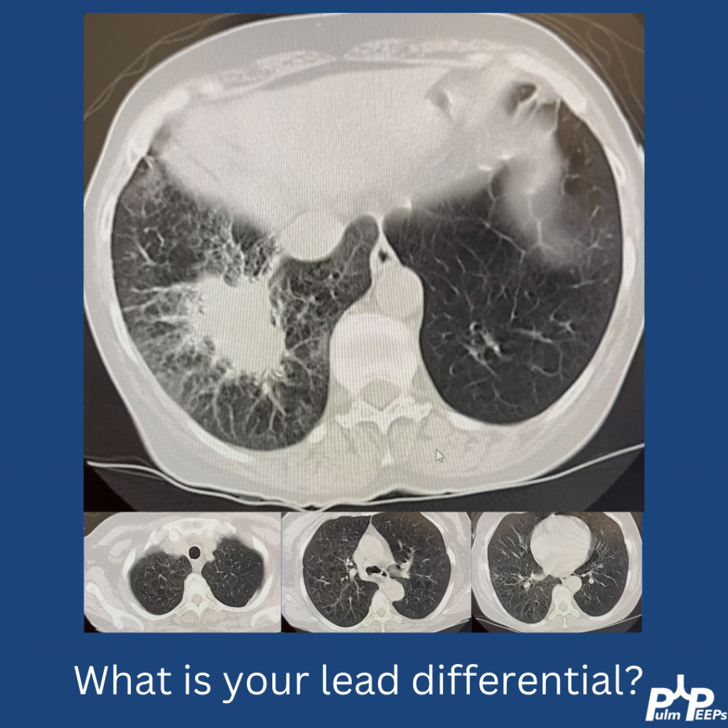
This week’s #RadiologyRounds is brought to you by our newest contributor, Nick Ghionni! We’ll dive into a classic Radiology Sign and talk about what it represents, and how it helps inform your differential.

In this case, the patient underwent a biopsy that showed no malignancy but grew NTM! Given that NTM can be superimposed on malignancy, repeated biopsies were done that corroborated. She is being treated with close monitoring.

Radiology Rounds – 3/8/22

We’re excited to bring you another Radiology Rounds today that combines pulmonary and critical care.

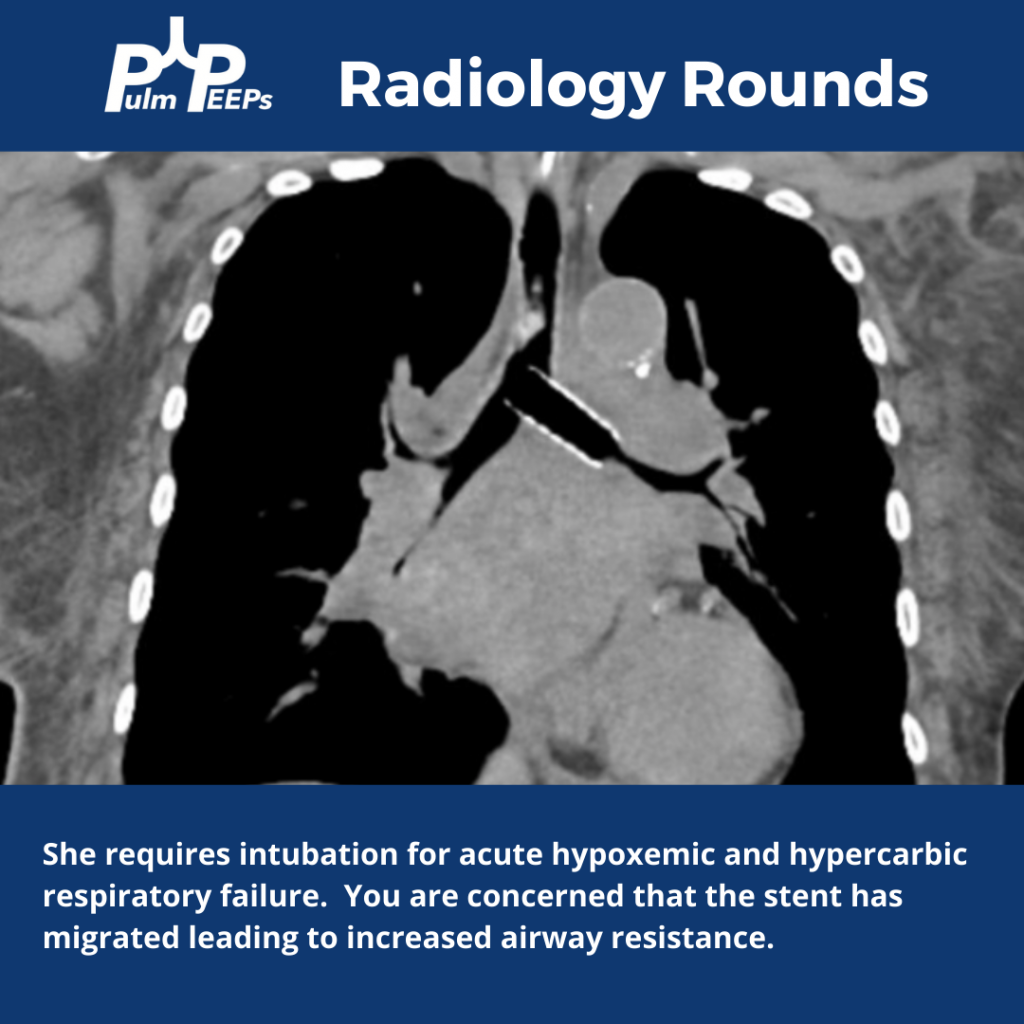
The patient is diagnosed with small cell lung cancer and requires a left bronchial stent. She develops acute hypoxemic and hypercapnic respiratory failure requiring intubation.

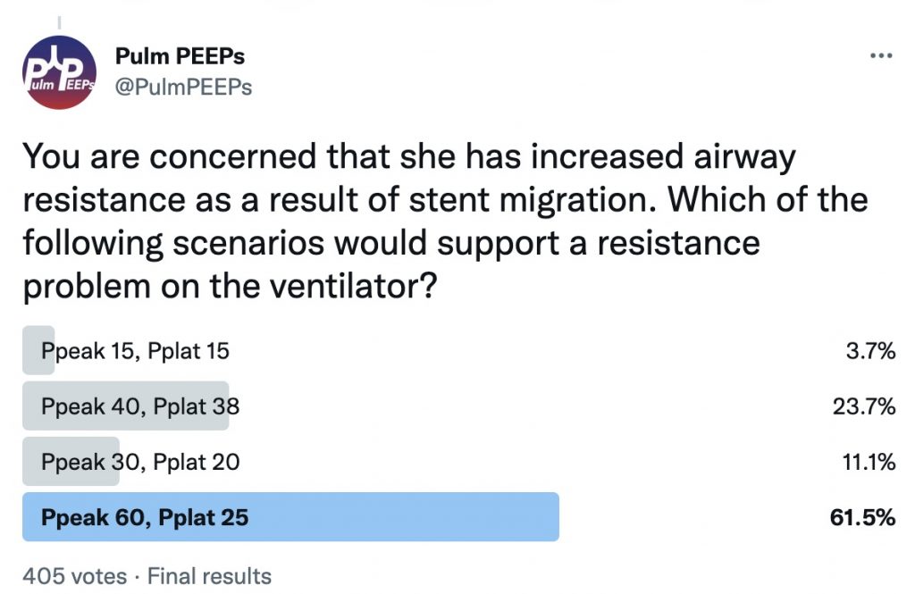
You are concerned that she has increased airway resistance as a result of stent migration. What would you expect to see on the ventilator if this is the case?

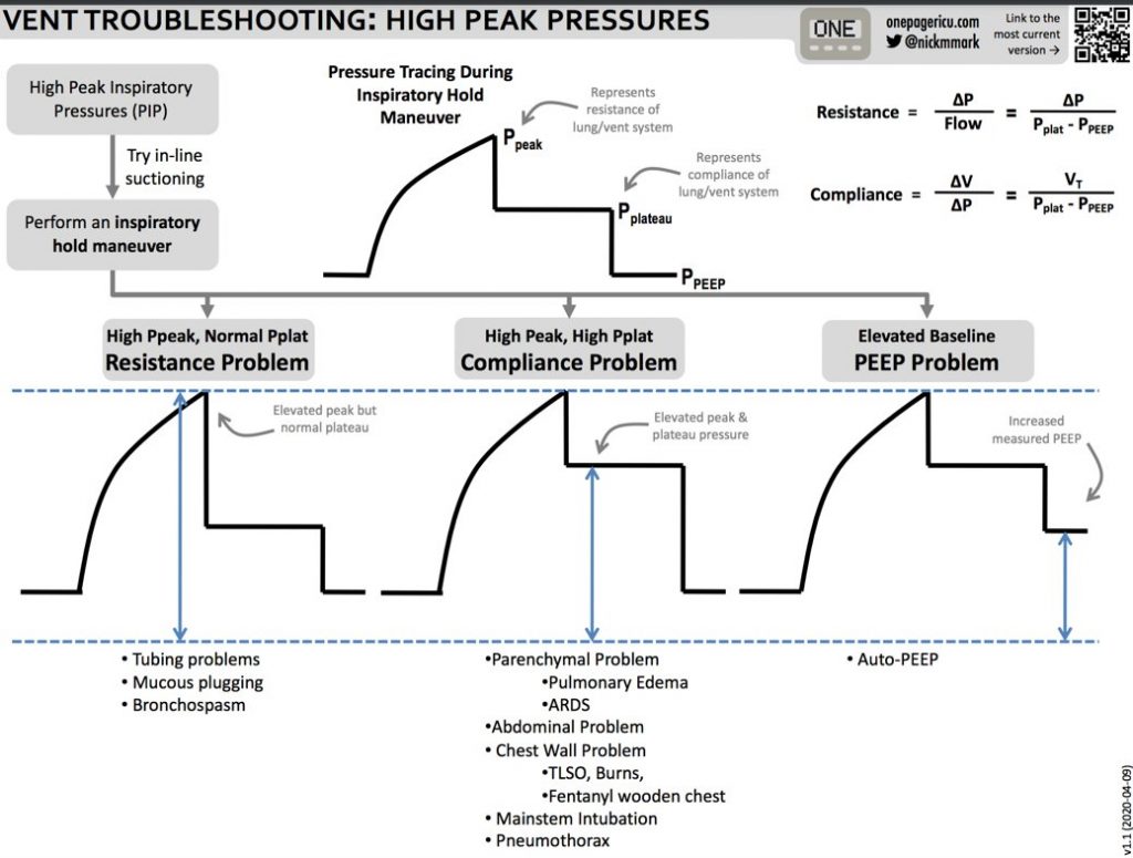
Here are some tips from ICU OnePager on interpreting high peak pressures on the ventilator

Radiology Rounds – 1/25/22

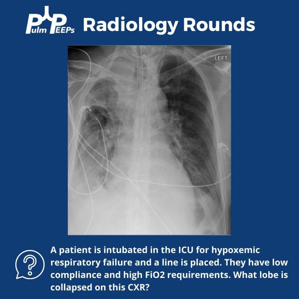
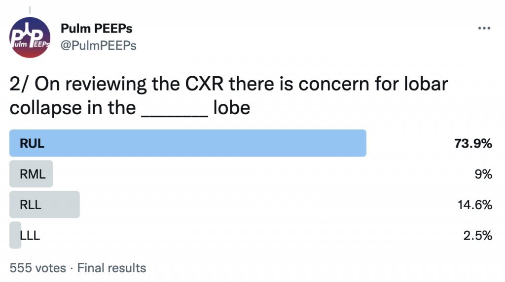
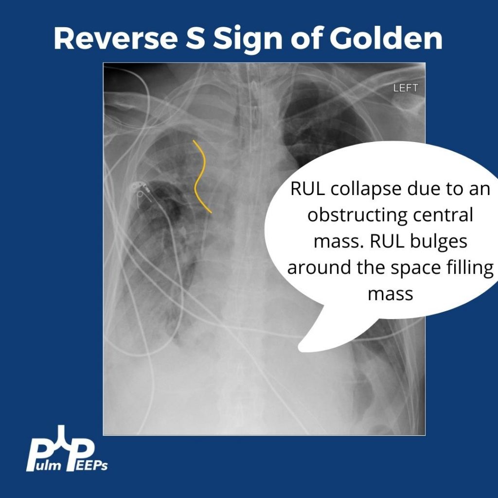
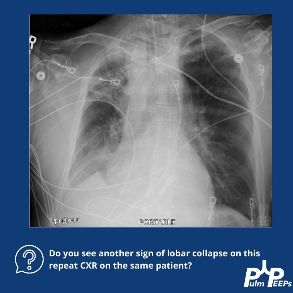
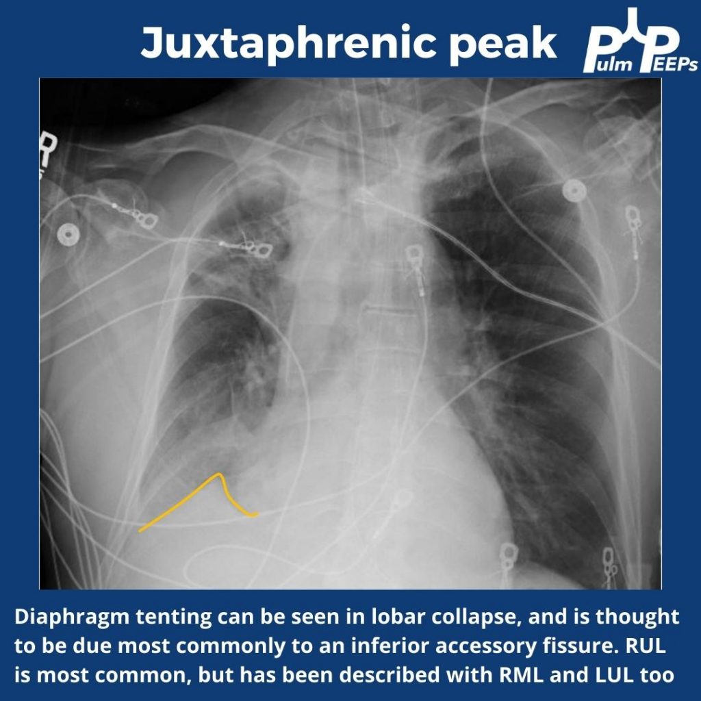
This week for radiology rounds we’re looking at some classic imaging signs of lobar collapse. Take a look, respond to our polls on Twitter, and make sure to subscribe to our podcast to get all of the Pulm PEEPs content!

References

  1. Kumaresh A, Kumar M, Dev B, Gorantla R, Sai PV, Thanasekaraan V. Back to Basics – ‘Must Know’ Classical Signs in Thoracic Radiology. J Clin Imaging Sci. 2015;5:43. doi:10.4103/2156-7514.161977
  2. Algın O, Gökalp G, Topal U. Signs in chest imaging. Diagn Interv Radiol. 2011;17(1):18-29. doi:10.4261/1305-3825.DIR.2901-09.1
  3. Gupta P. The Golden S Sign. Radiology. 2004;233(3):790-791. doi:10.1148/radiol.2333021407