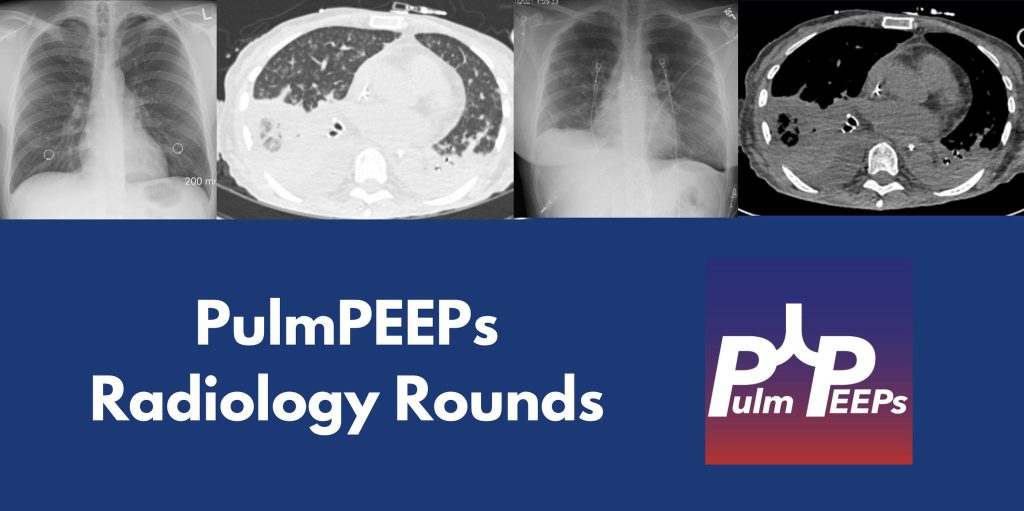
107. Fellows’ Case Files: University of Kansas Medical Center KUMC

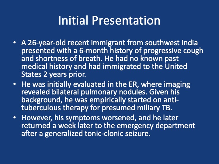
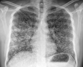
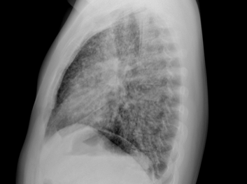
After a brief hiatus, we are excited to be back today with another Fellows’ Case Files! Today we’re virtually visiting the University of Kansas Medical Center (KUMC) to hear about a fascinating pulmonary presentation. There are some fantastic case images and key learning points. Take a listen and see if you can make the diagnosis along with us. As always, let us know your thoughts and definitely reach out if you have an interesting case you’d like to share.
Dr. Vishwajit Hegde completed his internal medicine residency at University of Kansas Medical Center where he stayed for fellowship and is currently a second year Pulmonary and Critical Care medicine fellow. 

Dr. Sahil Pandya is an Associate Professor of Medicine and Program Director of the PCCM Fellowship at KUMC.

1) Initial frame & diagnostic mindset

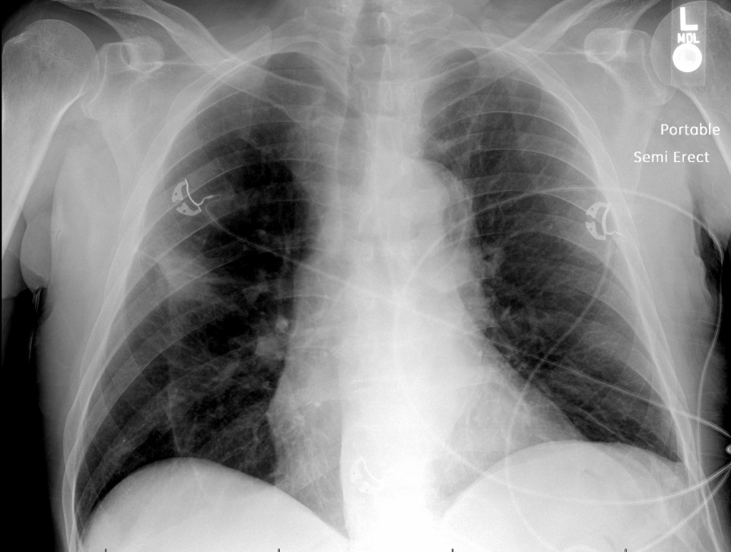
  • Young (26), subacute → chronic dyspnea/cough with diffuse pulmonary nodules; avoid premature closure on TB.
  • Use a Bayesian approach: combine pre-test probability (epidemiology, exposures, tempo) with targeted tests to decide next steps.
  • Always confirm TB when possible (micro/path + resistance testing); empiric RIPE may be reasonable but shouldn’t replace tissue when stakes are high.

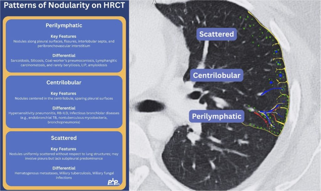
2) Imaging pearls—nodular pattern recognition

  • Ask three things: craniocaudal distribution, symmetry, central vs peripheral.
  • Centrilobular (spares pleura/fissures): airway-centered (e.g., NTM, bronchiolitis, tree-in-bud).
  • Perilymphatic (tracks fissures/pleura & septa): sarcoid, lymphangitic spread.
  • Random/diffuse (involves pleural surfaces): hematogenous spread → think miliary TB, disseminated fungal, septic emboli, metastatic disease.
  • Interval change matters: new cavitation and confluence can upweight infection or aggressive malignancy.

3) Neuro findings—ring-enhancing lesions

  • Differential: septic emboli/abscess, nocardia, fungal, TB, parasites, metastases, vasculitis, sarcoid.
  • Partner with neuroradiology for pattern nuances; treat seizures but keep searching for the unifying diagnosis.

4) Lab/serology strategy

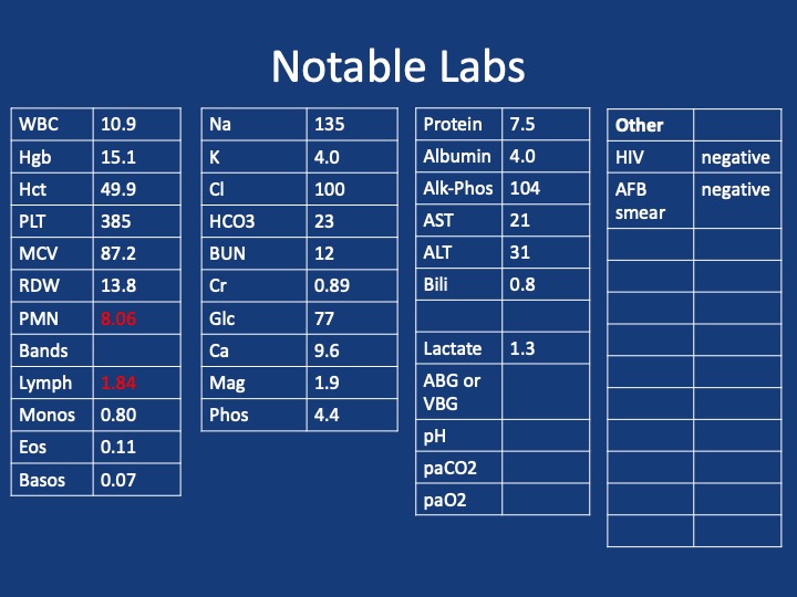
  • Broad infectious workup (AFB × multiple, fungal serologies), HIV and basic immune screen.
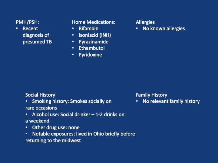
  • Negative/indeterminate tests don’t end the search—revisit history (e.g., Ohio travel → histo/blasto risk).

5) “Tissue is the issue”—choosing the procedure

  • For diffuse nodules with mediastinal adenopathy and stable patient: EBUS-TBNA + BAL, consider transbronchial or cryobiopsy.
  • Cryobiopsy pros: larger, less crush artifact, better for molecular testing; cons: ↑ bleeding/pneumothorax vs forceps.
  • VATS still best for certain ILD questions or if less invasive routes are non-diagnostic—but weigh patient preference and stage/likelihood of yield.
6) ROSE (rapid on-site evaluation) in bronchoscopy

  • Confirms adequacy in real time, steers you away from necrotic zones, helps decide when you’ve got enough for molecular studies, and when to pivot sites—reduces anesthesia time and repeat procedures.

7) Final diagnosis & management

  • Path: TTF-1+, CK7+, napsin A → pulmonary adenocarcinoma with a fusion driver.
  • Therapy: Targeted TKI (crizotinib) → dramatic radiographic response of miliary lung disease and CNS lesions.
  • Teaching point: even “miliary TB-like” lungs + CNS lesions in a 20-something can be driver-positive lung cancer—don’t let age or pattern blind you.

  1. Desai, S., Devaraj, A., Lynch, D., & Sverzellati, N. (2020). Webb, Müller and Naidich’s high-resolution CT of the lung (6th ed.). Lippincott Williams & Wilkins.
  2. Rajeswaran, G., Becker, J. L., Michailidis, C., Pozniak, A. L., & Padley, S. P. G. (2006). The radiology of IRIS (immune reconstitution inflammatory syndrome) in patients with mycobacterial tuberculosis and HIV co-infection: appearances in 11 patients. Clinical radiology, 61(10), 833-843
  3. Poletti, V., Ravaglia, C., & Tomassetti, S. (2016). Transbronchial  cryobiopsy in diffuse parenchymal lung diseases. Current opinion in pulmonary medicine, 22(3), 289-296.
  4. Norman, G. R., Monteiro, S. D., Sherbino, J., Ilgen, J. S., Schmidt, H. G., & Mamede, S. (2017). The causes of errors in clinical reasoning: cognitive biases, knowledge deficits, and dual process thinking. Academic Medicine, 92(1), 23-30.

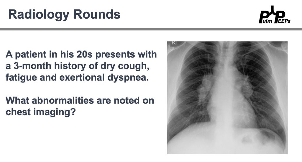
Radiology Rounds – 9/24/25

We’re back with another Pulm PEEPs Radiology Rounds!

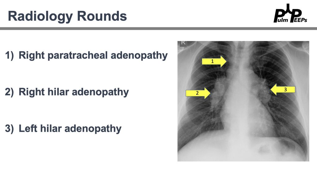
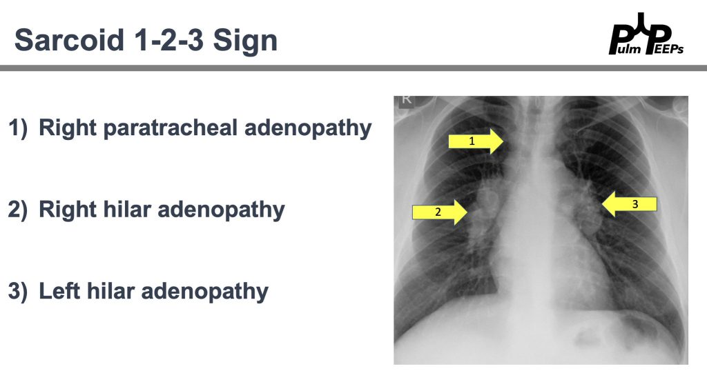
That’s right! All of the findings are present

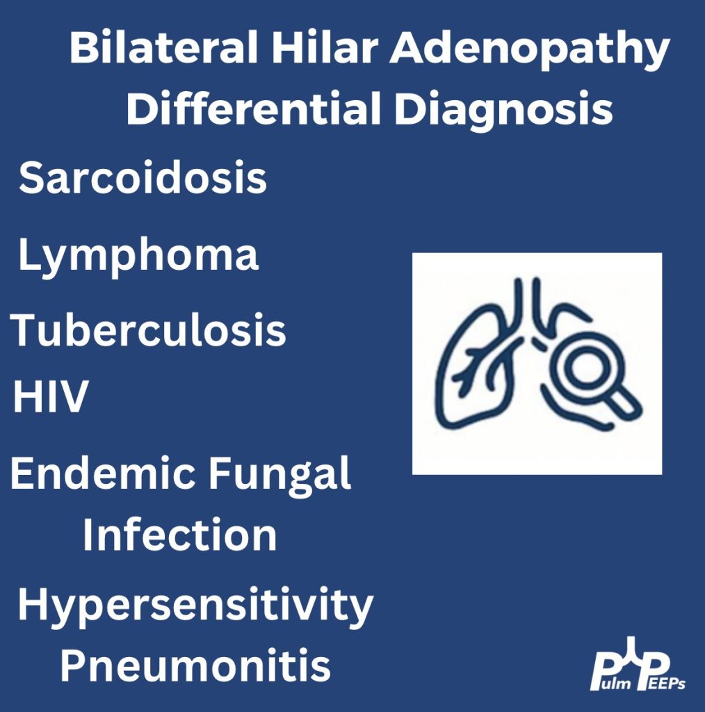
History and physical exam are essential. Here are a few can’t miss diagnoses when working up a young adult with bilateral hilar adenopathy.

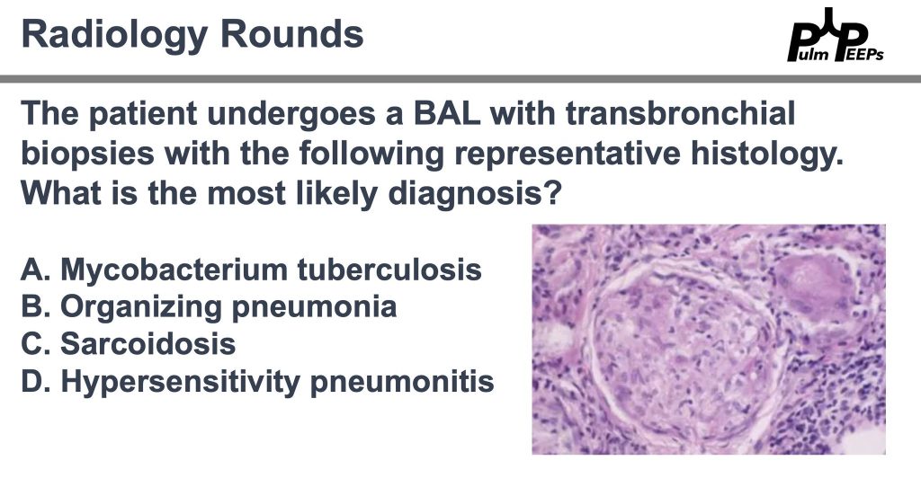
Pathology is consistent with a non-caseating granuloma. What is the most likely diagnosis?

Imaging and pathology were consistent with sarcoidosis and after negative work-up for alternative causes. The patient will follow-up with outpatient pulmonary for sarcoidosis management. Don’t forget about the Sarcoid 1-2-3 sign!

106. Pulm PEEPs Pearls: ICI Pneumonitis

We are so excited to be launching a new series here at Pulm PEEPs! We’ll be talking about high yield topics in 15 minutes or less. In this series, Furf and Monty will tackle core points and provide an overview, key points, and further reading. We’re starting with a key point review of Immune Checkpoint Inhibitor Pneumonitis. Let us know if there are other topics you want to hear about!

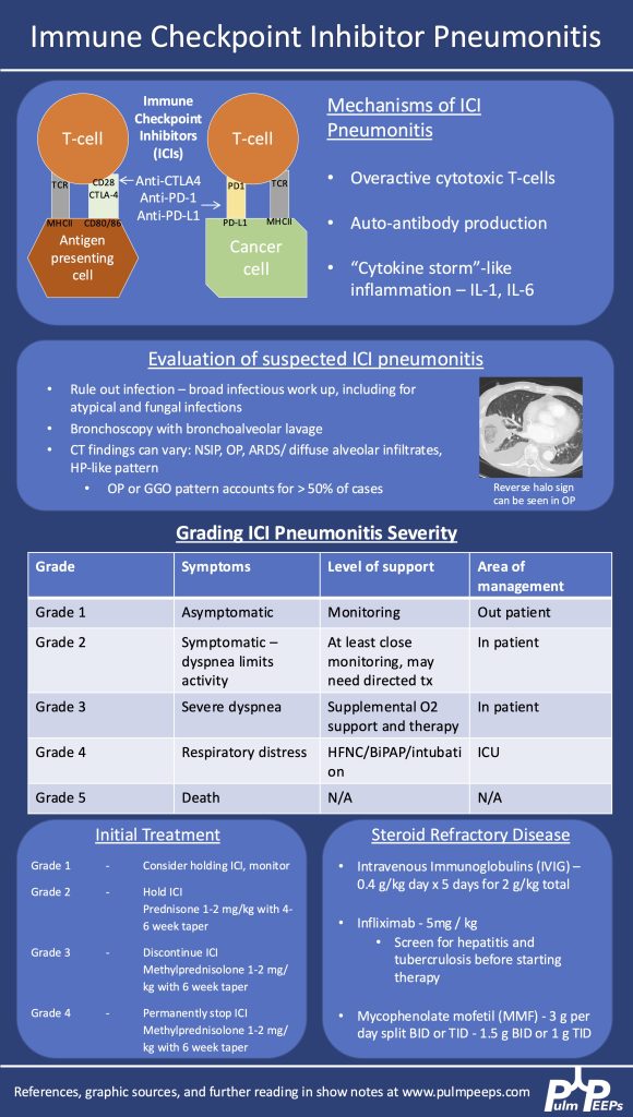
  1. Epidemiology & Pathophysiology
    • Increasingly common as immunotherapy use grows in oncology.
    • Caused by immune activation from PD-1, PD-L1, or CTLA-4 inhibitors.
    • Mechanisms:
      1. Overactive T cells
      2. Autoantibody production
      3. Cytokine-mediated inflammation (e.g., ↑IL-1, ↑IL-6)
  1. Clinical Suspicion & Diagnosis
    • Any new respiratory symptoms in a patient currently or previously on ICI → consider ICI pneumonitis.
    • CT findings are variable: can mimic organizing pneumonia, NSIP, ARDS, or diffuse ground glass opacities. Imaging pattern does not determine severity grade.
    • Diagnosis is of exclusion — infection and malignancy progression must be ruled out first.
    • Workup:
      • Broad infectious evaluation (cultures, viral panel, fungal markers).
      • Early bronchoscopy with BAL if feasible — typically lymphocyte-predominant in ICI pneumonitis.
      • Screen for TB and hepatitis early (in case infliximab is needed).
  1. Severity Grading (Symptom- & O₂-based, not imaging-based)
    • Grade 1: Asymptomatic → monitor, may hold ICI.
    • Grade 2: Symptomatic but not hypoxic → prednisone 1 mg/kg/day PO.
    • Grade 3–4: Hypoxemia or ICU-level care → methylprednisolone 1–2 mg/kg/day IV. Usually hold or permanently stop ICI.
  1. Steroid Management
    • Typical taper: over 6 weeks for grade ≥3.
      • Week 1: 1–2 mg/kg/day
      • Gradual step-down to 0.25 mg/kg/day by week 5, then stop week 6.
    • Chronic/recurrent cases may need slower tapers over months.
    • Add GI prophylaxis and PJP prophylaxis during prolonged steroid use.
  1. If Steroids Fail (no improvement after 48–72 hrs)
    • Consider adding:
      • IVIG (2 g/kg over 5 days)
      • Infliximab (TNF-α inhibitor — requires TB/hepatitis screening)
      • Mycophenolate mofetil (1–1.5 g/day BID or TID, start at effective dose quickly)
    • IVIG may have lower mortality in some series but comes with risks (volume overload, thrombosis, infusion reactions).
  1. Emerging Therapies
    • JAK inhibitors are under investigation as possible future options.
  1. Multidisciplinary Care
    • ICU management is a team sport — coordinate with oncology, critical care, infectious disease, and pharmacy.


  1. Managing Immune Checkpoint Inhibitor Pneumonitis in the ICU. Montemayor, Kristina et al.CHEST Critical Care, Volume 3, Issue 1, 100126
  2. Lavalle S, Masiello E, Valerio MR, Aliprandi A, Scandurra G, Gebbia V, Sambataro D. Immune checkpoint inhibitor therapy‑related pneumonitis: How, when and why to diagnose and manage (Review). Exp Ther Med. 2024 Jul 30;28(4):381. doi: 10.3892/etm.2024.12670. PMID: 39113908; PMCID: PMC11304171.
  3. Delaunay M, Prévot G, Collot S, Guilleminault L, Didier A, Mazières J. Management of pulmonary toxicity associated with immune checkpoint inhibitors. Eur Respir Rev. 2019 Nov 6;28(154):190012. doi: 10.1183/16000617.0012-2019. PMID: 31694838; PMCID: PMC9488507.

88. Fellows’ Case Files: NYU

We are joined today by two amazing educators from NYU for our latest Fellows’ Case Files Episode. Listen today as we go through a great case with some fantastic teaching points highlighted throughout the episode.

Dr. Jeremy Grossman completed his Medicine-Pediatrics residency at Stony Brook Medicine where he was also a Chief Resident. He is currently a second-year PCCM fellow at NYU.

Dr. Shari Brosnahan is an Assistant Professor of Medicine and one of the Assistant Program Directors for the NYU Langone’s Division of Pulmonary, Critical Care, and Sleep Medicine. Her clinical and research interests are focused on pulmonary embolism and thrombosis in critically ill patients.

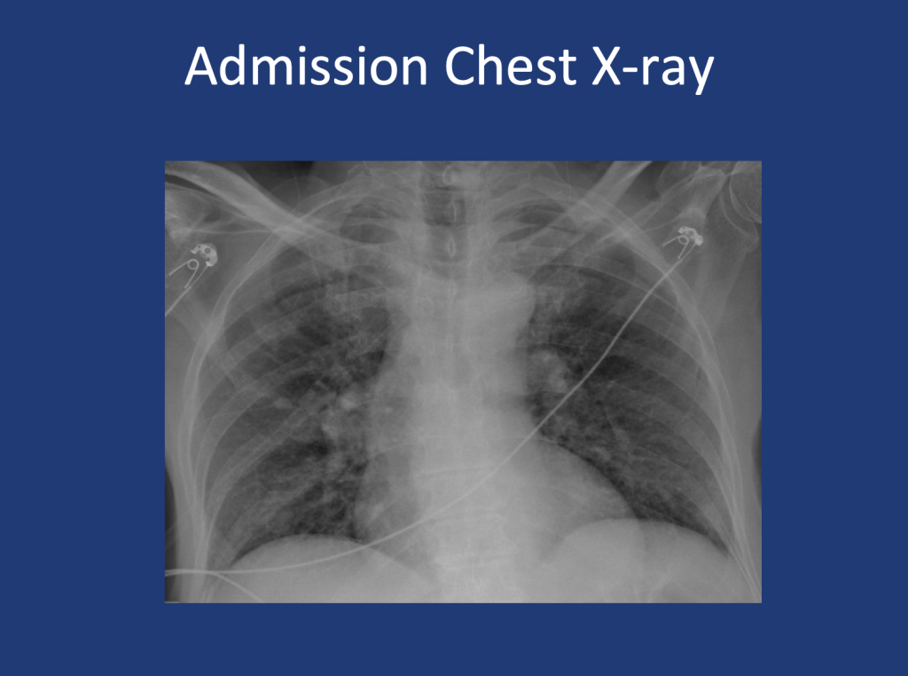
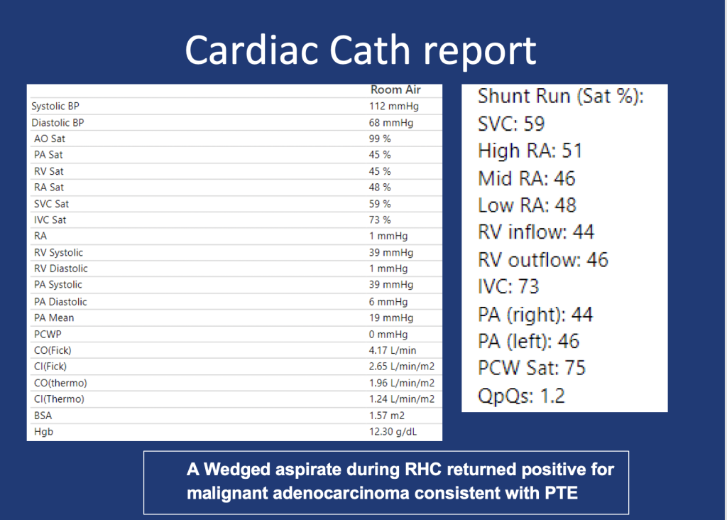
An 80-year-old male presents with shortness of breath. At home, his oxygen saturation was 82% on room air, improving only to 86% on 4L nasal cannula. Over the past month, he has experienced worsening symptoms, including a dry cough, fatigue, and difficulty speaking or ambulating due to shortness of breath at rest. He denies recent fever, sputum production, chest pain, or lower extremity swelling and presents to the ED for further evaluation.

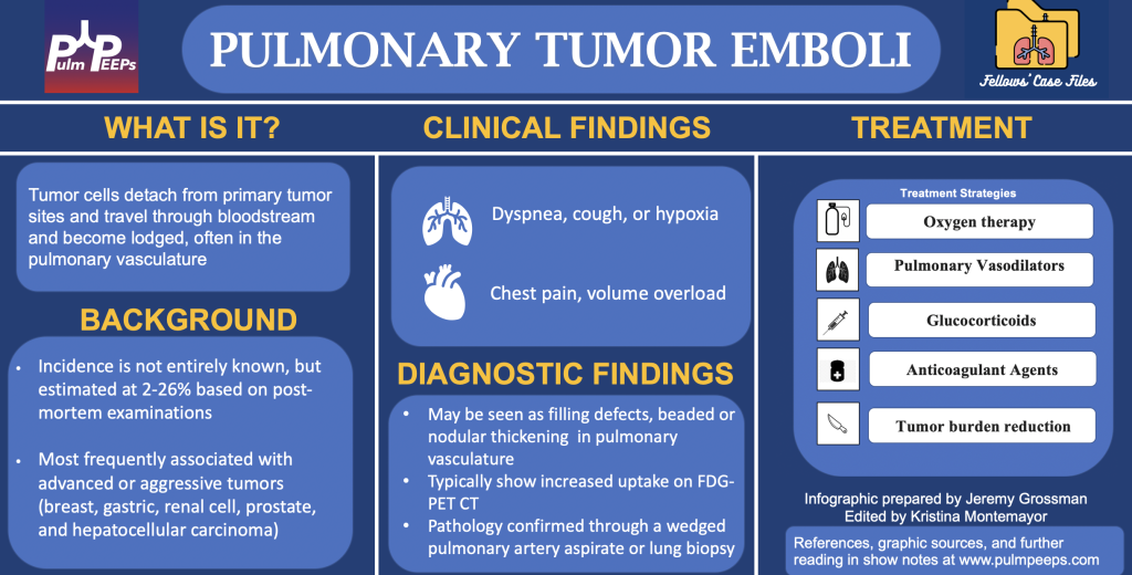
1.In any patient with a history of malignancy and hypoxia, clinicians should keep pulmonary tumor emboli (PTE) on the differential as early intervention may alter outcomes.

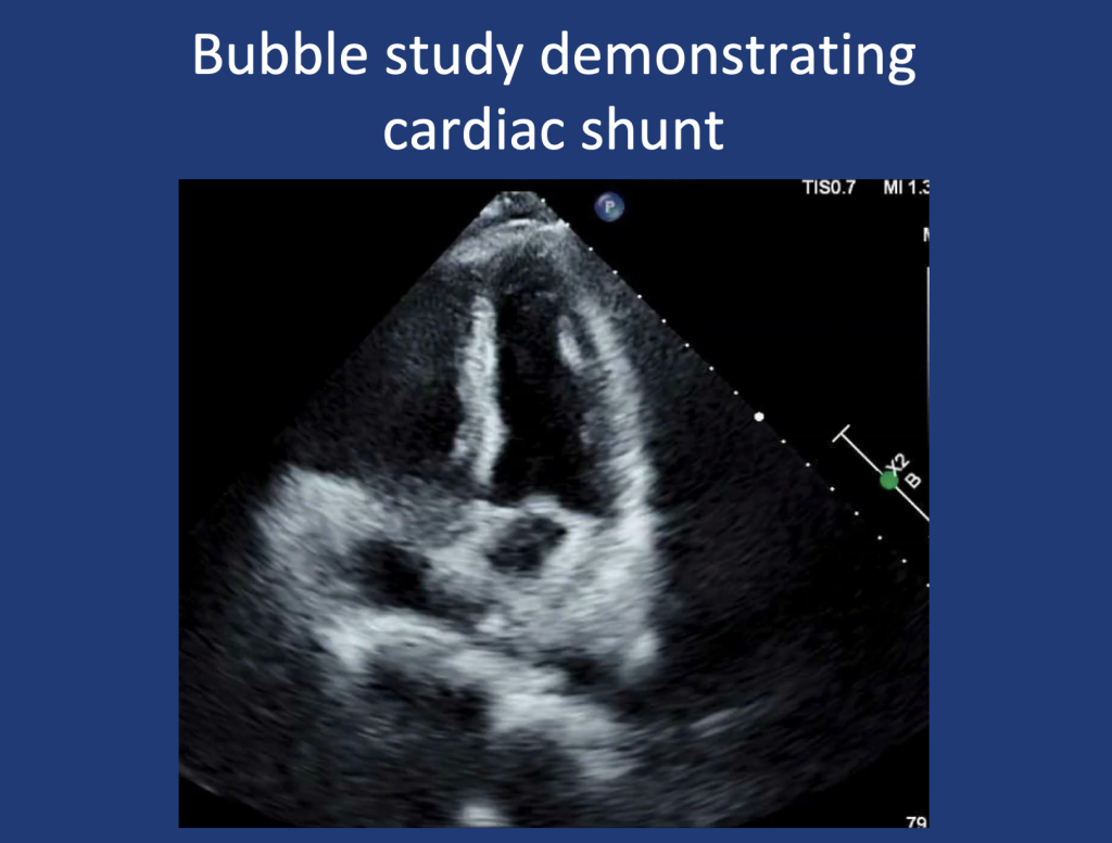
2.PTE contributes to hypoxia via mechanisms of mechanical obstruction of pulmonary arteries leading to shunting, VQ Mismatch, and in some cases pulmonary hypertension due to increased PVR.

3.A wedged aspirate can be used to diagnose PTE ante-mortem

Radiology Rounds – 12/19/23

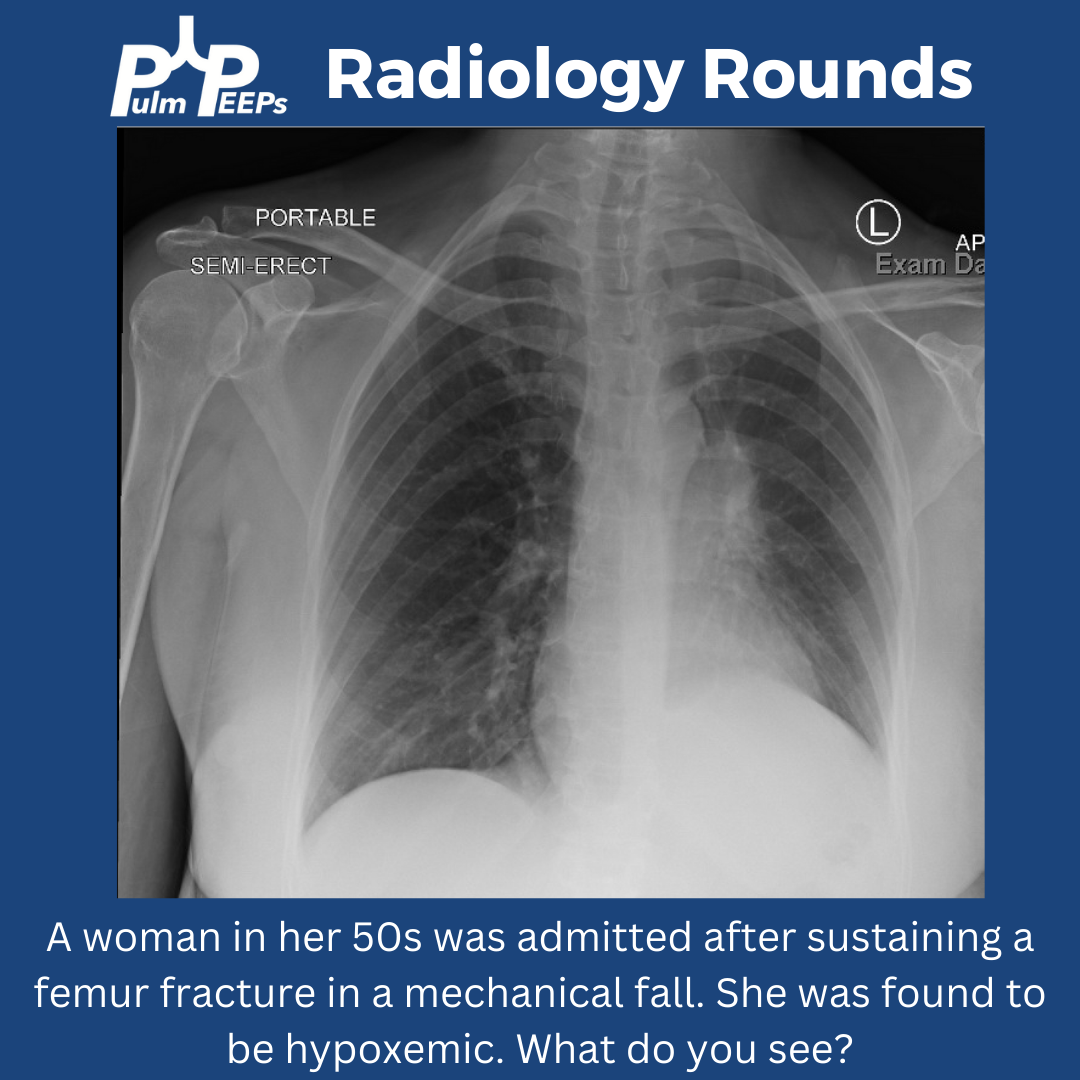
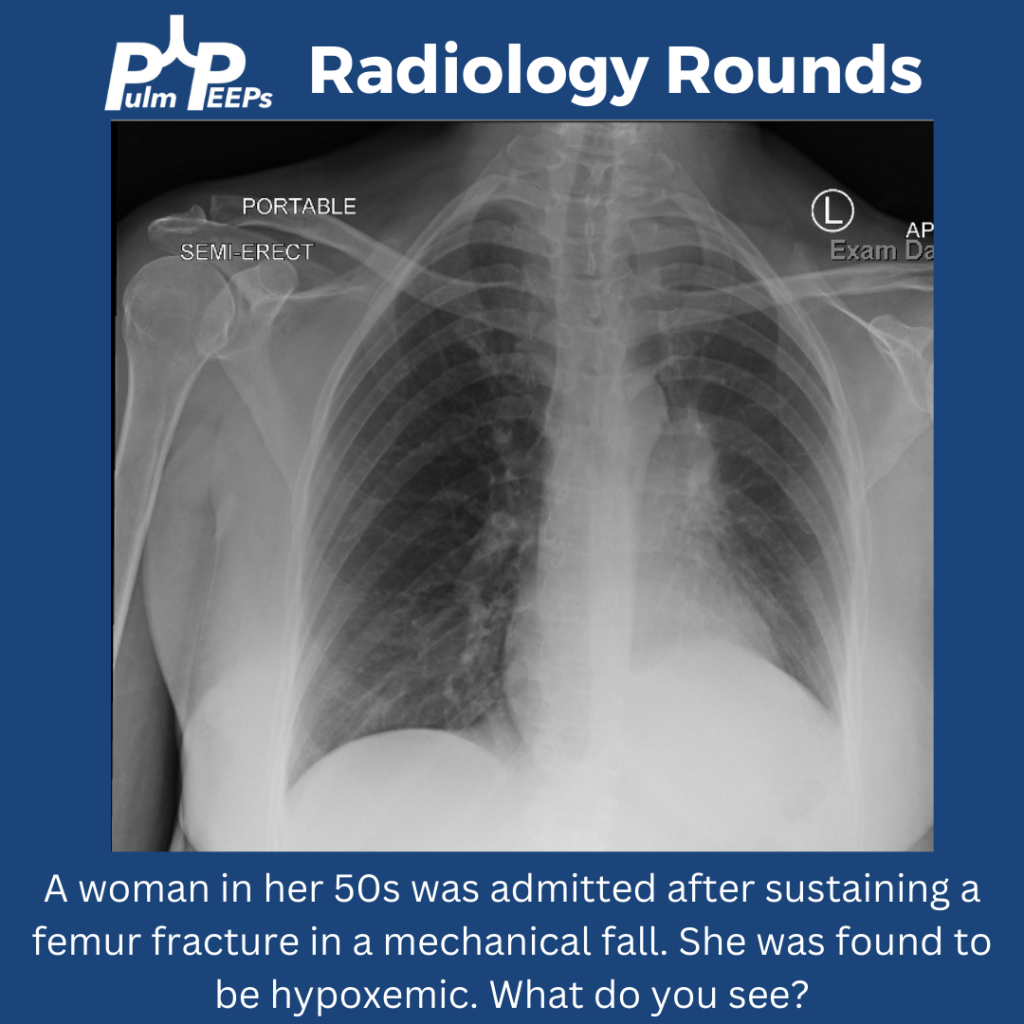
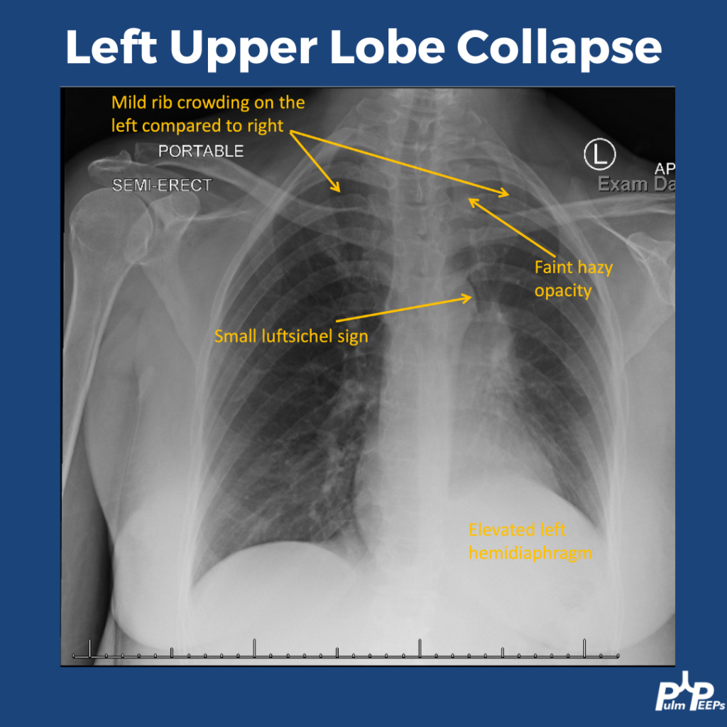
As we approach the end of the year, we know that the hospital and clinic keep on rolling, so here is a CXR and pulmonary differential based #RadiologyRounds if you’re on for the holiday blocks A woman in her 50s has hypoxemia after being admitted with a femur fracture

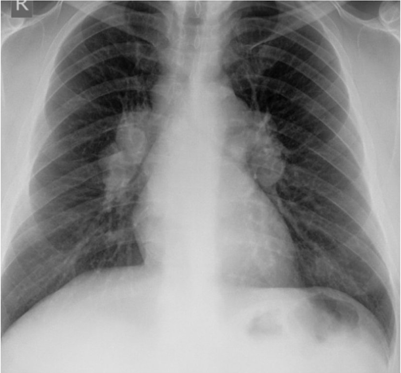
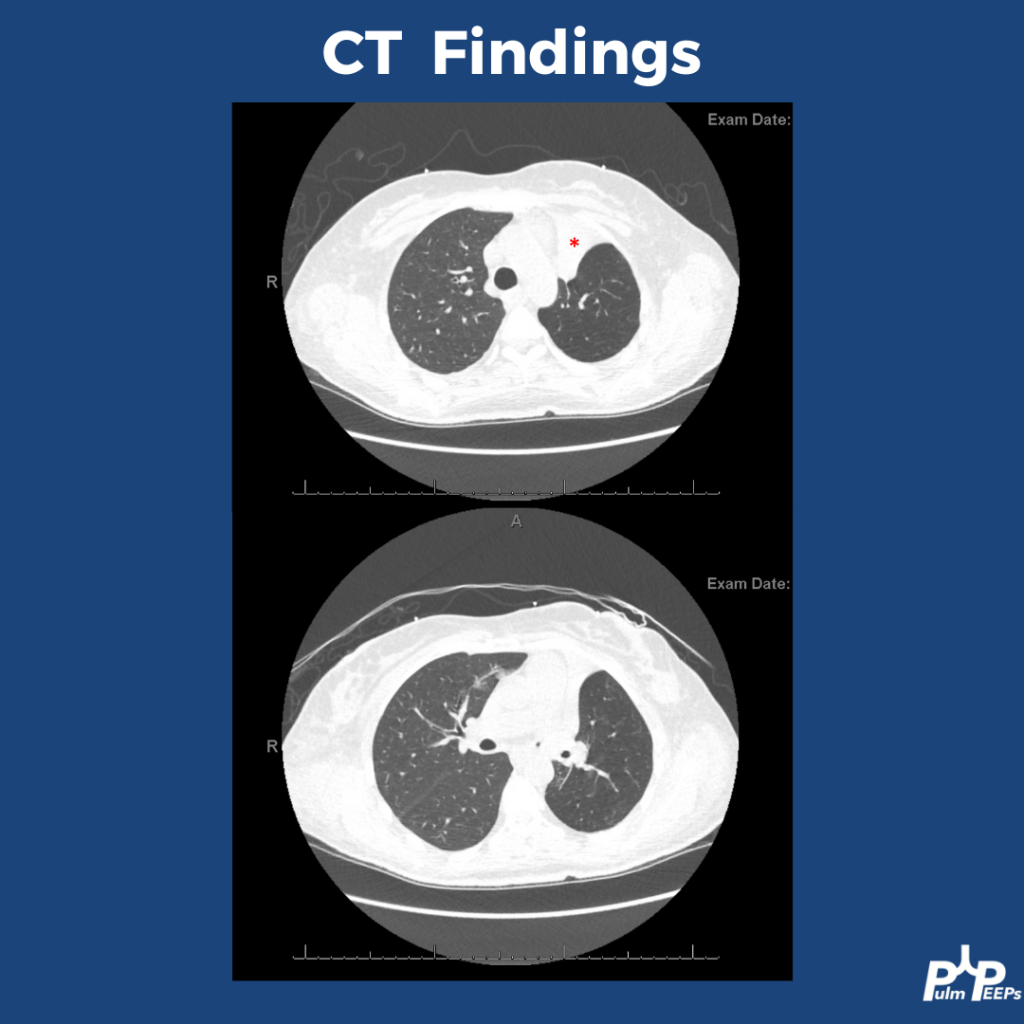
The CXR shows signs of a left upper lobe collapse. The upper lungs should be the most lucent, with increased opacity as you go down. LUL collapse can be subtle, but you can see increased haziness at the lung apex. There is also rib crowding and left hemidiaphragm elevation. You can also see a small luftsichel sign. Luftsichel sign is a thin strip of air that can be seen between the collapsed LUL and the superior mediastinum created by the still-aerated superior segment of the lower lobe. The collapse is better appreciated on CT.

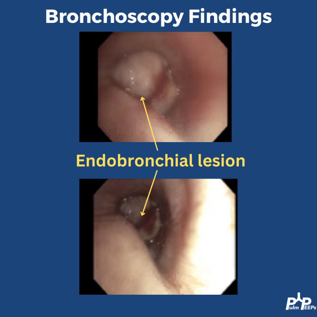
Bronchoscopy was performed to investigate the etiology of the lobar collapse. On the bronchoscopy there was an endobronchial lesion completely occluding the left upper lobe bronchus. The mass was biopsied and sent for histopathology

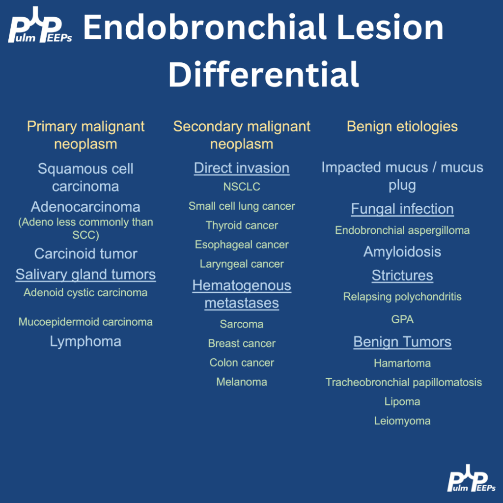
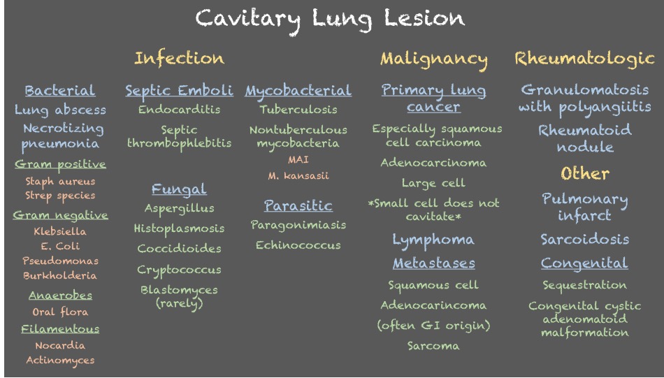
Endobronchial lesions can be malignant or benign, and within malignancy then can be primary or secondary. This patient had no clinical signs of pneumonia and no clear reason to mucus plug so malignancy was top of the differential. Here are differential considerations:

The pathology returned positive for a carcinoid tumor, which can typically arise as endbronchial lesions. PET/CT showed only local disease (femur fracture unrelated), and she is planned for rigid bronchoscopy with laser therapy and corecath ablation for definitive resection.

58. Implications of Race-Neutral PFTs on Lung Cancer Surgery

We are excited to bring you a fantastic episode today where we are joined by two guest experts to discuss the recent JAMA Surgery manuscript, “Clinical Implications of Removing Race-Corrected Pulmonary Function Tests for African American Patients Requiring Surgery for Lung Cancer

Meet our Guests

Sidra Bonner is a general surgery resident at Michigan Medicine. She completed her undergraduate education at Cornell University and medical education at the University of California-San Francisco. Sidra also has a Master’s in Public Health focused in Health Policy from Harvard and a Master’s in Science Health and Healthcare Research from the University of Michigan. She is interested in pursuing a career in general thoracic surgery with a research focus aimed at addressing the multi-level contributors to racial and ethnic inequities in access, quality, and outcomes of surgical care for patients with lung and esophageal cancer.

Tom Valley is an Associate Professor in the Division of Pulmonary and Critical Care Medicine at the University of Michigan. He completed his IM residency and chief residency at the University of Texas-Southwestern/Parkland Memorial Hospital and then joined the University of Michigan as a pulmonary and critical care fellow in 2013 and stayed on for faculty and is the physician-lead for the University of Michigan Schwartz Rounds for Compassionate Care. Tom’s research aims to understand and improve medical decision making in the intensive care unit.

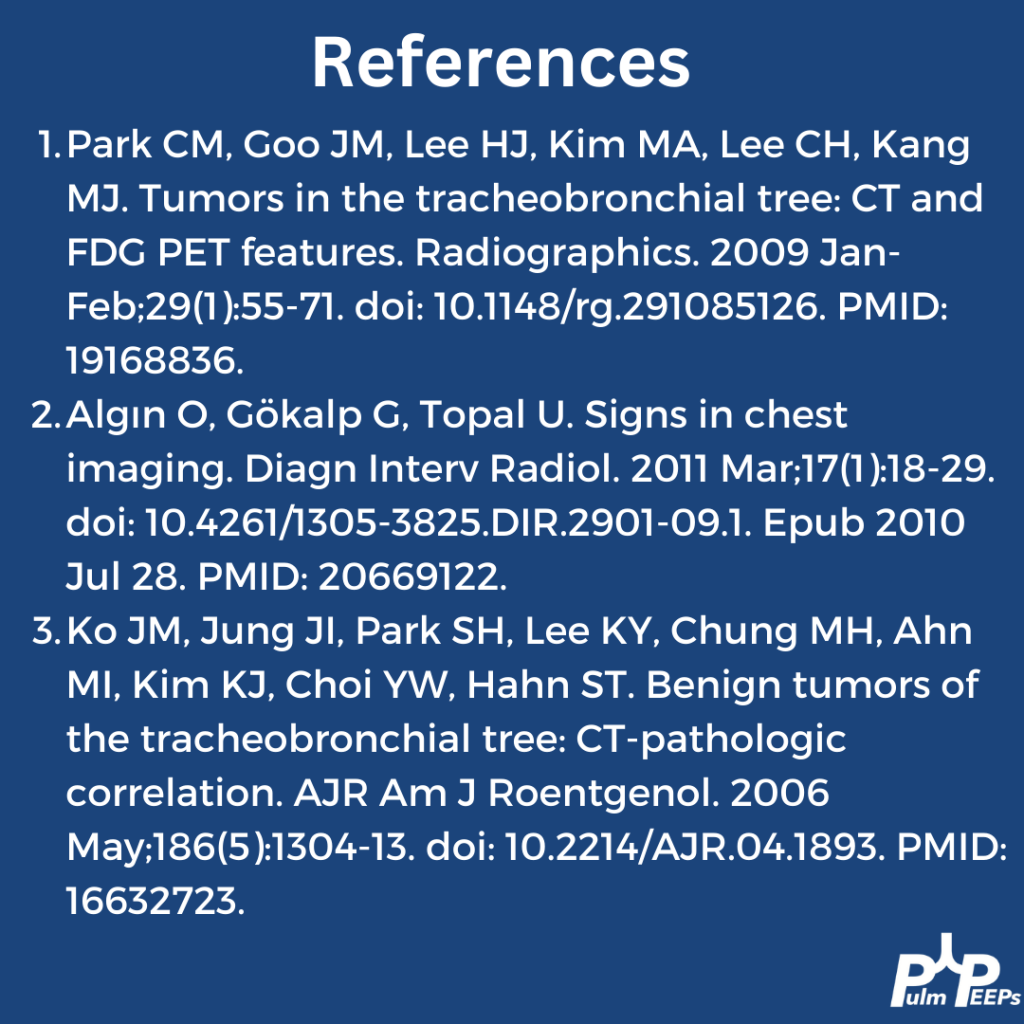
54. Top Consults: Solitary Pulmonary Nodule

We are thrilled to be back with another episode in our Top Consults series. We are talking about Solitary Pulmonary Nodules, which is something every pulmonologist will encounter in the clinic and on in-patient consults. We go through a number of cases and provide a framework for approaching these cases.

Meet our guests

Dr. Jessica Wang Memoli is board certified in pulmonary disease, critical care medicine and internal medicine. She is the Director of Bronchoscopy and Interventional Pulmonary, as well as the Associate Fellowship Program Director for Pulmonary Critical Care Medicine at the MedStar Washington Hospital Center. Dr. Wang Memoli received her medical degree from the University of Miami Miller School of Medicine. She completed her residency at MedStar Washington Hospital Center and her fellowship training at the Medical University of South Carolina in Charleston.

Dr. Nick Ghionni works at Union Memorial, Good Samaritan, and Franklin Square as an Intensivist and Pulmonologist. He completed his Internal Medicine residency at Mercy Catholic Medical Center in PA serving as Chief Internal Medicine resident. He was a fellow at MedStar Washington Hospital Center where he was the Chief Pulmonary Critical Care Fellow. His specific interests include mechanical ventilation, POCUS, and medical education.

Case Presentations

Case 1:

33 year old woman who came to the emergency department with acute onset of shortness of breath. She states that she had been in her normal state of health until this morning when she developed shortness of breath at rest, and chest pain. She does report a non-productive cough over the last few weeks which she feels may be contributing to her chest pain. She does report a history of asthma during childhood but without any exacerbations or maintenance therapies needed during her adulthood. She does report wheezing when she is sick with a cold but this is infrequent. The ED team sent off an initial work-up including a D-Dimer which was elevated, and she underwent a CTA of the chest for concern for possible PE. On the CT scan, there was no PE but the radiologist did call a “2 mm indeterminate right upper lobe pulmonary nodule.”

Case 2:

We have a 67-year-old male with a past medical history of ischemic cardiomyopathy, chronic systolic heart failure (LVEF 10-15%), s/p AICD, diabetes mellitus type 2, hyperlipidemia, hypertension, chronic kidney disease stage III, prostate cancer s/p seed implantation that was over 15 years ago who presented with acute decompensation of his heart failure and cardiogenic shock. He was successfully managed for that and is now being worked up by advanced HF and as a part of that workup got a chest CT, which found a RUL 6 mm nodule.

Case 3:

We have a 66-year-old male with a past medical history of HTN and drug abuse who presented to the ED with acute SOB, likely a COPD exacerbation. He was given bronchodilator and steroids as well as being started on Bipap. He eventually was able to be weaned off Bipap and was able to tolerate nasal cannula. As a part of his initial work up, the patient underwent CT scan for possible PE which demonstrated a new LUL spiculated nodule that is 1.3cm that is new since 2019.

Key Learning Points

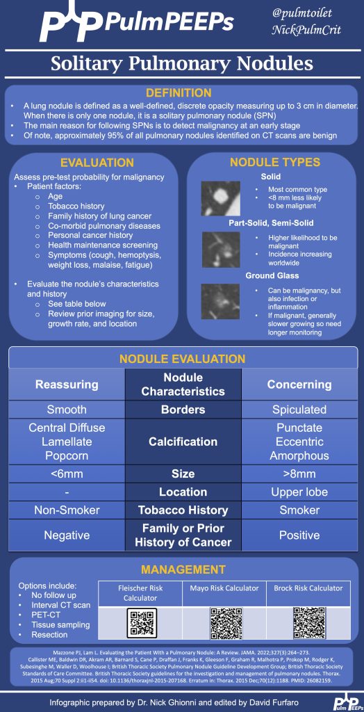
Approaching Pulmonary Nodules:

  1. A structured approach is essential due to the complexities of diagnosing pulmonary nodules.
  2. Patient history, including risk factors, past interventions, and imaging, plays a vital role.
  3. Nodules’ appearance, such as location, shape, or characteristics like calcification or spiculation, can provide diagnostic clues.
  4. The nodules history on serial imaging is a key predictive risk factor for determining the likelihood that the nodule represents cancer
  5. Tools like the Mayo Risk Calculator and Fleishner Society guidelines assist in risk assessment and guidance.
  6. It’s essential to assess patient risk, and nodule risk, and prioritize patient concerns and education. Periodic monitoring or follow-up might be necessary based on the nodule’s risk and size.
  7. A multidisciplinary approach involving various specialists ensures comprehensive care.

Key Discussion Points:

PET Scans:

  1. Useful in gauging a nodule or tumor’s metabolic activity.
  2. Large, hypermetabolic nodules are suspicious.
  3. Not every positive PET result means malignancy; other causes like inflammation or scars can produce positive results.

Evaluating Nodules:

  1. Consideration of nodule size, characteristics, patient history, and risk calculators is crucial.
  2. Tumor boards provide a collaborative expertise approach.

Tissue Sampling & Testing:

  1. The method of tissue sampling depends on resources and expertise.
  2. CT-guided biopsy offers a high diagnostic yield but with a risk of pneumothorax.
  3. Bronchoscopic biopsy provides a lower diagnostic yield than CT-guided biopsy but has a significantly reduced risk of complications.
  4. Advanced diseases now often require molecular testing on tissue samples.

Ground Glass Nodules:

  1. Different from solid nodules due to their slow growth rate.
  2. Monitoring is crucial due to the potential for transformations raising cancer suspicions.
  3. The approach for ground glass nodules typically involves more extended monitoring intervals than for solid nodules.

Holistic Evaluation:

  1. Consider the nodule’s characteristics, the patient’s history, and clinical intuition.
  2. Individualized patient assessment is as vital as evidence-based guidelines and clinical expertise.

See the infographic for a summary of key learning points:

References and further reading

Loverdos K, Fotiadis A, Kontogianni C, Iliopoulou M, Gaga M. Lung nodules: A comprehensive review on current approach and management. Ann Thorac Med. 2019 Oct-Dec;14(4):226-238. doi: 10.4103/atm.ATM_110_19. PMID: 31620206; PMCID: PMC6784443.

Mazzone PJ, Lam L. Evaluating the Patient With a Pulmonary Nodule: A Review. JAMA. 2022 Jan 18;327(3):264-273. doi: 10.1001/jama.2021.24287. PMID: 35040882.

MacMahon H, Naidich DP, Goo JM, Lee KS, Leung ANC, Mayo JR, Mehta AC, Ohno Y, Powell CA, Prokop M, Rubin GD, Schaefer-Prokop CM, Travis WD, Van Schil PE, Bankier AA. Guidelines for Management of Incidental Pulmonary Nodules Detected on CT Images: From the Fleischner Society 2017. Radiology. 2017 Jul;284(1):228-243. doi: 10.1148/radiol.2017161659. Epub 2017 Feb 23. PMID: 28240562.

Wahidi MM, Govert JA, Goudar RK, Gould MK, McCrory DC; American College of Chest Physicians. Evidence for the treatment of patients with pulmonary nodules: when is it lung cancer?: ACCP evidence-based clinical practice guidelines (2nd edition). Chest. 2007 Sep;132(3 Suppl):94S-107S. doi: 10.1378/chest.07-1352. PMID: 17873163.

Godoy MC, Sabloff B, Naidich DP. Subsolid pulmonary nodules: imaging evaluation and strategic management. Curr Opin Pulm Med. 2012 Jul;18(4):304-12. doi: 10.1097/MCP.0b013e328354a5f2. PMID: 22575798.

Hammer MM, Hatabu H. Subsolid pulmonary nodules: Controversy and perspective. Eur J Radiol Open. 2020 Sep 4;7:100267. doi: 10.1016/j.ejro.2020.100267. PMID: 32944597; PMCID: PMC7481135.

Radiology Rounds – 9/5/23

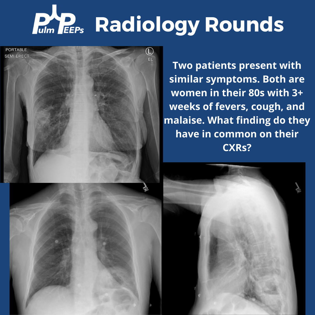
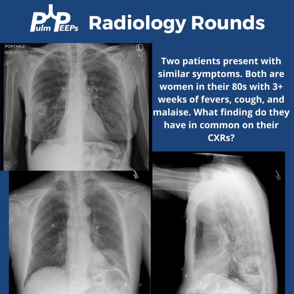
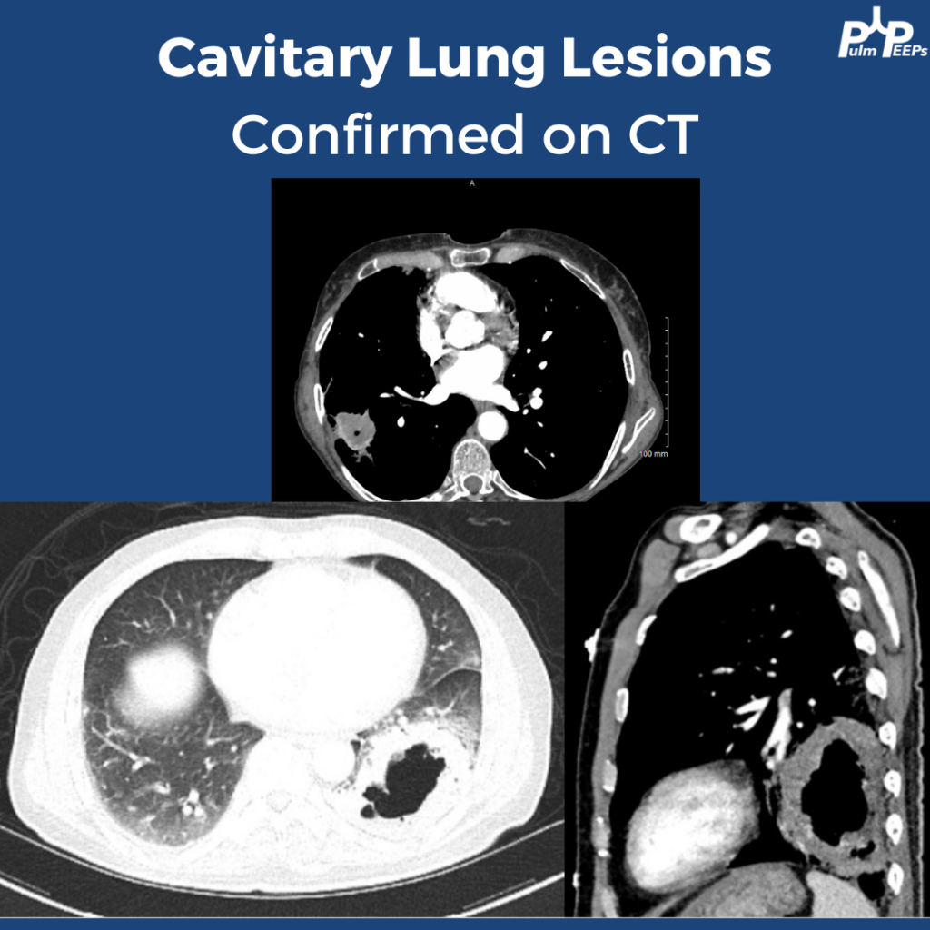
Tuesday is time for another #RadiologyRounds! Time for some CXR reading and a differential diagnosis mnemonic Two women presented to the hospital with similar presentations. They are both in their 80s with multiple weeks of cough, fever, and fatigue. Here are the CXRs

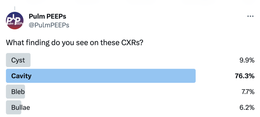
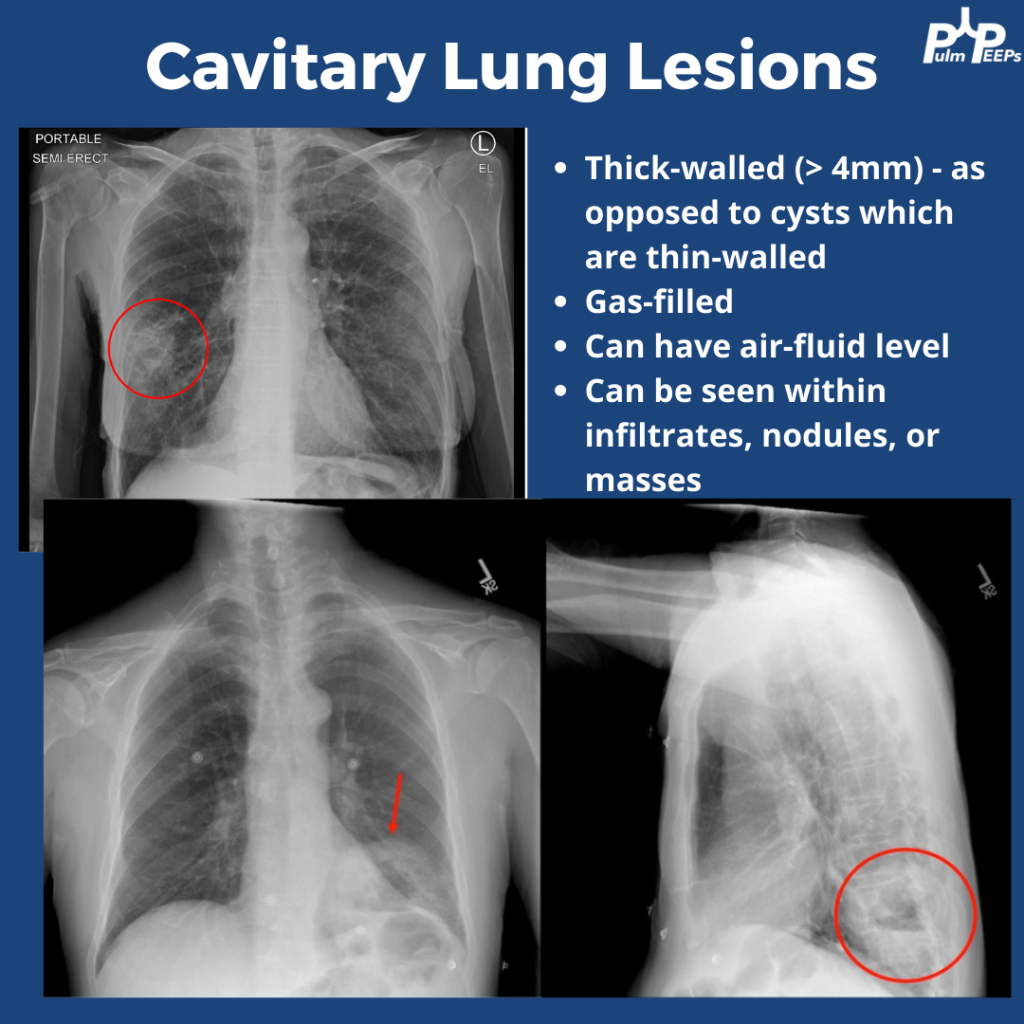
The CXRs both showed cavities. They are thick-walled (>4mm) and gas-filled. Cavitary lung lesions are seen within infiltrates, nodules, or masses. There can be an air-fluid level within the cavity. Cysts have thinner walls. The findings were confirmed on CT scan

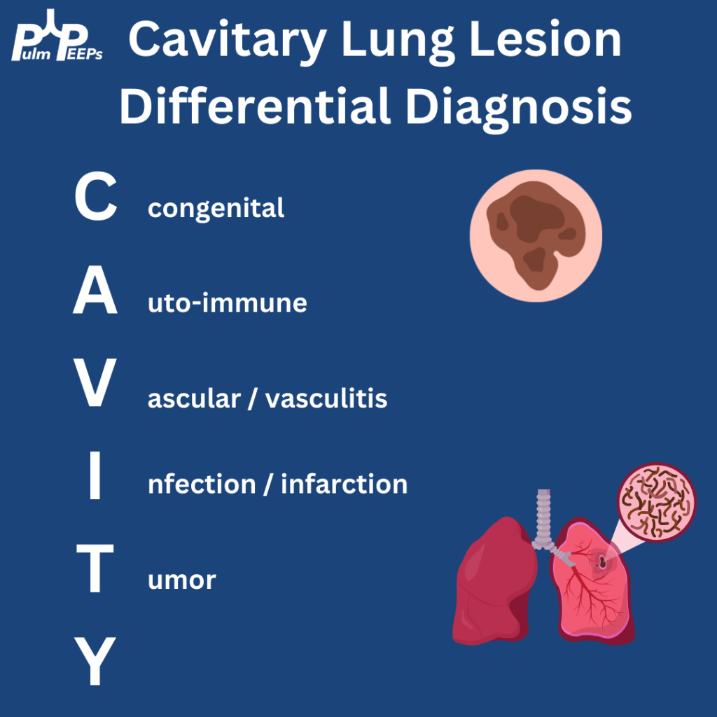
Cavitary lung lesions can have a broad differential so it is helpful to have a systematic approach. To make it easy, when you see this just remember: CAVITY

Bonus points to anyone who can fill in the Y

Both patients were ultimately diagnosed with pulmonary abscesses which improved with prolonged courses of antibiotics with anaerobic and gram-negative coverage.

52. Fellows’ Case Files: University of Pittsburgh

We’re excited to be back with another Fellows’ Case Files. Today, we’re visiting the University of Pittsburgh to meet a fantastic fellow and a dedicated educator, and to hear about a fascinating case. Let us know if you’ve ever had a similar case, and share your interesting cases with us!

Meet Our Guests

Rachel Wojcik obtained her B.S. in Biology from Mercyhurst University and a Master’s in Liberal Studies from the University of Denver in Global Affairs with a focus on Healthcare. She completed her MD at the University of Colorado before completing her residency and chief resident year at the University of Pittsburgh and has continued her training at Pitt for PCCM fellowship.

Dr. Stephanie Maximous is an Assistant Professor of  Medicine at the University of Pittsburgh School of Medicine and is the Clinical Education APD for the Pulmonary and Critical Care Fellowship program. She completed her fellowship at Pitt in addition to obtaining a Master’s Degree in Medical Education there. She teaches in and directs courses throughout the medical school, residency, and fellowship and was recently awarded the 2023 Outstanding Subspecialty Teaching Attending Award from the housestaff.

Case Presentation

Patient: A 70-year-old male with a history of idiopathic thrombocytopenia on chronic prednisone and a history of tobacco use disorder.

Presentation: Came to the hospital with 2-3 days of right-sided weakness and slurred speech.

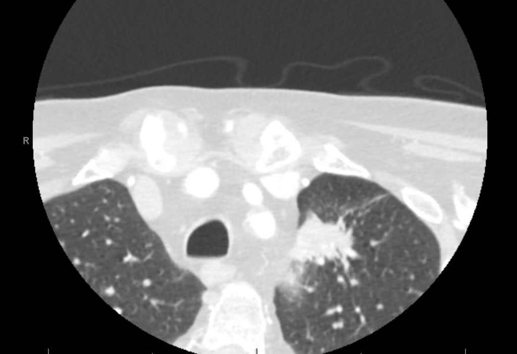
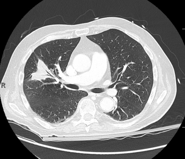
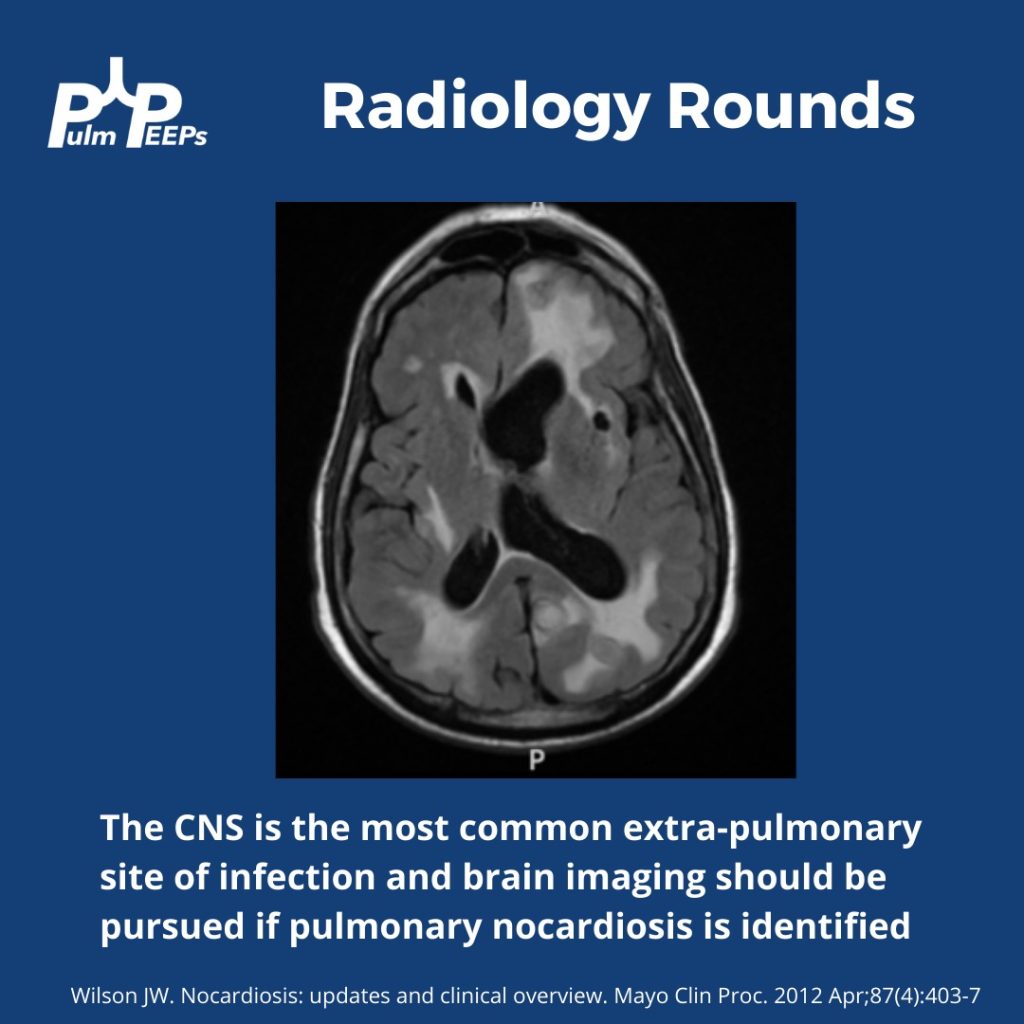
Findings: MRI showed a moderate-sized left pontine stroke. A CT angiogram of the neck showed no evidence of an occlusion, but a spiculated two-centimeter nodule at the apex of the left lung was found.

Additional Information: He requires a walker for mobility and needs help with activities like taking a shower and dressing. He had an unintentional 20-pound weight loss over six months, increased fatigue, and malaise.

Previous Investigations: A chest x-ray ordered two months prior by his hematologist was unremarkable, and a CT of the abdomen and pelvis showed no masses.

Key Learning Points

Bronchoscopy in Decision Making:

  • The decision to perform bronchoscopy in patients depends on a myriad of factors, including the location of any lesions, accessibility, potential risks, and the potential diagnostic yield.
  • Fiber optic bronchoscopy with BAL can rule out infections, and if no diagnosis is reached, more invasive methods like surgical biopsy might be necessary.
  • Consider the location of consolidated masses; navigational bronchoscopy might be needed for lesions without a clear airway leading into them.

Nocardia Insights:

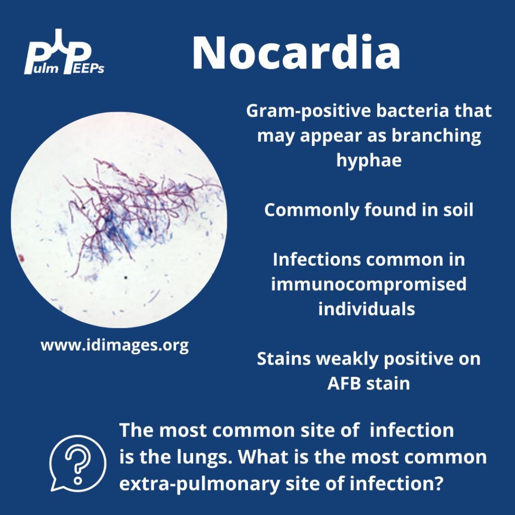
  • Nocardia is a gram-positive bacterium that stains weakly acid-fast.
  • It can be found in soil and certain water sources and can infect through the skin or by inhalation.
  • Two-thirds of patients with Nocardia are immunocompromised.
  • The dosage of Bactrim given for PJP prophylaxis doesn’t prevent Nocardia infections in immunocompromised individuals.
  • While the lungs are the most common infection site, Nocardia can manifest elsewhere, like the skin or CNS.

Treatment Approach:

  • Bactrim is the mainstay of treatment for Nocardia. If someone is allergic, desensitizing them can be crucial.
  • IV induction phases vary in length depending on the severity of the disease.
  • The overall treatment duration is protracted to prevent relapse.

Takeaway Points:

  • Bactrim for PJP prophylaxis doesn’t necessarily prevent Nocardia infections in immunocompromised individuals.
  • If someone is allergic to Bactrim, consider desensitizing them due to its importance in treating Nocardia.

References and Further Reading:

Menéndez R, Cordero PJ, Santos M, Gobernado M, Marco V. Pulmonary infection with Nocardia species: a report of 10 cases and review. Eur Respir J. 1997 Jul;10(7):1542-6. doi: 10.1183/09031936.97.10071542. PMID: 9230244.

Zia K, Nafees T, Faizan M, Salam O, Asad SI, Khan YA, Altaf A. Ten Year Review of Pulmonary Nocardiosis: A Series of 55 Cases. Cureus. 2019 May 26;11(5):e4759. doi: 10.7759/cureus.4759. PMID: 31363440; PMCID: PMC6663111.

49. Top Consults: Malignant Pleural Effusions

We’re diving back into some pleural fluid today (okay that is kind of gross to think about and we apologize). If you haven’t listened to our prior pleural effusion episodes and want to start from the top, then check out Episode 36 for a general approach to pleural effusions, and Episode 37 to hear about parapneumonic effusions. Malignant effusions are another common consult question and we’ll talk about everything from detection to monitoring, to definitive management.

Meet Our Guests

Dr. David DiBardino is an Assistant Professor of Medicine at the University of Pennsylvania Medicine and is the Associate Director for Clinical Research within the Section of Interventional Pulmonology. He is also the Program Director for the Interventional Pulmonary Fellowship there.

Dr. Jamie Bessich is an Assistant Professor of Medicine and Cardiothoracic Surgery at NYU Grossman School of Medicine. She is the Section Chief of Interventional Pulmonology and is the Director of Bronchoscopy at Tisch Hospital.

Dr. Van Holden is an Associate Professor of Medicine at the University of Maryland School of Medicine and is the Pulmonary and Critical Care Fellowship Program Director there. She was last on the show for our very first Fellows’ Case Files when we discussed a fascinating case of pulmonary alveolar proteinosis.

Case Presentation

The page: 72M smoker, new effusion, concern for malignancy, tap?

Further history: 72 year old man with PMH of GOLD B COPD, tobacco use (55 pack years), HTN, HLD, and diabetes. He presented to the ED with progressive dyspnea and fatigue. He is on LAMA/LABA for his COPD, and he does not frequently have exacerbations. He has no increased sputum production or wheezing, but he has been feeling progressively fatigued and lethargic. H Over the past few weeks he has had more dyspnea on exertion, and now has it at rest too. It is a bit worse when he lies flat. He has had no weight gain or edema in his legs and has actually lost 10-15 pounds in the last 3 months. In the ED, he is newly requiring 3-4L NC, has decreased breath sounds on the right, and a CXR shows a large right-sided pleural effusion, as well as a large apical nodule with some spiculation, both of which are new from prior.

Key Learning Points

Causes of malignant pleural effusion

–Lung cancer is the most common in men

–Breast cancer is the most common cause in women

–Lung and breast cancer account for > 50% of all malignant pleural effusions

–Other less common causes are lymphoma, GU or GI tract cancer

–Remember to consider mesothelioma

Prognosis of MPE

–Malignant pleural effusion means the cancer is advanced and stage 4 by definition

–The average life expectancy after a diagnosis of MPE is 3-12 months, depending on the patient and the malignancy

Imaging and MPE

–Make sure to get a CT scan after drainage so no lesions are missed

–Ultrasound can be helpful to look for disruptions of the pleural line, loculated fluid, or pleural nodules

Pleural fluid analysis

–Make sure to send common labs (gram stain, culture, pH)

–Cell count is very important as most MPE are lymphocyte-predominant

–Triglycerides can be helpful as well (more on chylothorax in future episodes)

–Cytology is essential and makes the diagnosis. The sensitivity is 65 – 75 percent so repeat taps may be needed and you need to send at least 60 cc of fluid (often more)

–If you have high suspicion and no diagnosis after two taps, pleuroscopy and pleural biopsy is warranted

Management of MPE

–First you need to determine if the MPE is recurrent. This requires drainage and then monitoring

— The main options are repeat thoracenteses, pleurodesis, or indwelling pleural catheter. A combination can often be used, and shared decision making is essential to the determining the best option

–The three things to consider with recurrent malignant pleural effusion are:

  1. Did the patient feel better after drainage?
  2. Did the lung fully re-expand?
  3. What is the best option for this specific patient to optimize quality of life and time outside of the hospital?

–In AMPLE, pleural cetehters and talc pleurodesis were compared, and both are reasonable options with equivalent outcomes on quality of life; although pleural catehters had fewer hospital days overall.

References and Further Reading

  1. Thomas R, Fysh ETH, Smith NA, Lee P, Kwan BCH, Yap E, Horwood FC, Piccolo F, Lam DCL, Garske LA, Shrestha R, Kosky C, Read CA, Murray K, Lee YCG. Effect of an Indwelling Pleural Catheter vs Talc Pleurodesis on Hospitalization Days in Patients With Malignant Pleural Effusion: The AMPLE Randomized Clinical Trial. JAMA. 2017 Nov 21;318(19):1903-1912. doi: 10.1001/jama.2017.17426. PMID: 29164255; PMCID: PMC5820726.
  2. Iyer NP, Reddy CB, Wahidi MM, Lewis SZ, Diekemper RL, Feller-Kopman D, Gould MK, Balekian AA. Indwelling Pleural Catheter versus Pleurodesis for Malignant Pleural Effusions. A Systematic Review and Meta-Analysis. Ann Am Thorac Soc. 2019 Jan;16(1):124-131. doi: 10.1513/AnnalsATS.201807-495OC. PMID: 30272486.
  3. Wahidi MM, Reddy C, Yarmus L, Feller-Kopman D, Musani A, Shepherd RW, Lee H, Bechara R, Lamb C, Shofer S, Mahmood K, Michaud G, Puchalski J, Rafeq S, Cattaneo SM, Mullon J, Leh S, Mayse M, Thomas SM, Peterson B, Light RW. Randomized Trial of Pleural Fluid Drainage Frequency in Patients with Malignant Pleural Effusions. The ASAP Trial. Am J Respir Crit Care Med. 2017 Apr 15;195(8):1050-1057. doi: 10.1164/rccm.201607-1404OC. PMID: 27898215.
  4. Feller-Kopman DJ, Reddy CB, DeCamp MM, Diekemper RL, Gould MK, Henry T, Iyer NP, Lee YCG, Lewis SZ, Maskell NA, Rahman NM, Sterman DH, Wahidi MM, Balekian AA. Management of Malignant Pleural Effusions. An Official ATS/STS/STR Clinical Practice Guideline. Am J Respir Crit Care Med. 2018 Oct 1;198(7):839-849. doi: 10.1164/rccm.201807-1415ST. PMID: 30272503.