120. Pulm PEEPs & Irish Thoracic Society: Understanding Refractory Chronic Cough

We’re excited today to launch our first episode in collaboration with the Irish Thoracic Society and their podcast series. The Irish Thoracic Society represents respiratory professionals throughout Ireland and is dedicated to championing excellence in the prevention, diagnosis, and clinical care of respiratory disease through its work in advocacy, education and research.

In today’s episode, we explore the complex and often overlooked world of refractory chronic cough — a condition that can significantly impact patients’ quality of life but is frequently misunderstood or underdiagnosed. With insights from leading respiratory specialists in Ireland and the United States, we discuss the latest thinking on diagnosis, management, and emerging treatments aimed at improving outcomes for patients and helping clinicians navigate this challenging area of respiratory medicine.

Joining us are renowned experts Professor Lorcan McGarvey and Professor Brendan Canning, both internationally recognised leaders in respiratory medicine and cough research. Together, they share their perspectives on the neurobiology of chronic cough, the considerable morbidity experienced by patients, and how clinicians can approach diagnostic investigations more effectively.

We also explore current treatment strategies and promising new therapies on the horizon as chronic cough increasingly gains recognition as a disease in its own right — rather than simply a symptom. Whether you’re a clinician, researcher, or simply interested in advances in respiratory medicine, this episode offers valuable insights into a condition that is finally receiving the attention it deserves.

The definitions and classifications of chronic cough, including unexplained, refractory, and unexplained refractory cough

The importance of a thorough clinical history and focused diagnostics over exhaustive testing

Common causes of chronic cough

The role of personalized, multidisciplinary management—combining pharmacologic, speech therapy, and psychological support—to improve quality of life for even the most challenging patients.

The concept of cough hypersensitivity syndrome and its role in refractory cases

Evidence-based approach to treatment, including pharmacologic and non-pharmacologic options

Emerging therapies on the horizon, including novel receptor modulators and neuromodulatory agents and ongoing clinical trials in this rapidly evolving field

The impact of chronic cough on mental health, social life, and overall quality of life

The importance of reframing chronic cough as a disease entity in its own right

Chung KF, Pavord ID. Prevalence, pathogenesis, and causes of chronic cough. Lancet. 2008;371(9621):1364-1374.

Gibson PG, Vertigan AE. Management of chronic refractory cough. BMJ. 2015;351:h5590.

Matsumoto H, Kanemitsu Y, Ohe M, Tanaka H, Terada K, Nishi K, et al. Real-world usage and response to gefapixant in refractory chronic cough. ERJ Open Res. 2025;11(4):01037-2024. doi:10.1183/23120541.01037-2024.

McGarvey LP, Birring SS. Cough hypersensitivity syndrome: a novel paradigm for understanding cough. Lancet Respir Med. 2014;2(8):647-656.

Morice AH, Millqvist E, Bieksiene K, Birring SS, Dicpinigaitis P, Ribas CD, et al. ERS guidelines on the diagnosis and treatment of chronic cough in adults and children. Eur Respir J. 2020;55(1):1901136.
Parker SM, Smith JA, Birring SS, Chamberlain-Mitchell S, Gruffydd-Jones K, Haines J, et al. British Thoracic Society clinical statement on chronic cough in adults. Thorax. 2023;78(Suppl 1):S3-S19.

Smith JA, Woodcock A. Chronic cough. N Engl J Med. 2006;354(2):136-144.

Song WJ, Dupont L, Birring SS, Chung KF, Dąbrowska M, Dicpinigaitis P, et al. Consensus goals and standards for specialist cough clinics: the NEUROCOUGH international Delphi study. ERJ Open Res. 2023;9(6):00618-2023. doi:10.1183/23120541.00618-2023.

Song WJ, McGarvey L, Cho PSP, Mazzone SB, Chung KF, editors. Chronic cough. Sheffield: European Respiratory Society; 2025.

99. Fellows’ Case Files: Rutgers – Robert Wood Johnson Medical School

We’re back with another edition of Fellows’ Case Files! Today, we’re virtually visiting Rutgers University, Robert Wood Johnson Medical School to work through a fascinating pulmonary case. Enjoy, and let us know your thoughts.

Khalil El Gharib completed his residency training at Northwell at Staten Island University Hospital Program and is currently a first year fellow at Rutgers Robert Wood Johnson Medical School.

Sabiha Hussain completed her residency training at Robert Wood Johnson Medical School and her fellowship training at Columbia Presbyterian Medical Center in New York. She is currently a Professor of Medicine and the fellowship Program Director.

  • Patient: 28-year-old male with Asperger’s syndrome and IgA nephropathy.
  • Symptoms: 3-month history of progressive dry cough and dyspnea on exertion; later developed mild hemoptysis.
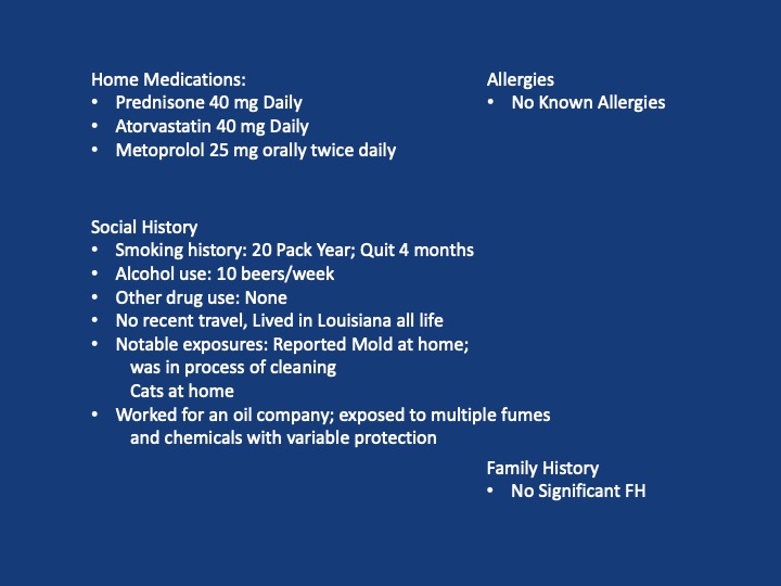
  • Notable exposure: Questionable black mold in the patient’s apartment.

Initial Workup and Diagnostic Reasoning

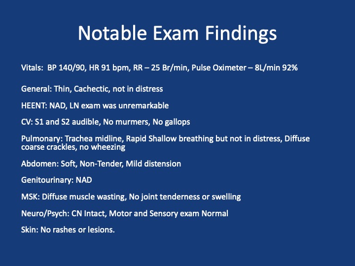
  • Vital signs: Hypoxemia (SpO₂ 91% on room air).
  • Exam: Inspiratory crackles.
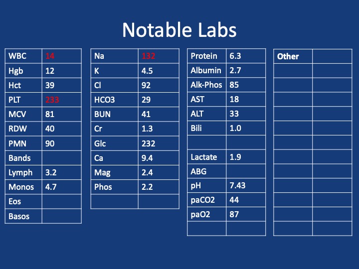
  • ABG findings: Elevated A–a gradient (~50), indicating a gas exchange problem.
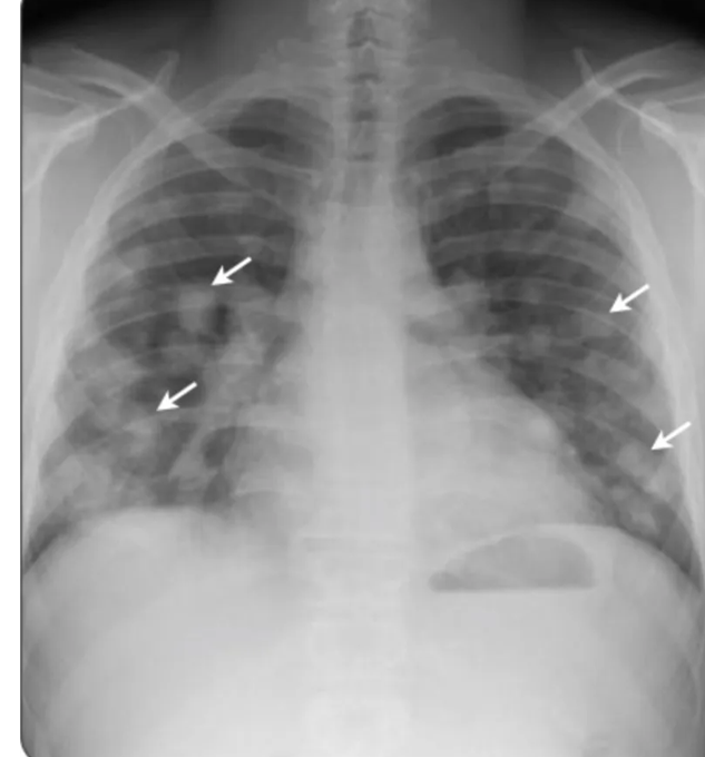
  • Chest X-ray: Bilateral, patchy infiltrates without specific lobar preference.
  • Initial management: Discharged with empiric antibiotics for presumed multifocal pneumonia.

Re-Presentation and Further Testing

  • Symptoms worsened; now with blood-tinged sputum.
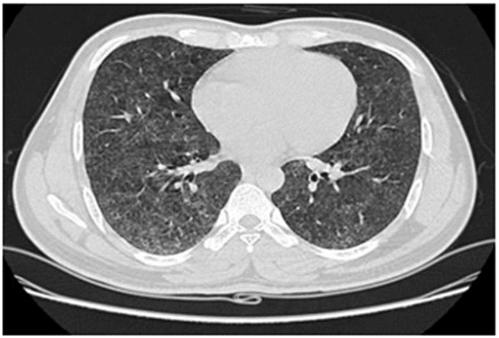
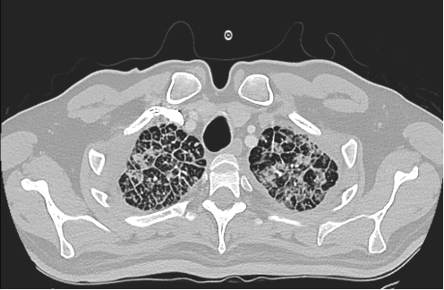
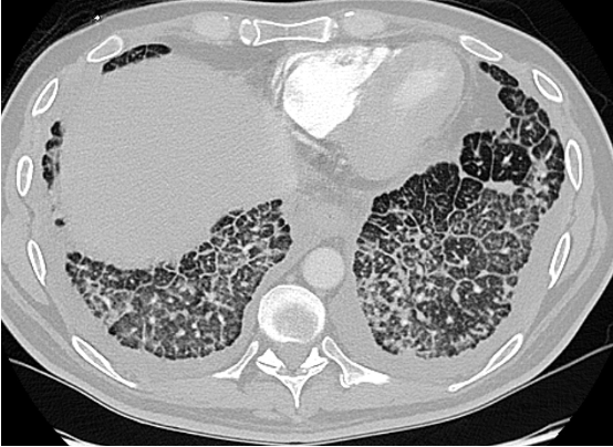
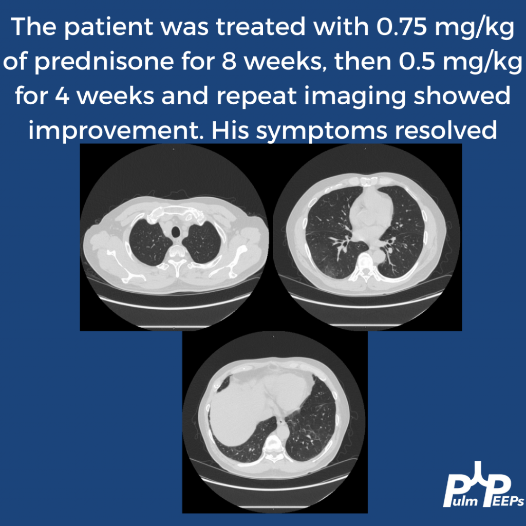
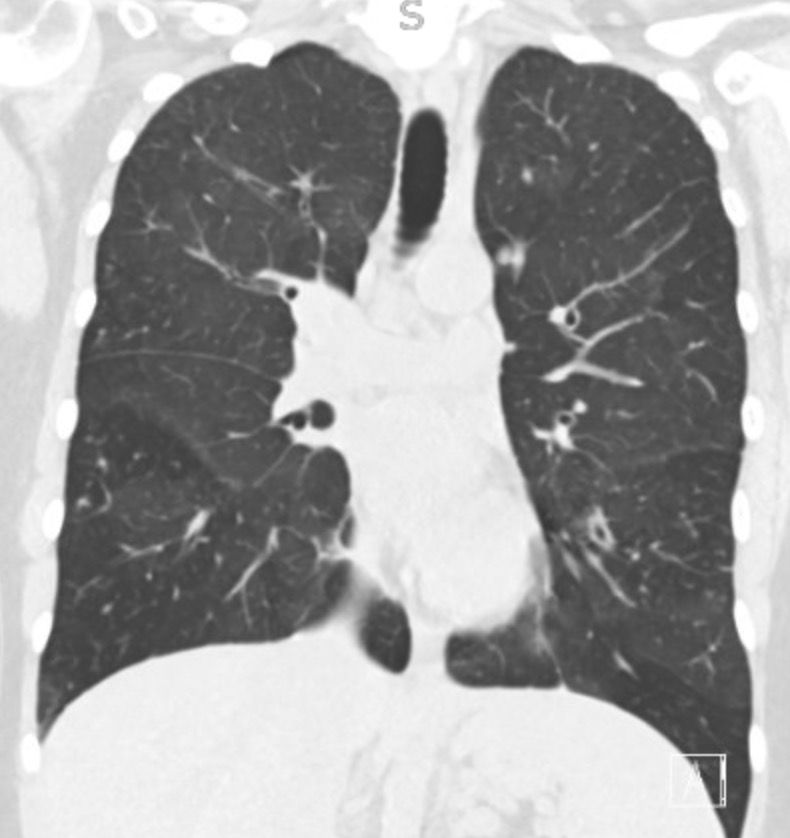
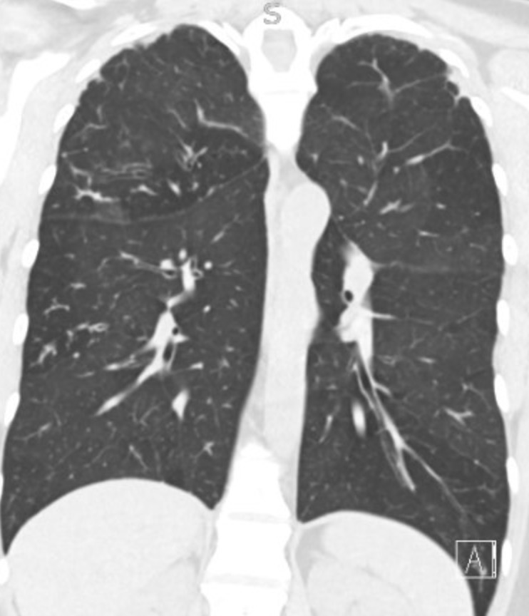
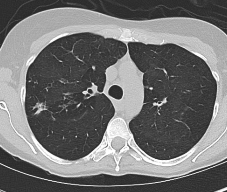
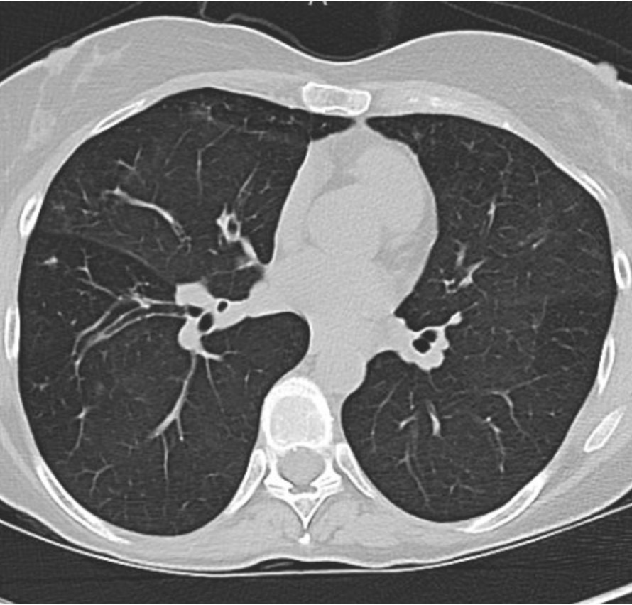
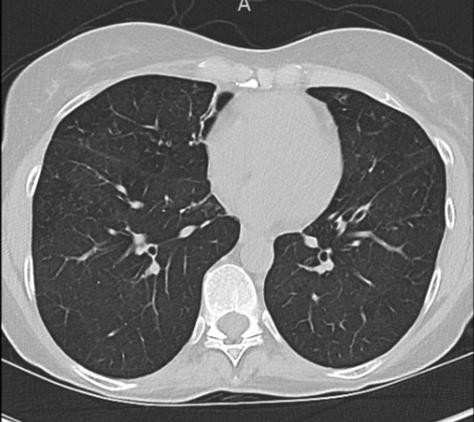
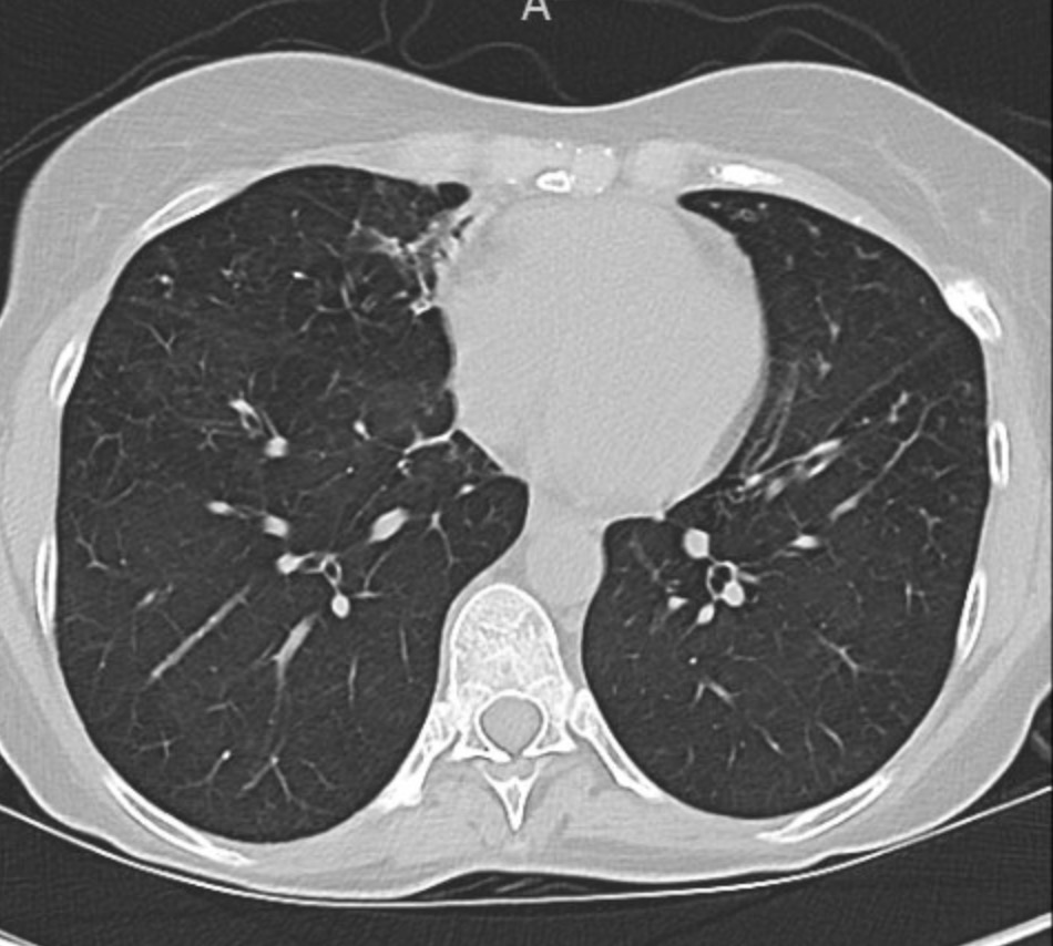
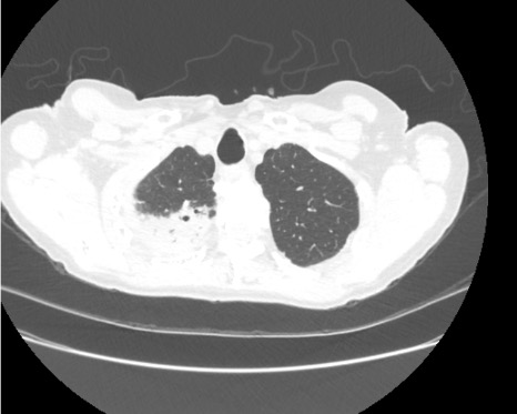
  • Chest CT: Showed diffuse ground-glass opacities (GGOs) without fibrosis, consolidation, or lymphadenopathy.

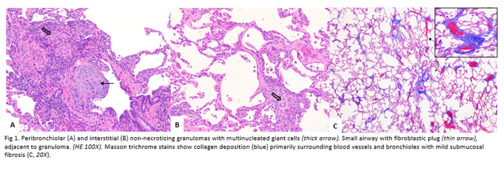
Pathology images a courtesy to Dr Isago Jerrett, pathology resident at RWJMS

Diagnostic Framework for Hypersensitivity Pneumonitis (HP)

  • New classification: Based on fibrotic vs. non-fibrotic phenotype (not acute/chronic).
  • CT features of HP:
    • GGOs with lobular air trapping.
    • “Three-density sign” (normal lung, low-density air-trapping, and ground-glass opacities).
  • BAL: Typically shows lymphocytic predominance in chronic HP, neutrophilic in early stages.
  • Serum IgG testing: Helps identify antigen exposure but doesn’t confirm disease alone.
  • Lung biopsy (VATS): Revealed poorly formed granulomas and airway-centered inflammation—consistent with HP.

Differential Diagnosis of Granulomatous Disease

  • Infectious: TB, fungal (must rule out with stains/cultures).
  • Non-infectious: Sarcoidosis, HP, granulomatosis with polyangiitis.
  • Key pathology clues for HP: Loosely formed granulomas, airway inflammation, giant cells.

Management and Outcome

  • Primary treatment: Antigen avoidance (patient moved out of mold-exposed apartment).
  • Adjunct therapy: Oral prednisone with a slow taper.
  • Outcome: Symptomatic and radiographic improvement over six months.

Teaching Pearls

  • Always take a detailed environmental and occupational exposure history.
  • Hypoxemia with an elevated A–a gradient in a young adult should trigger concern for interstitial/parenchymal lung disease.
  • CT and history are often enough to diagnose HP—biopsy is reserved for uncertain cases.
  • Remember evolving terminology: think fibrotic vs. non-fibrotic HP, not acute/chronic.

68. Fellows’ Case Files: Mount Sinai Morningside

We’re back with another Case Files episode from Mt. Sinai Morningside. Listen in to hear another great case and some key learning points along the way.

Dr. Sara Luby is a third-year Internal Medicine resident and rising chief resident at Mt. Sinai Morningside/West and planning on applying to Pulmonary and Critical Care fellowship this upcoming year.

Dr. Javier Zulueta is the  Chief of the Division of Pulmonary, Critical Care, and Sleep Medicine at Mount Sinai Morningside. He completed residency training at St. Luke’s Medical Center/Case Western in Cleveland and fellowship in Pulmonary/Critical Care at Tufts Medical Center in Boston. His research focuses on lung cancer screening and incidental lung findings.

 Dr. Mirna Mohanraj is the Associate Program Director for the Pulmonary and Critical Care Fellowship at Mt. Sinai Morningside / Beth Israel and an associate professor of medicine and medical education at the Icahn School of Medicine at Mount Sinai. She completed residency training at University of Chicago and fellowship training at Mt. Sinai Hospital.

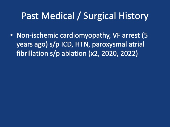
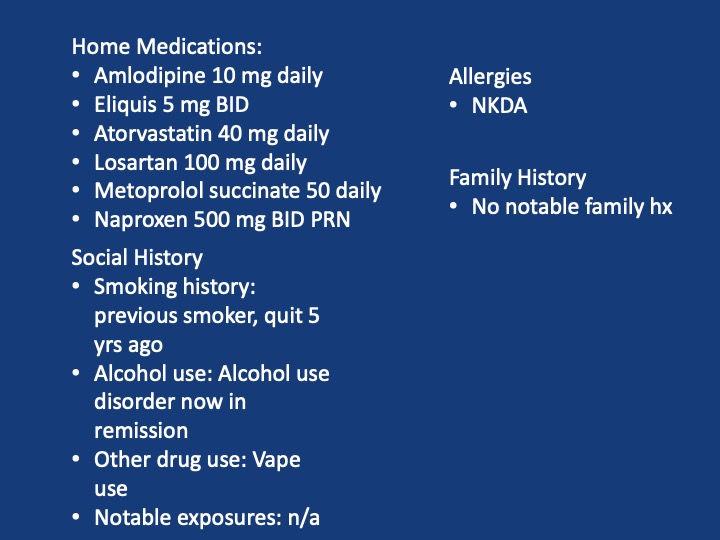
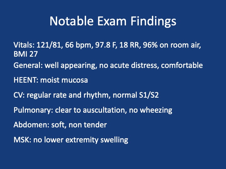
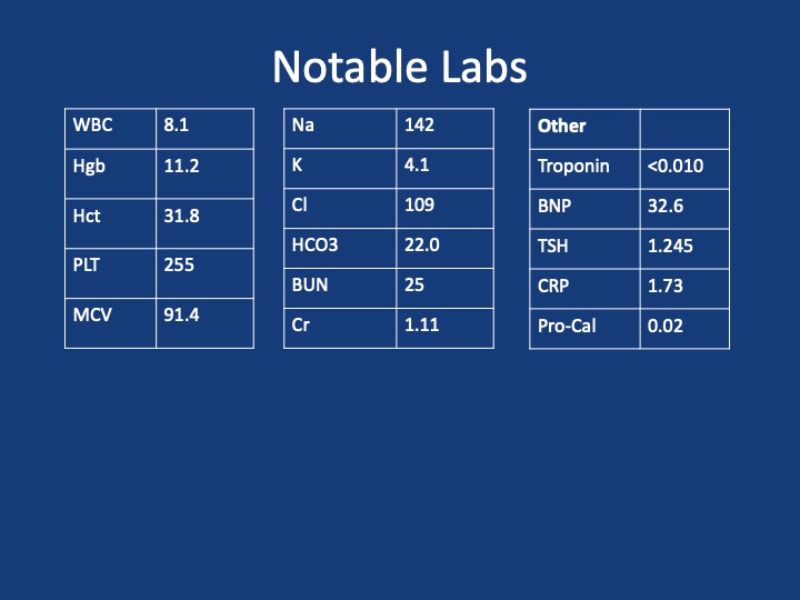
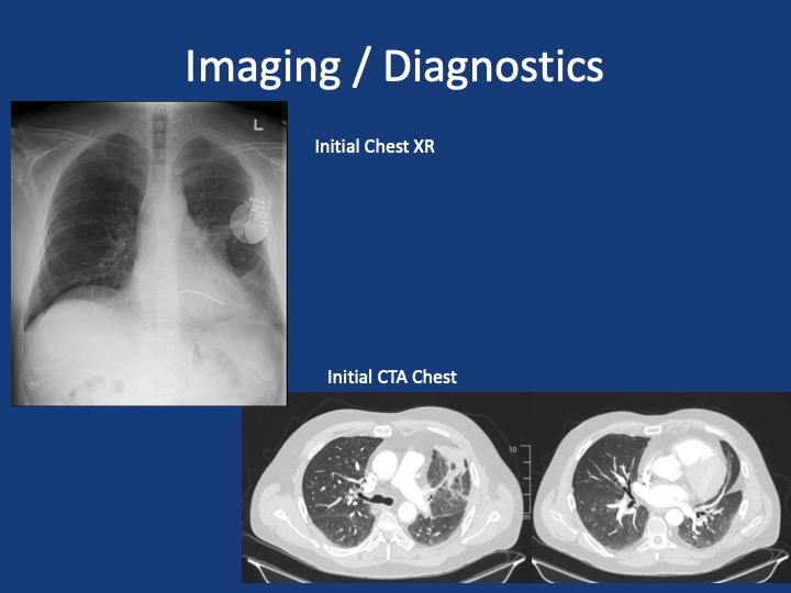
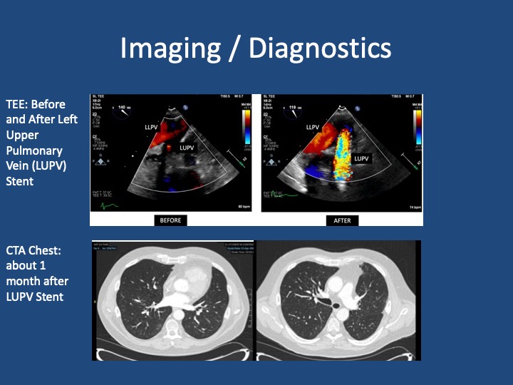
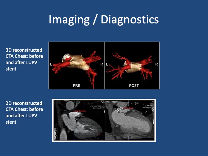
A 51 year old male presents with two days of acute on chronic chest pain and shortness of breath, worsening over the last month. His initial vitals: 143/ 100, pulse 85, temperature 36.5 °C (97.87°F), RR 16, SpO2 97 % on room air, BMI 29.8

Shroff N, Choi W, Villanueva-Meyer J, Palacio DM, Bhargava P. Pulmonary vein occlusion: A delayed complication following radiofrequency ablation for atrial fibrillation. Radiol Case Rep. 2021;16(12):3666-3671. doi:10.1016/j.radcr.2021.09.015 

Fender EA, Widmer RJ, Hodge DO, et al. Assessment and Management of Pulmonary Vein Occlusion After Atrial Fibrillation Ablation. JACC: Cardiovascular Interventions. Vol 11(16); 2018. doi:10.1016/j.jcin.2018.05.020 

López-Reyes R, García-Ortega A, Torrents A, et al. Pulmonary venous thrombosis secondary to radiofrequency ablation of the pulmonary veins. Respir Med Case Rep. 2018;23:46-48. doi:10.1016/j.rmcr.2017.11.008

Mizuno A, Mauler-Wittwer S, Muller H, Noble S. Recurrent pneumonia post atrial fibrillation ablation: do not forget to look for pulmonary vein stenosis. BMJ Case Rep. 2022;15(12):e250896. doi:10.1136/bcr-2022-250896

38. Fellows’ Case Files: Houston Methodist

Join us as we head to the Texas Medical Center and are joined by Dr. Bitar and Dr. Gotur from Houston Methodist Pulmonary and Critical Care Medicine.

Meet our Guests

Dr. Mohamad Bitar is a Pulmonary and Critical Care Fellow at the Houston Methodist Pulmonary and Critical Care Program in Houston, Texas. Dr. Bitar completed his medical school at Misr University for Science and Technology and his Internal Medicine residency at Good Samaritan Hospital.

Dr. Deepa Gotur is an Associate Professor of Clinical Medicine at Weill Cornell Medical College and an Adjunct Associate professor at Texas A&M. She is also the Pulmonary and Critical Care Program Director at Houston Methodist. She is a sepsis and ARDS researcher, as well as a dedicated educator.

Case Presentation

A 40s-year-old male presents with 3 months of progressive shortness of breath. He was diagnosed with COVID and given treatment with steroids, and antibiotics with no response.

He presented to the hospital 3 months later after not being able to carry out daily activities and was found to have low oxygen saturation

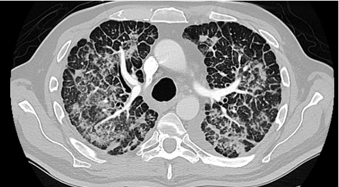
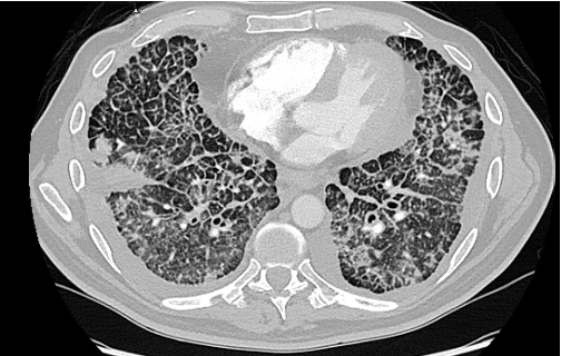
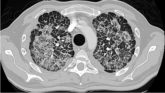
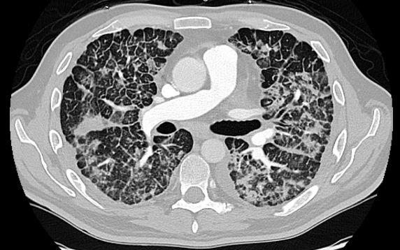
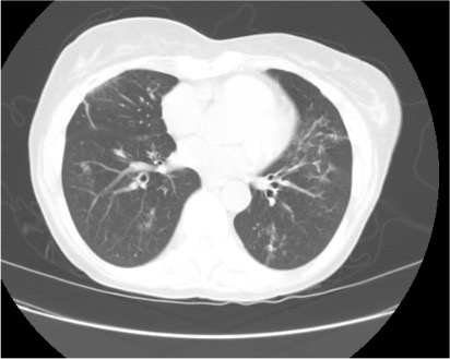
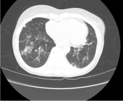
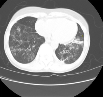
Admitted, initial CT Chest – showed “Non-specific patchy ground-glass bilateral pulmonary infiltrates with Coarsened interstitial markings that could be related to COVID-19/atypical infection”

References and further reading

1. Silva, C. Isabela S. MD, PhD; Müller, Nestor L. MD, PhD. Idiopathic Interstitial Pneumonias. Journal of Thoracic Imaging 24(4):p 260-273, November 2009.

2.Gruden JF, Naidich DP, Machnicki SC, Cohen SL, Girvin F, Raoof S. An Algorithmic Approach to the Interpretation of Diffuse Lung Disease on Chest CT Imaging: A Theory of Almost Everything. Chest. 2020 Mar;157(3):612-635.

3. Ryu JH, Daniels CE, Hartman TE, Yi ES. Diagnosis of interstitial lung diseases. Mayo Clin Proc. 2007 Aug;82(8):976-86.

Radiology Rounds – 1/18/23

We are back with our first #RadiologyRounds of 2023!

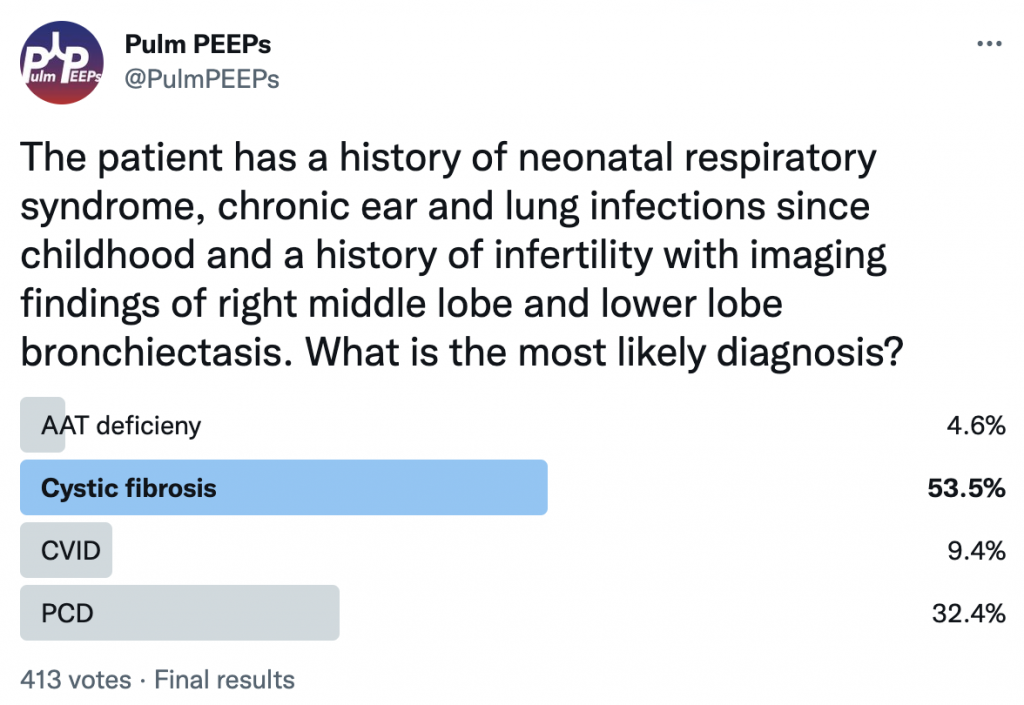
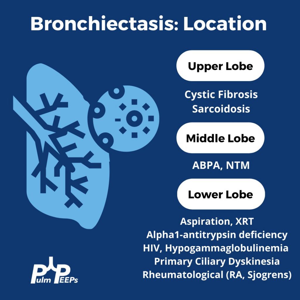
While chronic lung infections and infertility are overlapping symptoms for CF and Primary Ciliary Dyskinesia, the history of neonatal respiratory syndrome, ear infections and lower lobe bronchiectasis are most consistent with PCD

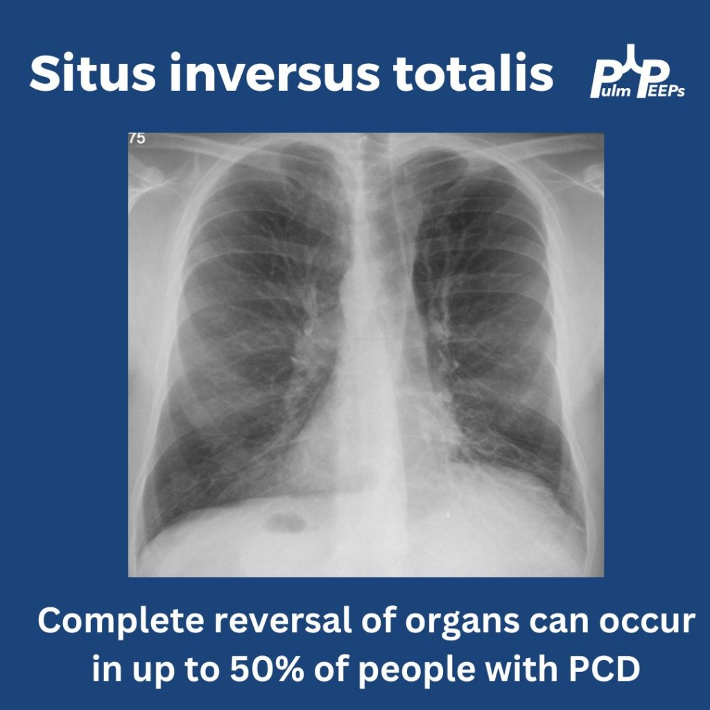
In up to 50% of people with PCD, you can get complete reversal of thoracic and abdominal organs. In this film you can see the heart in the right hemithorax, the gastric bubble on the right with the liver on the left resulting in elevation of the left hemidiaphragm

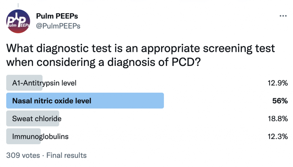
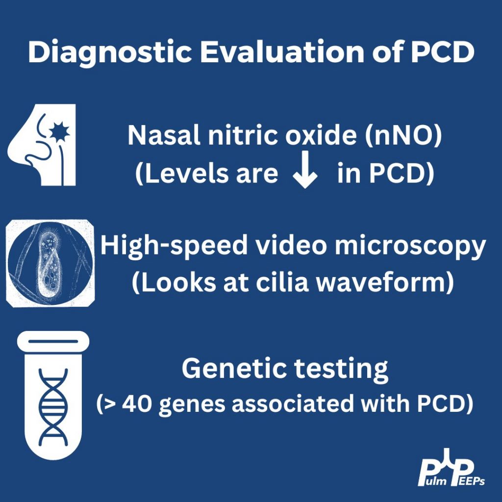
In patients with clinical symptoms and two decreased levels of nasal nitric oxide these findings suggest a PCD diagnosis but evaluation of the cilia structure and function as well as genetic testing are other diagnostic evaluations to confirm a diagnosis of PCD

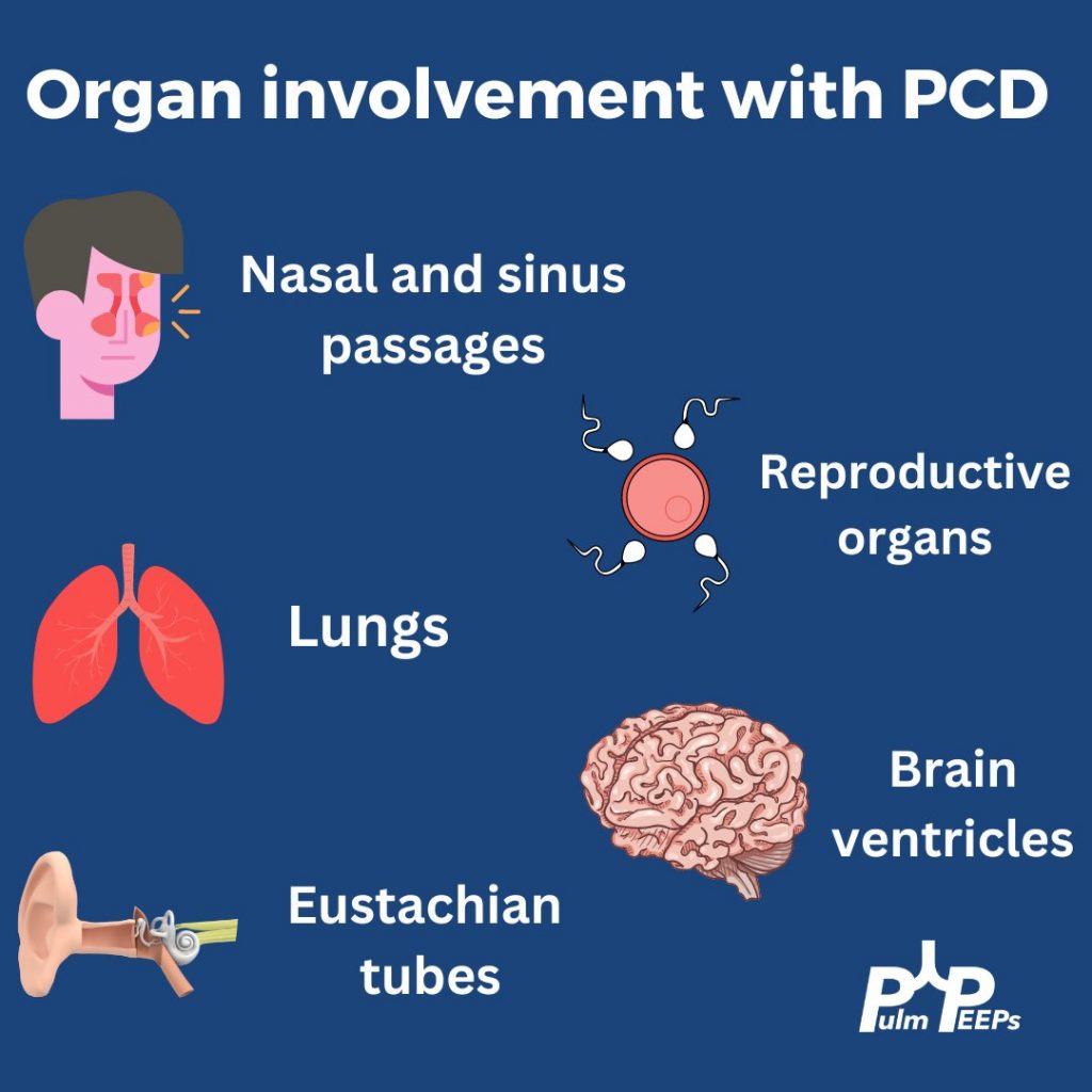
Primary ciliary dyskinesia is a autosomal recessive disorder that results in motile ciliary dysfunction and clinical manifestations can vary depending on which organs are involved

24. Fellows’ Case Files: Baylor College of Medicine

Welcome to another episode in our Pulm PEEPs Fellows’ Case Files series! The purpose of this series is to highlight and amplify the incredible clinical work that is done by pulmonary and critical care fellows, share fascinating cases, and assemble a diverse network of pulmonary and critical care educators. Today we’re headed to Baylor College of Medicine to hear about a fascinating case. Tune in, let us know what you think on Twitter, and let us know if you have a great case to share!

Meet Our Guests

Benjamin Moss completed his internal medicine residency training at Baylor College of Medicine in Houston, Texas, and is currently a senior pulmonary and critical care fellow there.

Philip Alapat is an Assistant Professor of Medicine and the Program Director of the Pulmonary and Critical Care Fellowship at Baylor. He completed his residency and fellowship training all at Baylor.

Patient Presentation

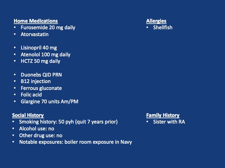
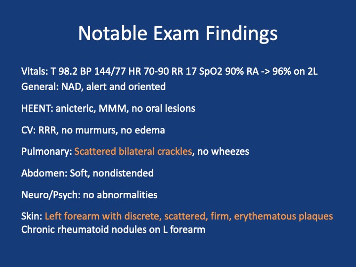
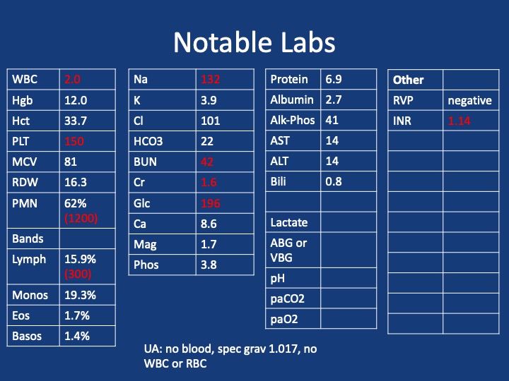
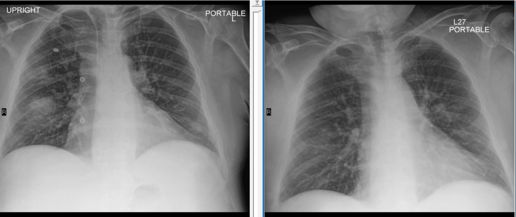
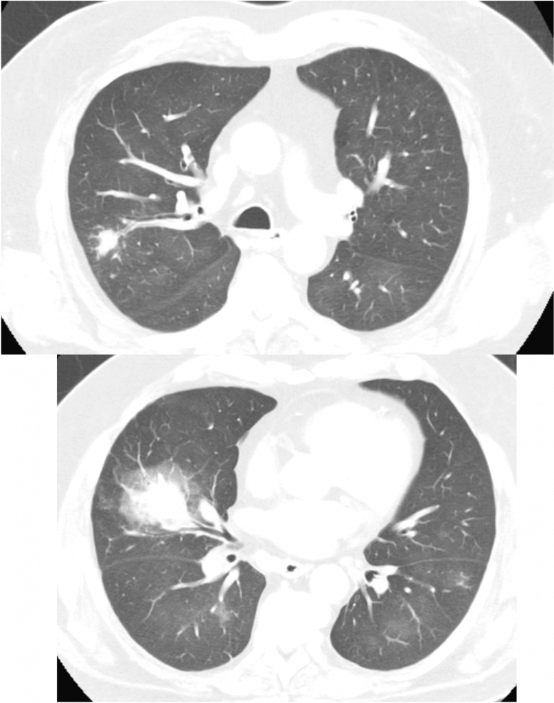
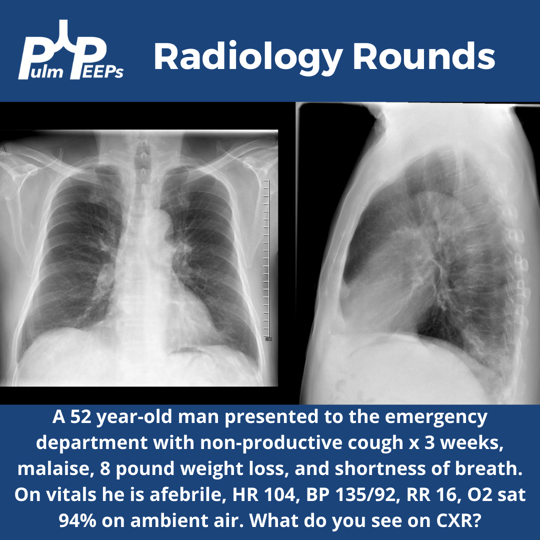
A 60s-year-old man with seropositive RA on Rituximab presents with dyspnea and cough, and overall “not feeling well.”  For the past week, he has had malaise, body aches, and subjective fever. For the past 3 days, he has had acutely worsening dyspnea that is worse with exertion, but present at rest and a cough with scant sputum production. He had been on Methotrexate previously but within the last year developed pancytopenia and MTX was stopped and he was switched to adalimumab/Humira. His pancytopenias did not resolve, and he was ultimately diagnosed with Felty syndrome (a triad of RA, neutropenia, and splenomegaly) and switched to rituximab every 6 months with his last dose being 4 months ago. During the last week, he tried taking prednisone 10 mg a day but his symptoms did not improve.

X-ray on presentation (L) X-ray 8 months ago (R)

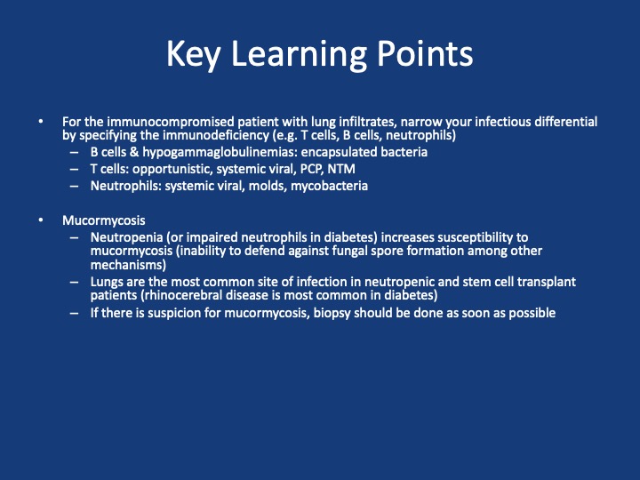
Key Learning Points

**Spoilers Ahead** If you want to think through the case on your own we advise listening to the episode first before looking at these points.

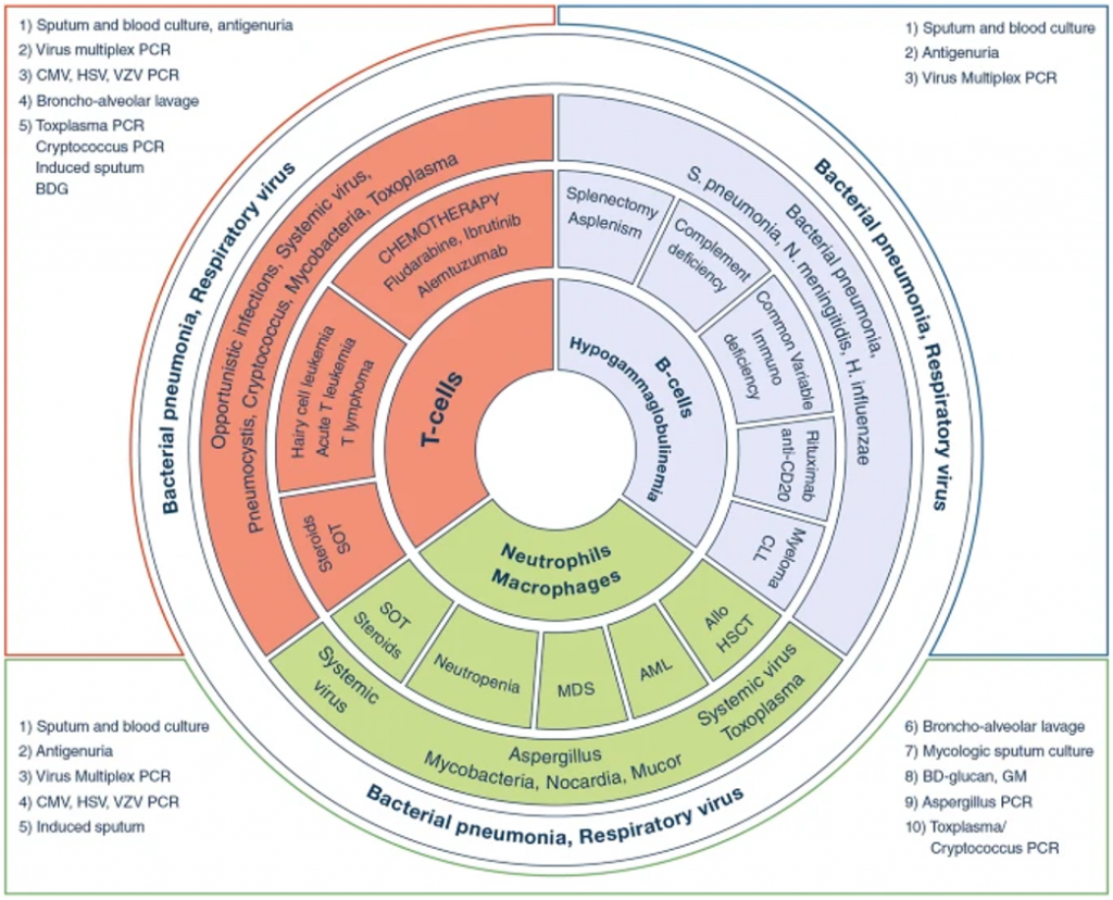
Image Source: Reference 1: https://doi.org/10.1007/s00134-019-05906-5

References and Further Reading

  1. Azoulay E, Russell L, Van de Louw A, et al. Diagnosis of severe respiratory infections in immunocompromised patients. Intensive Care Med. 2020;46(2):298-314. doi:10.1007/s00134-019-05906-5
  2. Cornely OA, Alastruey-Izquierdo A, Arenz D, et al. Global guideline for the diagnosis and management of mucormycosis: an initiative of the European Confederation of Medical Mycology in cooperation with the Mycoses Study Group Education and Research Consortium. Lancet Infect Dis. 2019;19(12):e405-e421. doi:10.1016/S1473-3099(19)30312-3
  3. Ibrahim AS, Spellberg B, Walsh TJ, Kontoyiannis DP. Pathogenesis of mucormycosis. Clin Infect Dis. 2012;54 Suppl 1:S16-22. doi:10.1093/cid/cir865

5. A Case of Chronic, Productive Cough

The Pulm PEEPs are joined again by Natalie West to discuss a patient who presented with a chronic, productive cough. Listen in today as we work through our differential diagnosis, interpret basic pulmonary testing, and share our clinical reasoning along the way. We have some fantastic diagnostic and treatment teaching points, so once you’ve solved the case check out the takeaways and infographics below. Please let us know any additional insights you have on Twitter!

Patient Presentation

A 50-year-old woman, who is a never smoker, with a past medical history of recurrent pancreatitis presents to the pulmonary clinic with a chronic, productive cough. Her cough has been present for 3 years and has increased in frequency to now being present daily. In the last three months, the cough has also worsened and is productive of small amounts of yellow to green sputum. She has a history of chronic post-nasal drip and sinus infections, and uses intranasal steroids, but has not noted changes in these symptoms. There is no significant family history of pulmonary disease, and an exposure history review of symptoms is negative.

On physical exam, she was a thin woman who appeared her stated age and was breathing comfortably on room air. Her exam was notable for mild expiratory wheezing, primarily on auscultation of the right posterior lung field. She had no cyanosis, clubbing, evidence of volume overload, or abdominal tenderness.

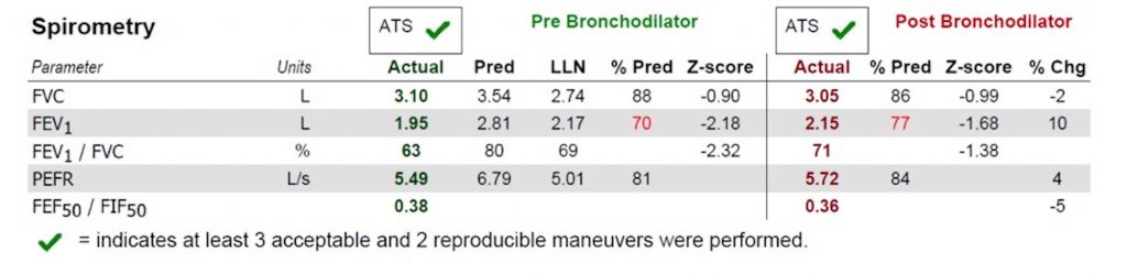
Basic Spirometry Values
Chest X-ray

Key Learning Points

**Spoilers Ahead** If you want to think through the case on your own we advise listening to the episode first before looking at the infographics below.

Differential Diagnosis of Chronic Cough

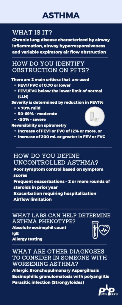
Three most common causes: upper airway cough syndrome, GERD, cough variant asthma

Additional etiologies to consider: chronic bronchitis, post-infectious after a respiratory tract infection, bronchiectasis, ACE inhibitors, lung cancer, eosinophilic bronchitis, interstitial lung disease


Imaging Pearl


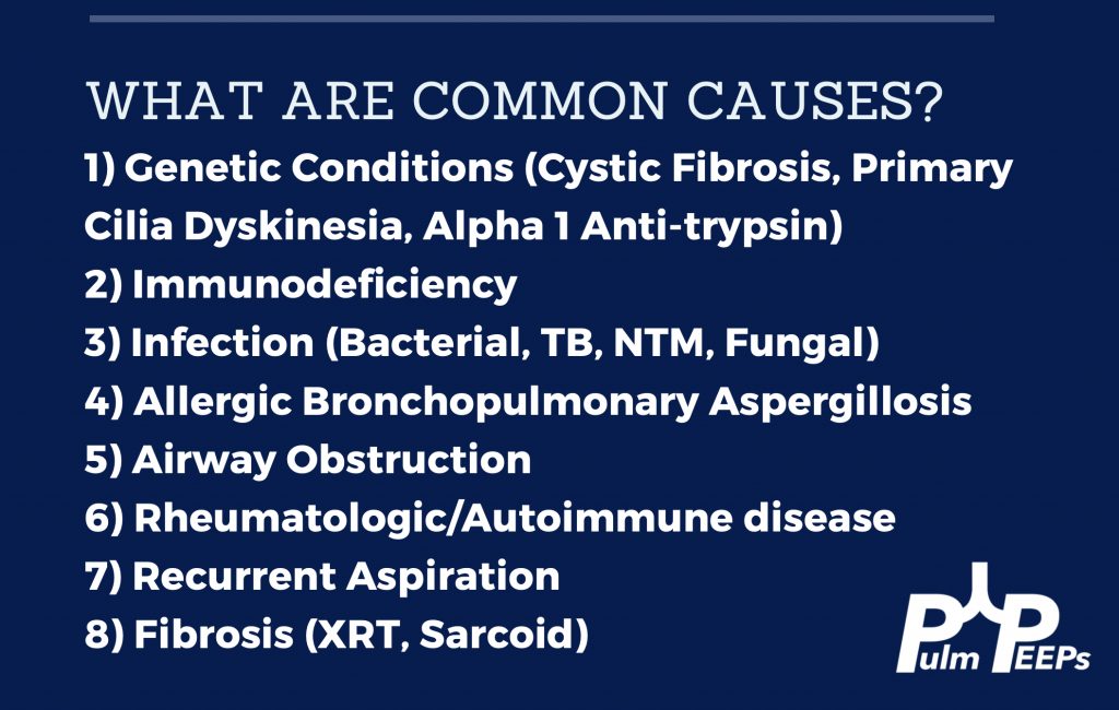
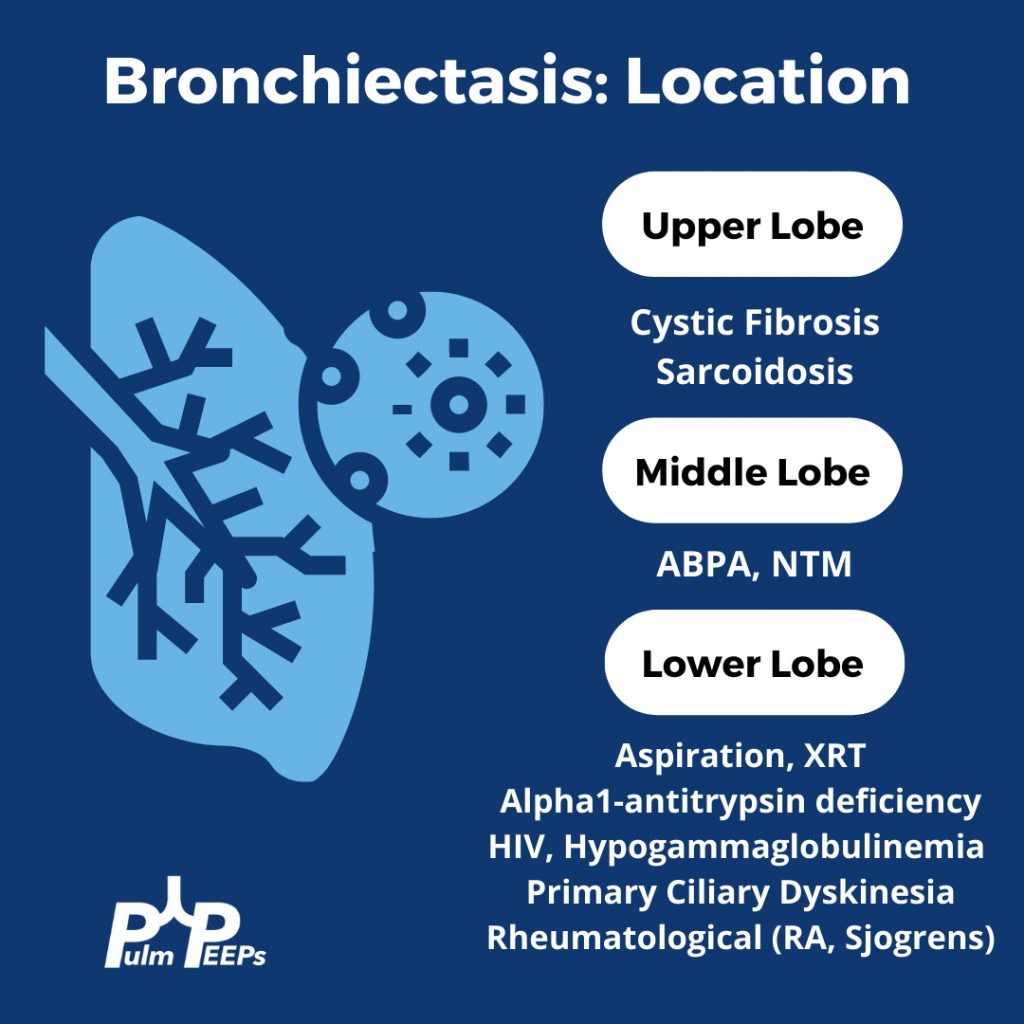
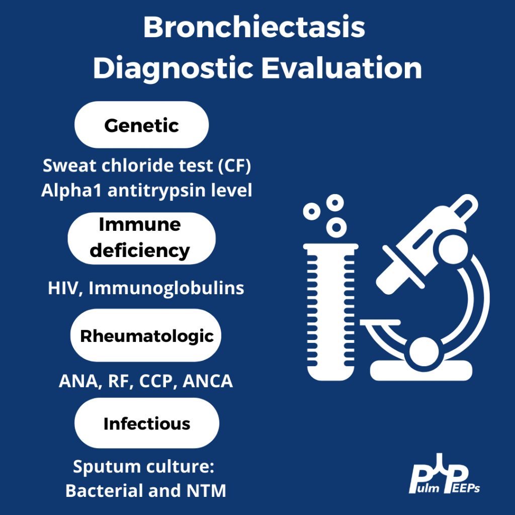
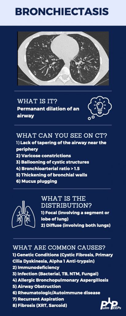
Evaluating Bronchiectasis


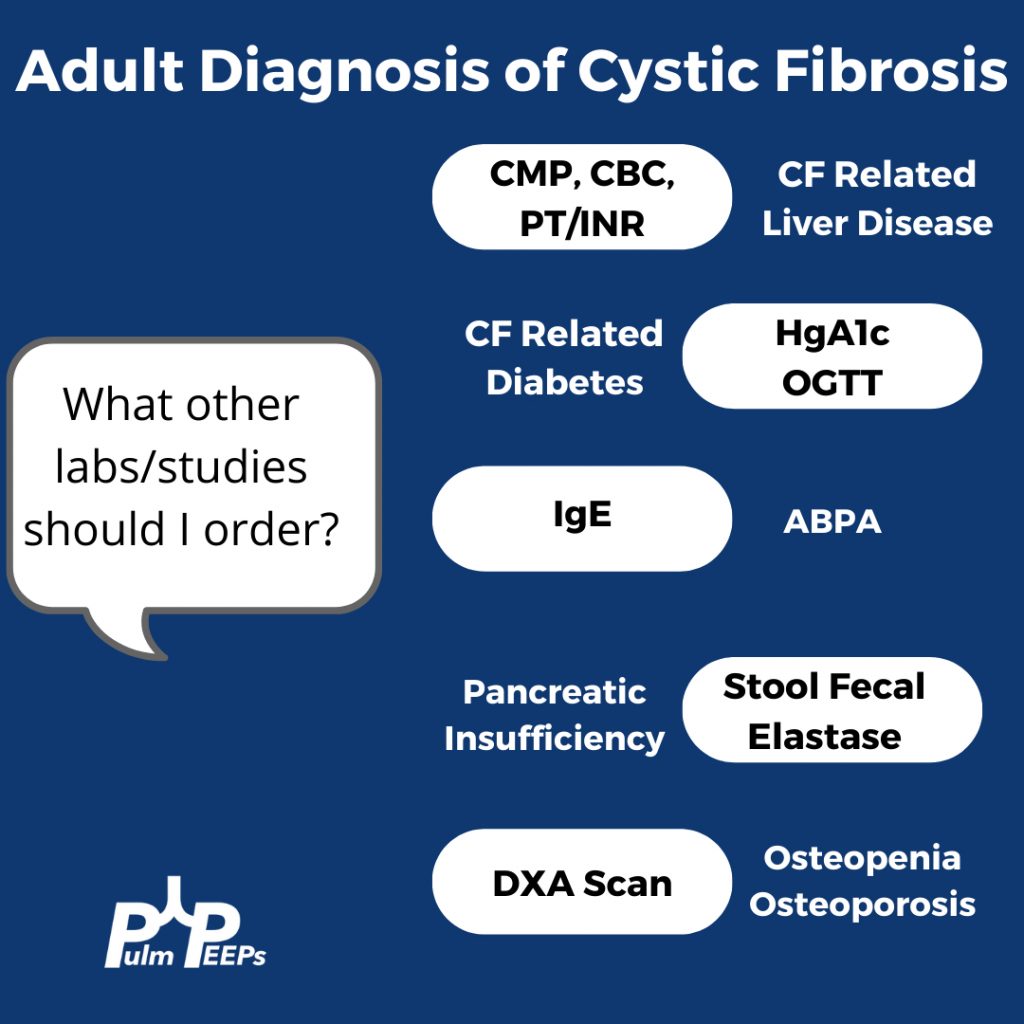
Making a New Diagnosis of Cystic Fibrosis in an Adult

Sweat testing

Sweat testing should be done in CF accredited center. Inform patients that there are no needles involved. Pilocarpine and electrical stimulation are applied to the arm or leg to stimulate the sweat gland, and then sweat is collected on filter paper, a gauze, or a plastic coil. From there, the amount of chloride in the sweat is calculated

Results

< 30 normal

31 – 60 indeterminate

> 60 is positive and Cystic Fibrosis is likely

What do you do with an Indeterminate test?

Patients with milder phenotypes of Cystic Fibrosis can have a normal or indeterminate sweat chloride level, and 10% of adults diagnosed with CF have a normal sweat chloride. If the sweat chloride test is indeterminate or normal, but suspicion is high for CF, then genetic testing for the whole array of mutations should be performed

References and links for further reading

  1. Morice AH, Millqvist E, Bieksiene K, et al. ERS guidelines on the diagnosis and treatment of chronic cough in adults and children. European Respiratory Journal. 2020;55(1). doi:10.1183/13993003.01136-2019
  2. Barker AF. Bronchiectasis. New England Journal of Medicine. 2002;346(18):1383-1393. doi:10.1056/NEJMra012519
  3. Bronchiectasis: a case-based approach to investigation and management | European Respiratory Society. Accessed November 23, 2021. https://err.ersjournals.com/content/27/149/180016
  4. Rowe SM, Miller S, Sorscher EJ. Cystic Fibrosis. New England Journal of Medicine. 2005;352(19):1992-2001. doi:10.1056/NEJMra043184
  5. Shteinberg M, Haq IJ, Polineni D, Davies JC. Cystic fibrosis. The Lancet. 2021;397(10290):2195-2211. doi:10.1016/S0140-6736(20)32542-3
  6. Jain R. Diagnosing Cystic Fibrosis in Adults: Better Late Than Never. Annals ATS. 2018;15(10):1140-1141. doi:10.1513/AnnalsATS.201806-432ED

4. Top Consults: Hemoptysis

Pulm PEEPs hosts, Kristina Montemayor and David Furfaro, bring our first episode in our Top Consults series. In this series, we will bring in experts to work through the most common pulmonary and critical care consults. Whether you are the consulting physician, or a pulmonologist responding to the page, these episodes are geared to give you all the information you need to care for your patients!

Today, we are joined by Chris Kapp and Matthew Schimmel, two interventional pulmonologists, to discuss hemoptysis. Chris and Matt will help us work through two hemoptysis consults, and together we’ll provide a framework for thinking about hemoptysis, outline some key components of the evaluation, and delve into treatment options.

Key Learning Points

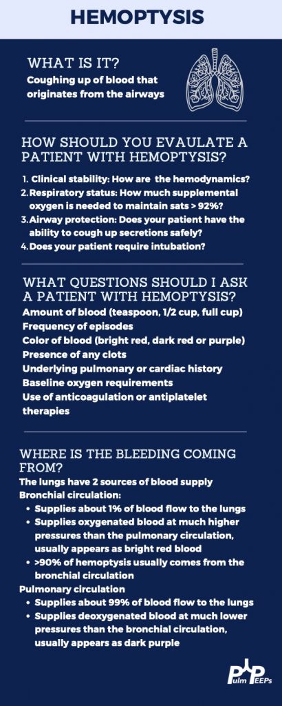
Hemoptysis Evaluation

Hemoptysis Management

Life-Threatening or Large Volume Hemoptysis

  1. Stabilize the patient! Make sure the airway is protected either by the patient coughing themselves, or intubation if needed. Provide hemodynamic support with IVF, blood products, and pressors if needed. If it is known which lung has the bleeding the patient can be positioned so the lung with the bleeding is down. This protects the non-bleeding lung.
  2. Correct any bleeding diathesis If the patient is on anti-coagulation, or has any reversible bleeding diathesis, these should be corrected immediately to reduce further bleeding.
  3. Localize the bleed If the patient is stable, they should undergo a CTA to localize the bleeding. If they are not stable to make it to a CT scan, a bronchoscopy should be performed.
  4. Bronchoscopic treatment In addition to clearing blood from the airway, bronchoscopy can localize the bleeding. With available expertise, bronchoscopic treatments can be performed such as ice saline, topical epinephrine, or balloon tamponade to isolate the bleed.
  5. Definitive therapy with arteriography and embolization Patients with life-threatening hemoptysis should ultimately undergo arteriography and embolization of any bleeding vessel. If this is not possible, then surgery can be needed in some cases.
  6. A note on diffuse hemoptysis If there is not one distinct bleeding lesion, then localizing and treating the bleed becomes more difficult. For diffuse alveolar hemorrhage, evaluation should be performed for if it is primary, and due to an immunologic cause and capillaritis, or secondary to a systemic disease and / or bleeding diathesis. These investigations will guide available treatment options. Capillaritis from an immunologic cause, such as lupus or vasculitis, can be treated with systemic glucocorticoids and an additional immunosuppressive agent such as cyclophosphamide or rituximab.

Non-life-threatening or Small Volume Hemoptysis

  1. Monitor for clinical worsening Patient’s should be monitored, either in the in-patient or out-patient setting, for increased volume or frequency of hemoptysis and for any clinical worsening, such as desaturations or decreased ability to clear the airway.
  2. Correct any bleeding diathesis If the patient is on anti-coagulation, or has any reversible bleeding diathesis, these should be corrected immediately to reduce further bleeding. In pattients with non-life-threateneing hemoptysis this requires careful consideration of balancing the risk of bleeding vs the benefits for continuing anti-coagulation.
  3. Evaluate for underlying cause Patient’s should undergo imaging and evaluation for the underlying cause of the hemoptysis. This may be evidence of an underlying infection, a pulmonary embolism, or new lung lesions making the patient at risk. If the source can’t be found on non-invasive imaging, and there is no clear systemic source such as an infection, a bronchoscopy is warranted. Any underlying cause should be treated and investigated further.
  4. Inhaled Tranexamic Acid Nebulized tranexamic acid is well tolerated and can help resolve hemopytysis without invasive procedures.

References and links for further reading

  1. Gagnon S, Quigley N, Dutau H, Delage A, Fortin M. Approach to Hemoptysis in the Modern Era. Can Respir J. 2017;2017:1565030. doi:10.1155/2017/1565030
  2. Radchenko C, Alraiyes AH, Shojaee S. A systematic approach to the management of massive hemoptysis. J Thorac Dis. 2017;9(Suppl 10):S1069-S1086. doi:10.21037/jtd.2017.06.41
  3. Davidson K, Shojaee S. Managing Massive Hemoptysis. Chest. 2020;157(1):77-88. doi:10.1016/j.chest.2019.07.012
  4. Lara AR, Schwarz MI. Diffuse Alveolar Hemorrhage. CHEST. 2010;137(5):1164-1171. doi:10.1378/chest.08-2084
  5. Wand O, Guber E, Guber A, Epstein Shochet G, Israeli-Shani L, Shitrit D. Inhaled Tranexamic Acid for Hemoptysis Treatment: A Randomized Controlled Trial. Chest. 2018;154(6):1379-1384. doi:10.1016/j.chest.2018.09.026