22. Fellows’ Case Files: University of Maryland

Case Files Maryland

This week we are absolutely thrilled to be launching a new series here at Pulm PEEPs. This is the first episode in our new Fellows’ Case Files series. The purpose of this series is to highlight the incredible clinical work that is done by pulmonary and critical care fellows everywhere, share fascinating cases from across the world, and assemble a diverse network of pulmonary and critical care educators. For each episode, we will visit a different institution, and be joined by a current fellow and the Pulmonary and Critical Care Fellowship Program Director. Our aim is to learn from them, amplify some incredible teaching points, and hear about their program. We hope you enjoy it, and if you have a case you want to bring on the series reach out to us on Twitter or at our email pulmpeeps@gmail.com.


Meet Our Guests

Fahid Alghanim is a senior pulmonary and critical care fellow at the University of Maryland. He attended medical school at the Lebanese American University Gilbert and Rose-Marie Chagoury School of Medicine and completed his internal medicine residency at Johns Hopkins Bayview. He has published on topics ranging from lung transplants to patient navigators in the ICU.

Dr. Van Holden is an Associate Professor of  Medicine at the University of Maryland School of Medicine and the Pulmonary and Critical Care Fellowship Program director. Clinically, she specializes in interventional pulmonology. She is also an accomplished educator and is very active with the American Thoracic Society. She helped write the 2021 Critical Care Core Curriculum and helped coordinate the 2022 Resident Boot Camp.

Patient Presentation

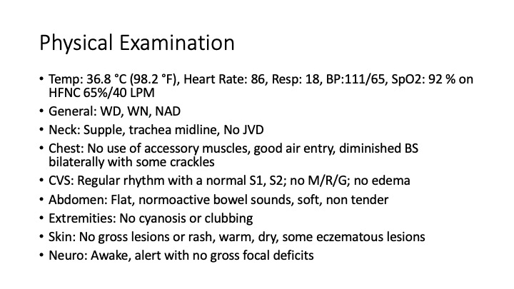
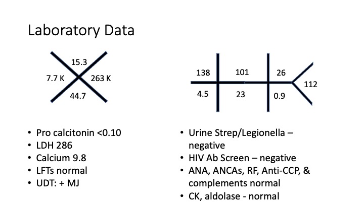
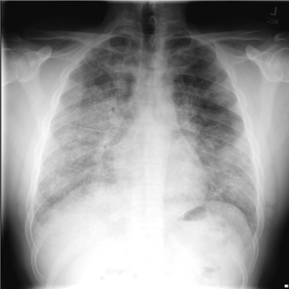
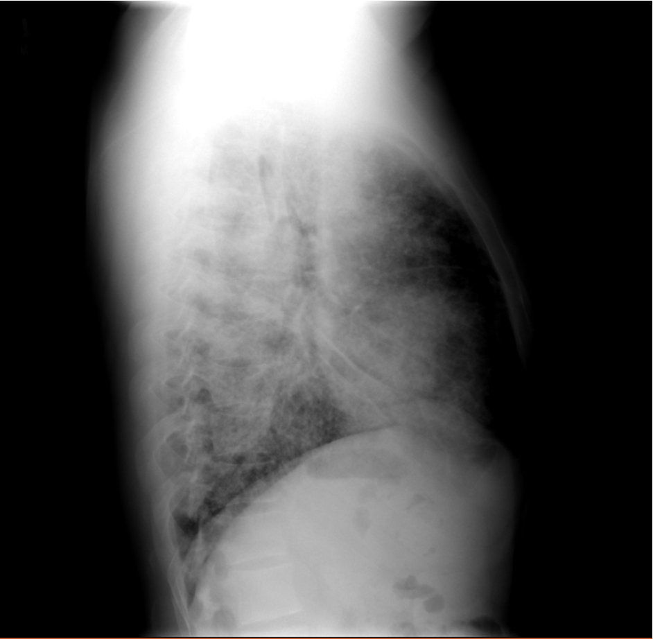
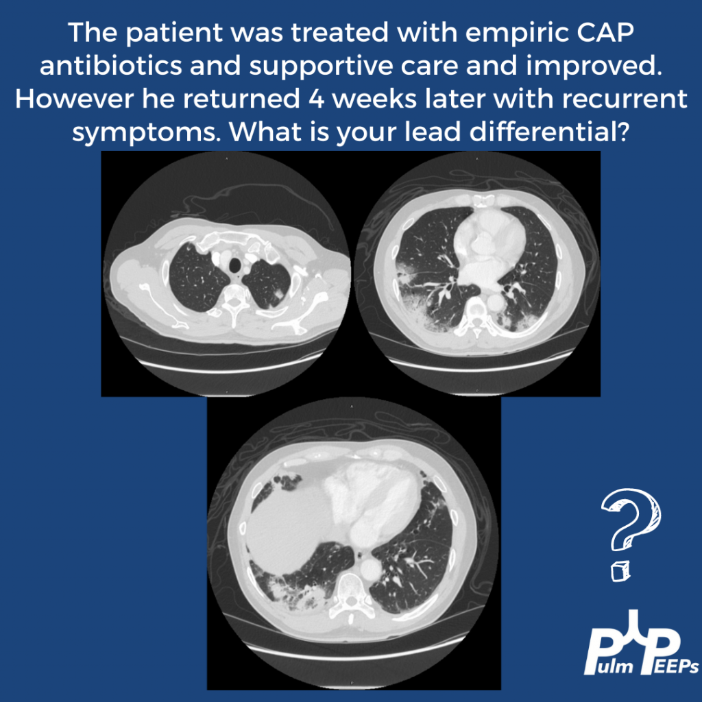
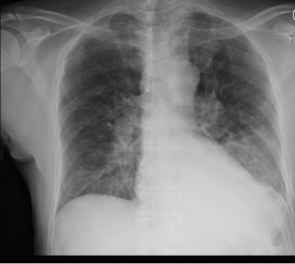
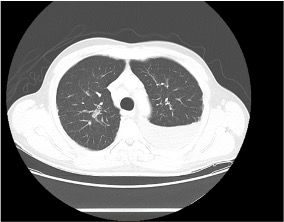
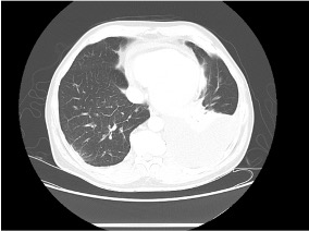
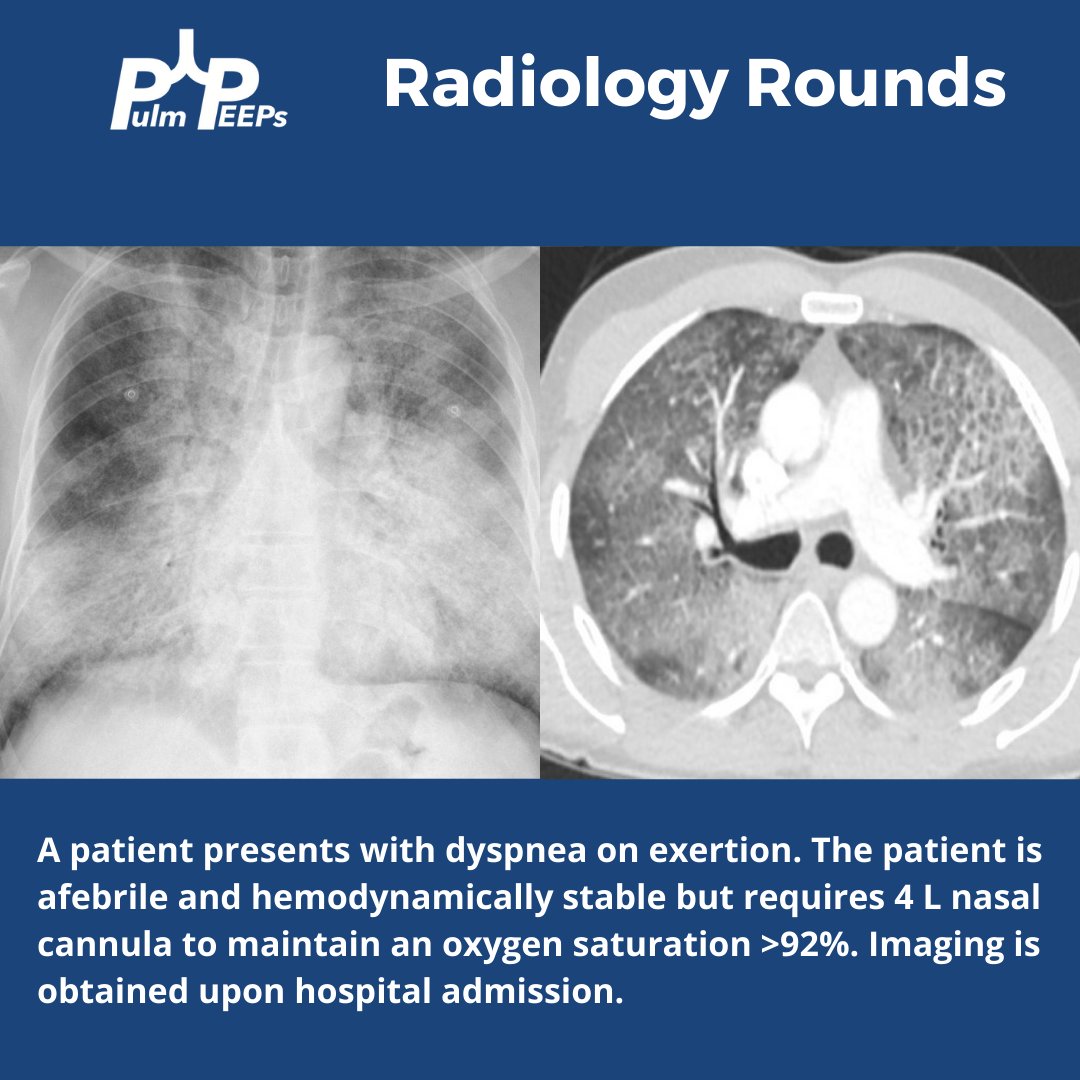
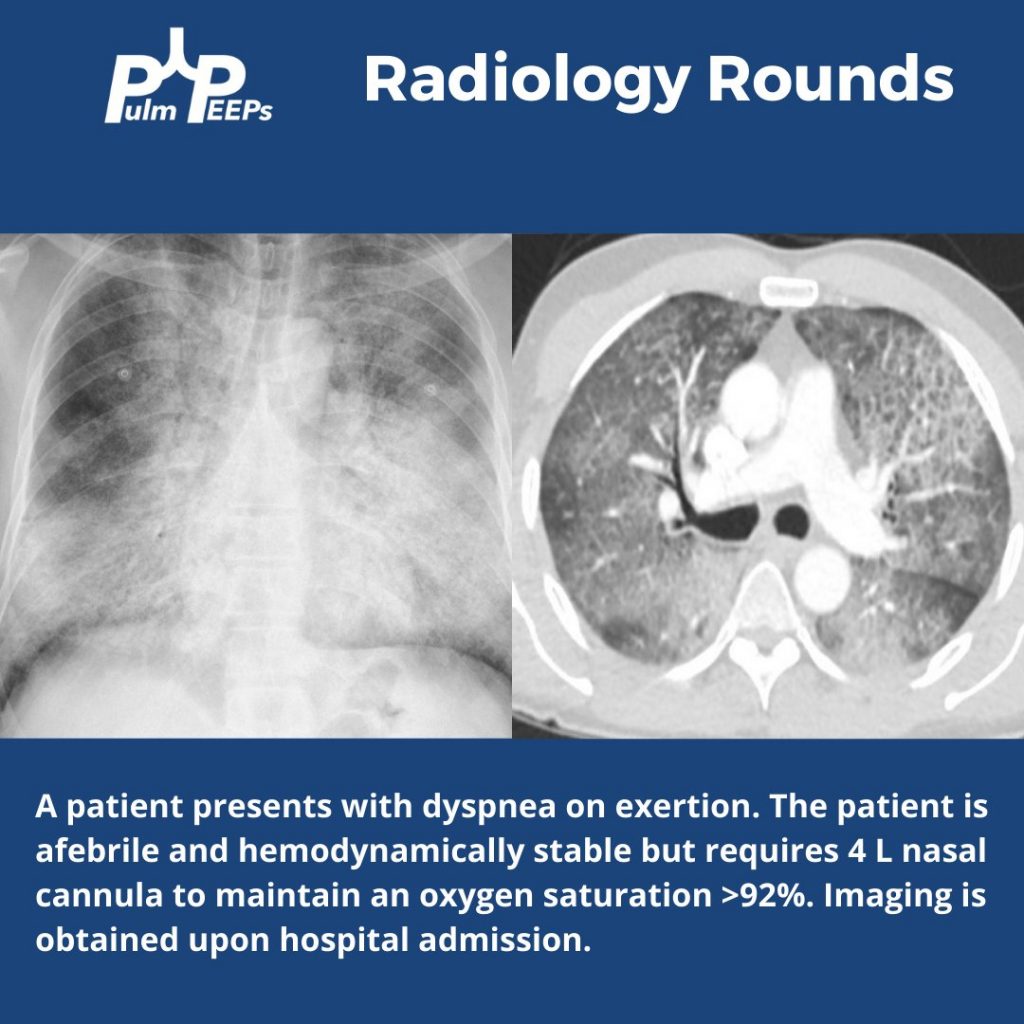
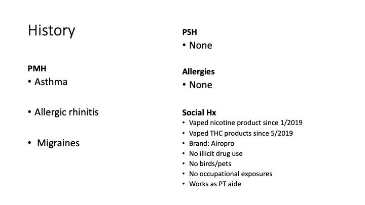
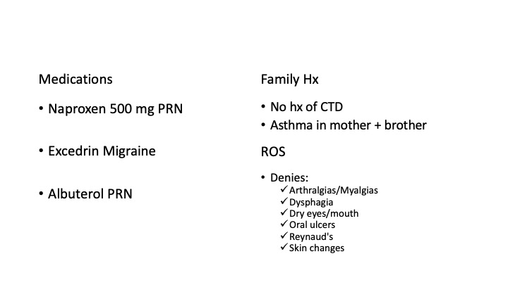
A 26-year-old man presents to his primary care doctor with 1.5 months of intermittent dyspnea, cough, chest tightness, and fatigue. His dyspnea was initially exertional, and he noticed he could do less at the gym. However, in the past 3-4 weeks it has progressed to being even with mild movement. His brother was recently diagnosed and treated for acute bronchitis so he thought this could be similar. In the office, he is noted to be tachypneic with an oxygen saturation of 83% breathing ambient air. A chest X-ray is obtained and he is sent urgently to the emergency department.
Key Learning Points

**Spoilers Ahead** If you want to think through the case on your own we advise listening to the episode first before looking at the infographics below

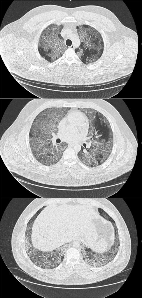
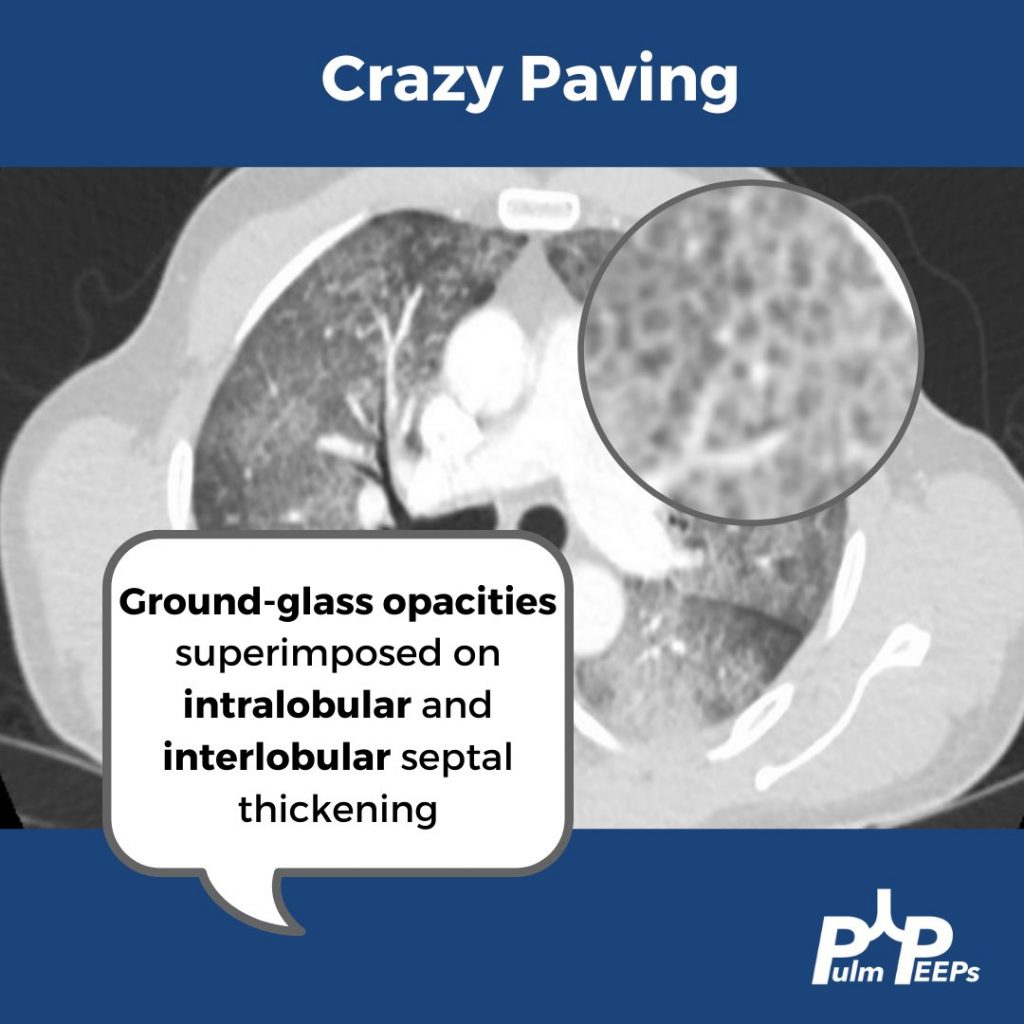
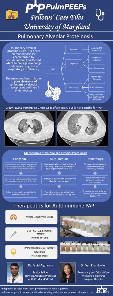
  1. Crazy Paving is a radiological term describing ground glass opacities with superimposed interlobular septal thickening. The differential diagnosis is broad and includes infectious, neoplastic, and autoimmune processes. It is not limited to just Pulmonary alveolar proteinosis (PAP) but is suggestive in an appropriate clinical setting.
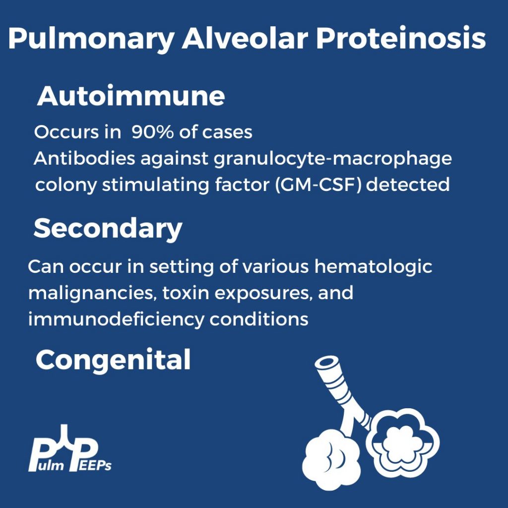
  2. PAP is a disorder of surfactant production or clearance and its etiology is divided into three major subgroups. Primary or autoimmune; Secondary such as from toxic inhalations, hematological disorders, or medications; and Congenital
  3. PAP is diagnosed by positive Periodic acid-Schiff (PAS) staining of lipo-proteinaceous material in the distal bronchioles and alveoli on lung biopsy. The diagnosis can be made with PAS-positive BAL staining, but this has limited sensitivity and lung biopsy is necessary for the diagnosis in up to 30 – 35% of cases.
  4. It is important not to anchor on a diagnosis when a patient presents to you for re-evaluation even if seen by a prior expert. This was pivotal in this case!
  5. Please don’t put anything in your lung. Any toxic inhalation exposure could result in significant damage to lung parenchyma and morbidity as a result.
References and Further Reading

  1. Borie R, Danel C, Debray MP, et al. Pulmonary alveolar proteinosis. Eur Respir Rev. 2011;20(120):98-107. doi:10.1183/09059180.00001311
  2. Carey B, Trapnell BC. The molecular basis of pulmonary alveolar proteinosis. Clin Immunol. 2010;135(2):223-235. doi:10.1016/j.clim.2010.02.017
  3. Inoue Y, Trapnell BC, Tazawa R, et al. Characteristics of a large cohort of patients with autoimmune pulmonary alveolar proteinosis in Japan. Am J Respir Crit Care Med. 2008;177(7):752-762. doi:10.1164/rccm.200708-1271OC
  4. Kavuru MS, Malur A, Marshall I, et al. An open-label trial of rituximab therapy in pulmonary alveolar proteinosis. Eur Respir J. 2011;38(6):1361-1367. doi:10.1183/09031936.00197710
  5. Michaud G, Reddy C, Ernst A. Whole-lung lavage for pulmonary alveolar proteinosis. Chest. 2009;136(6):1678-1681. doi:10.1378/chest.09-2295
  6. Smith BB, Torres NE, Hyder JA, et al. Whole-lung Lavage and Pulmonary Alveolar Proteinosis: Review of Clinical and Patient-centered Outcomes. J Cardiothorac Vasc Anesth. 2019;33(9):2453-2461. doi:10.1053/j.jvca.2019.03.047
  7. Tazawa R, Ueda T, Abe M, et al. Inhaled GM-CSF for Pulmonary Alveolar Proteinosis. New England Journal of Medicine. 2019;381(10):923-932. doi:10.1056/NEJMoa1816216
  8. Tung AH, Grace J, O’Kane GM, Kumar K. Transbronchial lung biopsy (TBLB) in diagnosing pulmonary alveolar proteinosis (PAP): forgotten role in Australia? Respirology Case Reports. 2015;3(4):145-147. doi:10.1002/rcr2.129
  9. Werner AK, Koumans EH, Chatham-Stephens K, et al. Hospitalizations and Deaths Associated with EVALI. New England Journal of Medicine. 2020;382(17):1589-1598. doi:10.1056/NEJMoa1915314

Radiology Rounds 17: Lymphangioleiomyomatosis

1

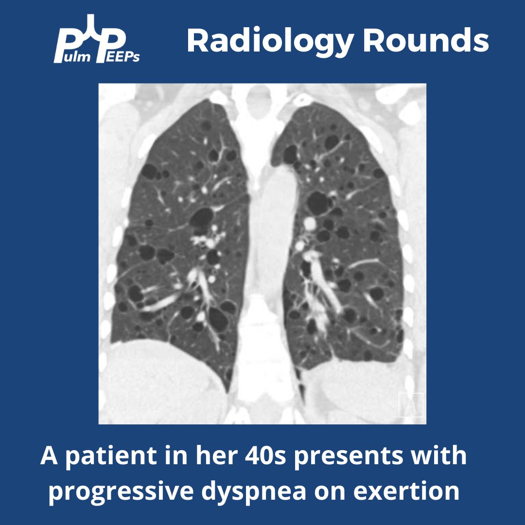
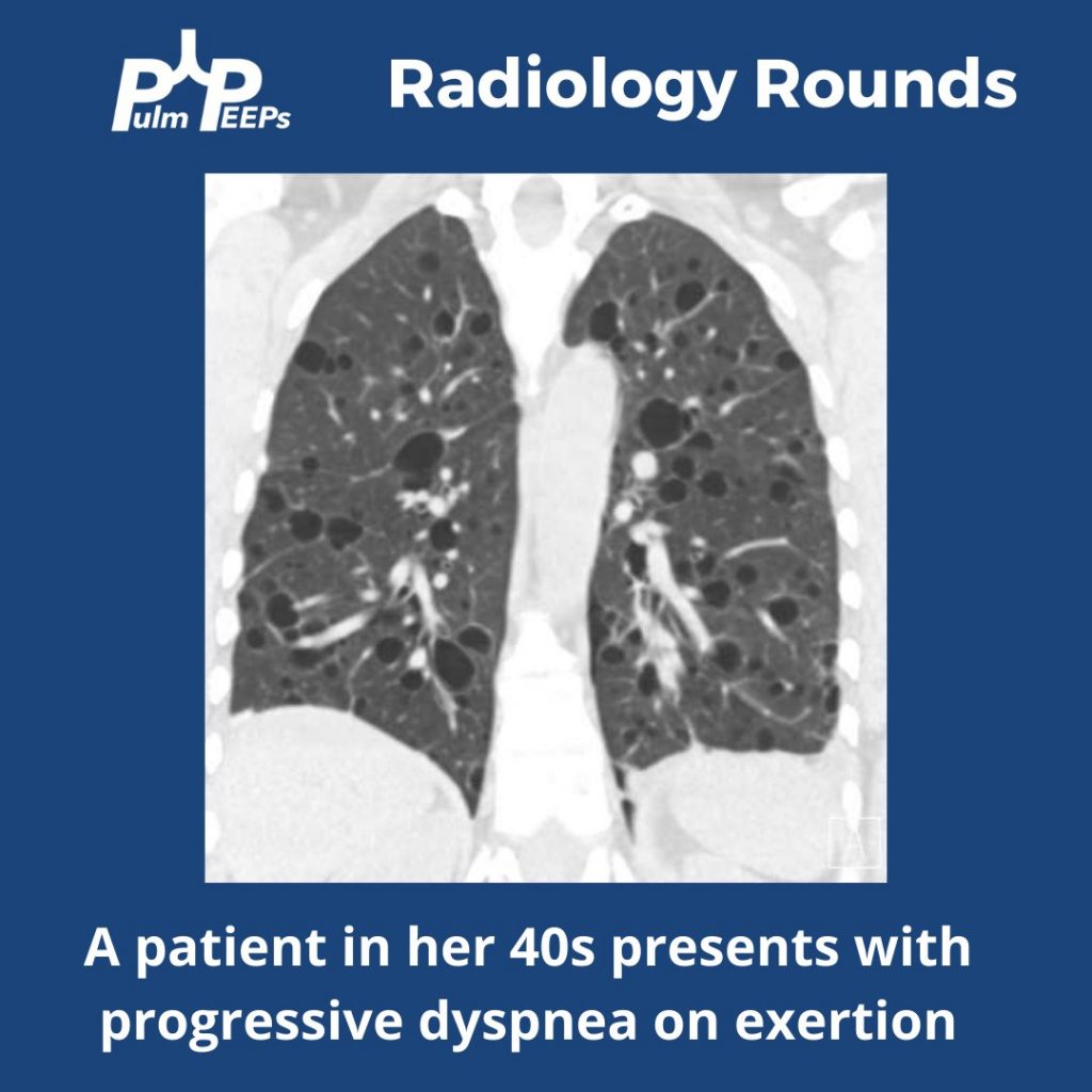
Time for another #RadiologyRounds!! This week we’re looking at the coronal CT scan views, which can be extremely helpful and are often under-utilized. Follow us on Twitter to work through Radiology Rounds cases as they come out.

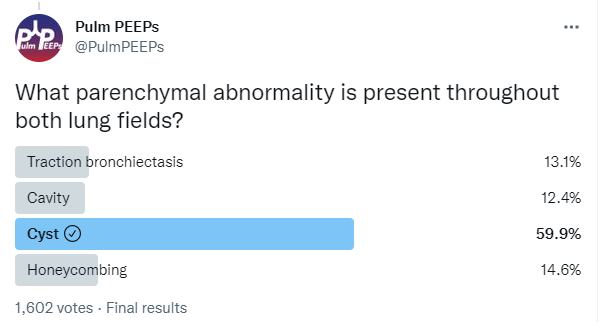
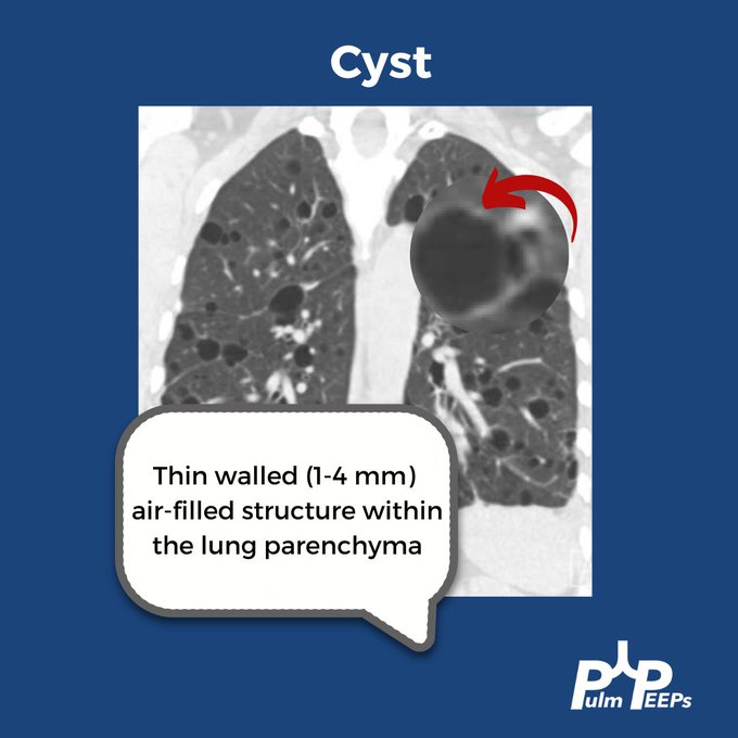
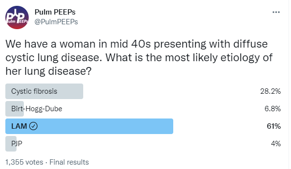
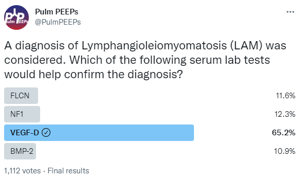
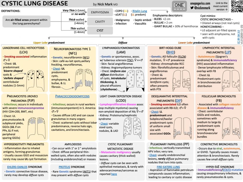
Our patient had an elevated VEGF-D level, a renal angiomyolipoma identified on CT abdomen, and imaging with diffuse cystic lung disease confirming her diagnosis of LAM. Make sure to check out the ICUOnePager made by Dr. Nick Mark.

Radiology Rounds 15: Usual Interstitial Pneumonia (UIP)

1

This week on #RadiologyRounds we are extremely excited to share a case brought to you by one of our new Associate Editors, Leon Mirson! Enjoy, and follow us on Twitter and Instagram for content delivered to you weekly!

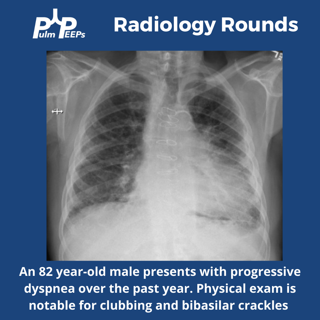
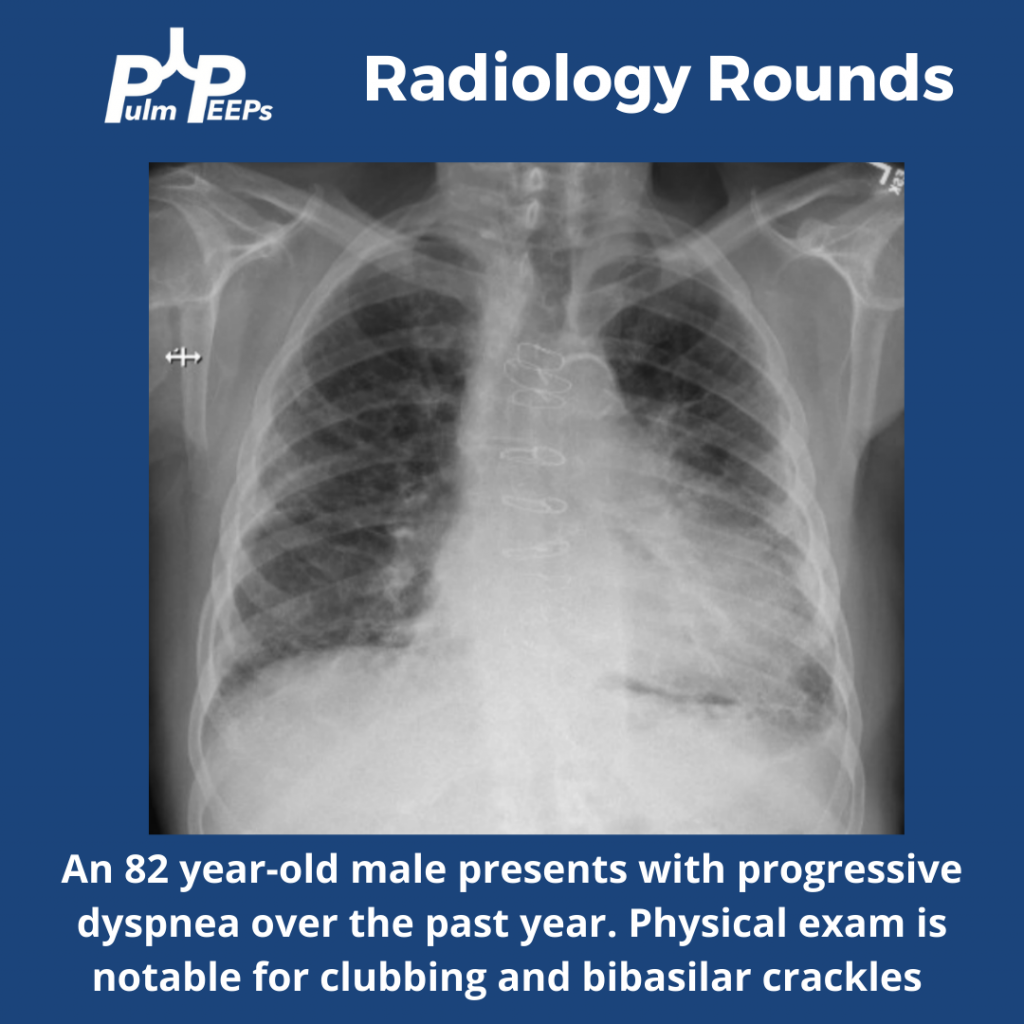
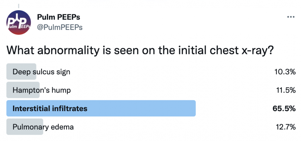
What abnormalities do you see on this CXR to help explain the patient’s presentation?

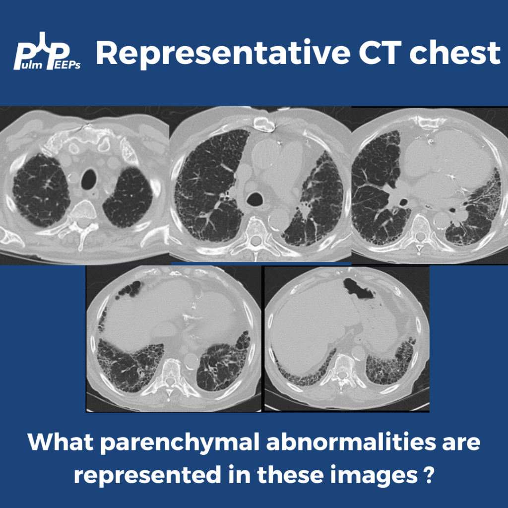
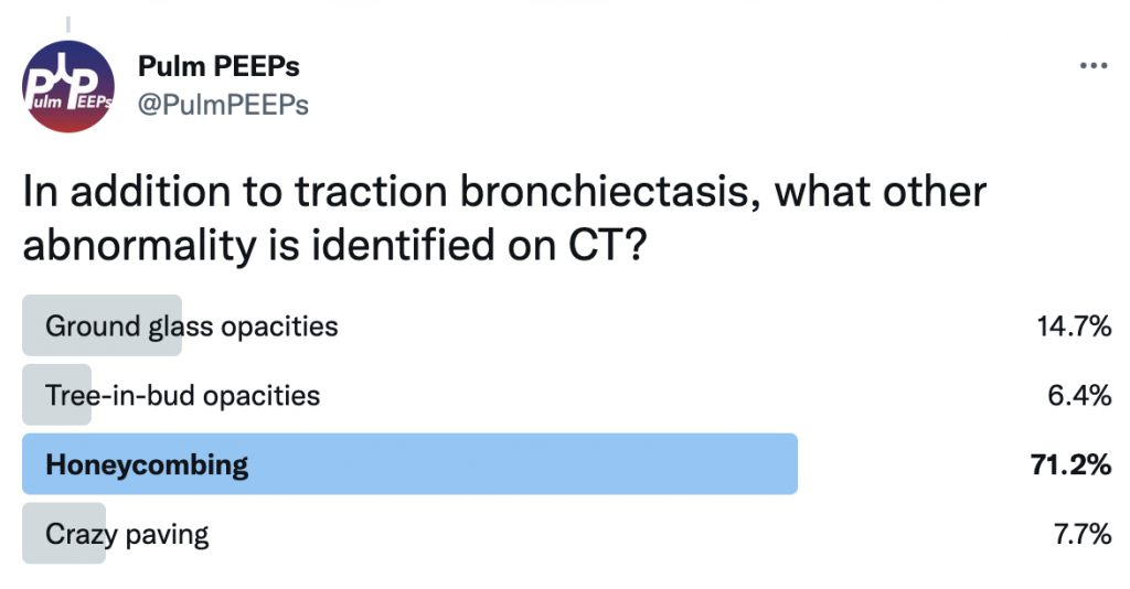
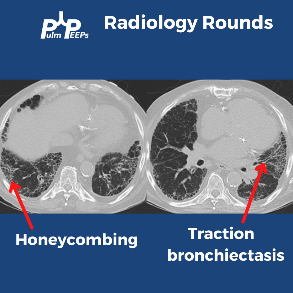
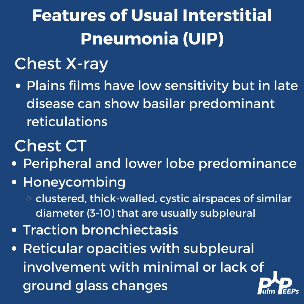
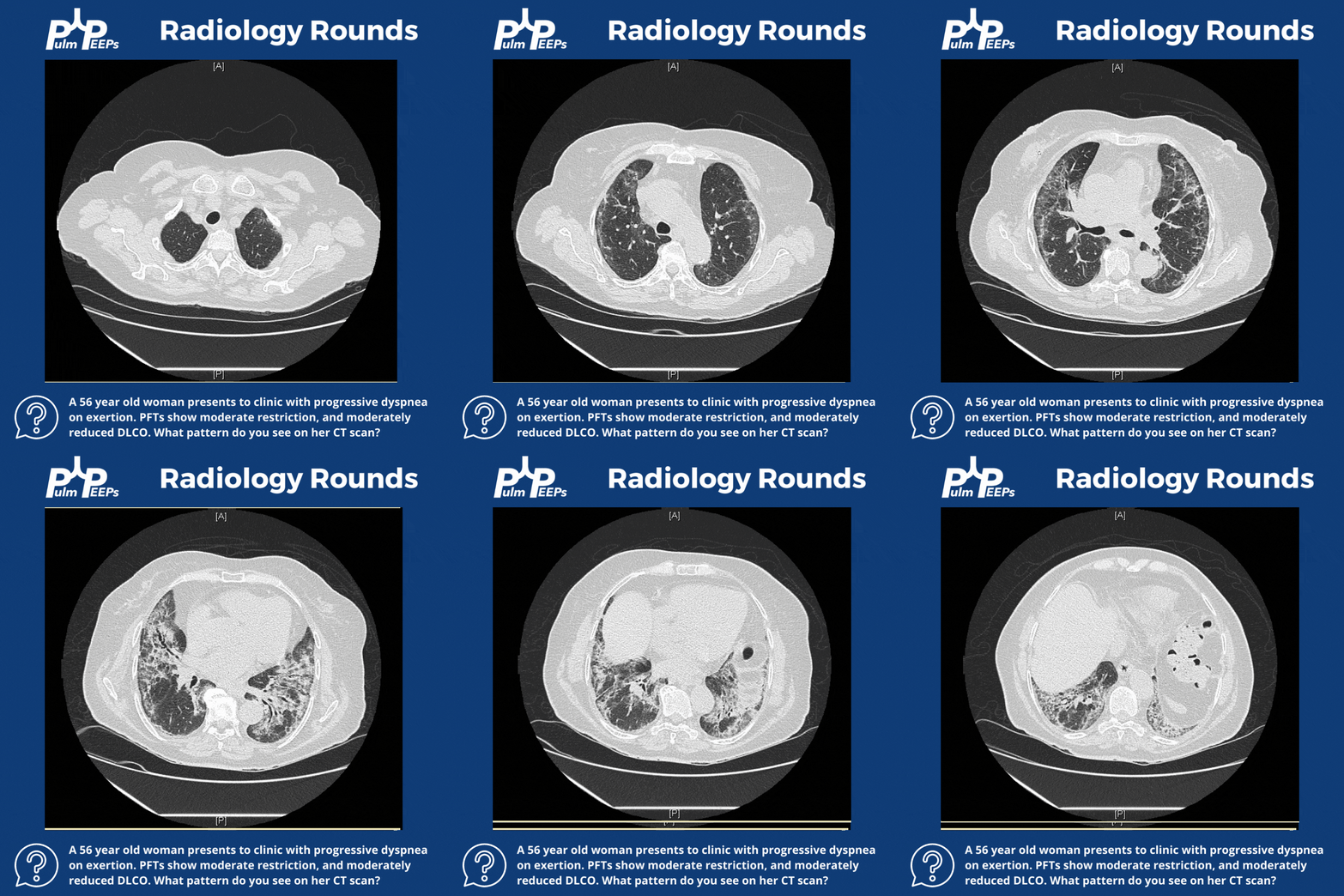
The CT scan has reticular changes consistent with interstitial lung disease and there are multiple features that help us define the pattern of the ILD. His CT notably has very few ground-glass opacities, there is traction bronchiectasis, and honeycombing with a basilar and peripheral / sub-pleural predominance.

Taking all these features together, the patient’s radiographic presentation is consistent with Usual Interstitial Pneumonia (UIP)

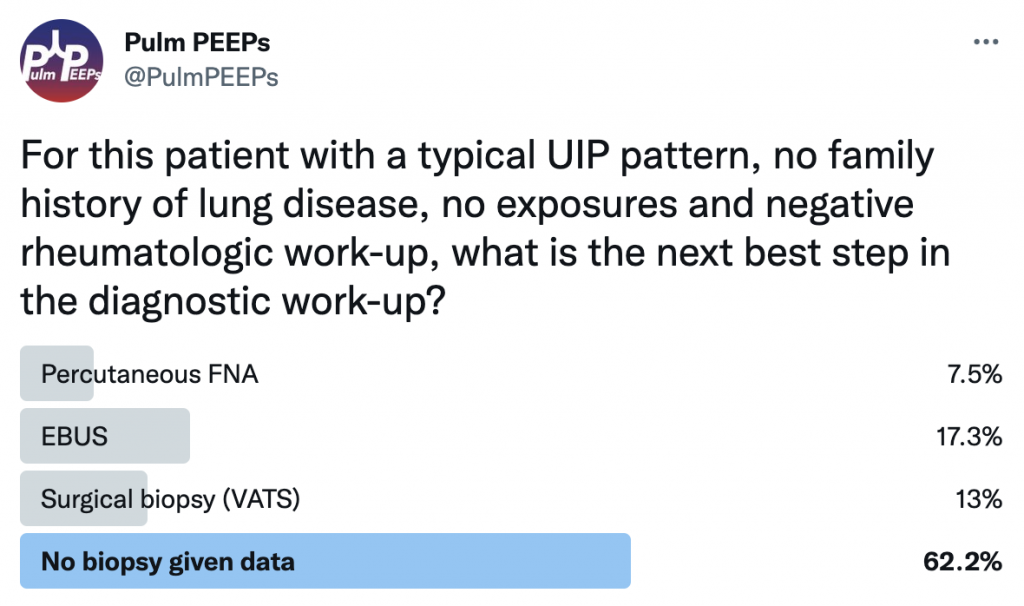
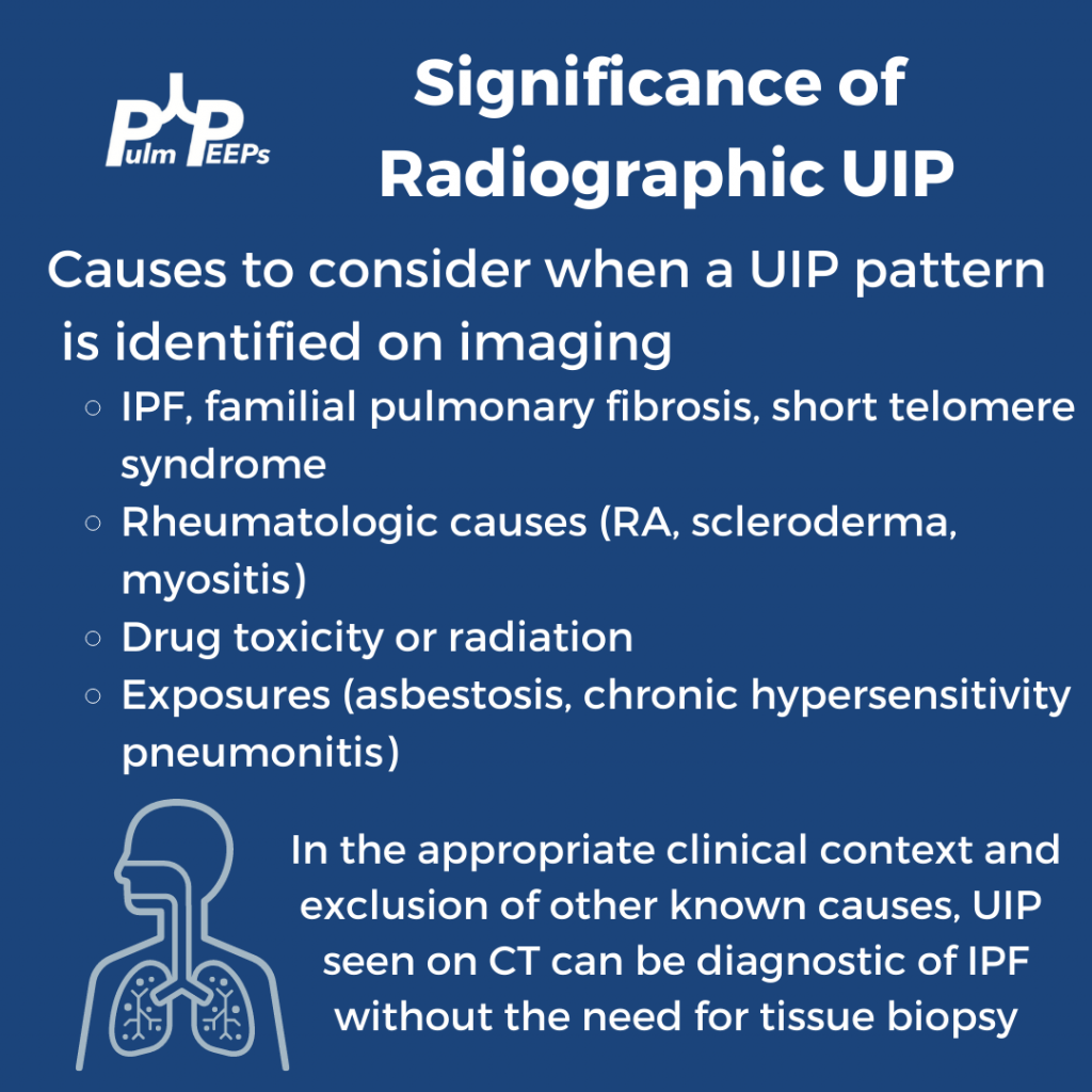
This patient had a thorough history taken and he had no prior smoking and no occupational or environmental exposures of significance. He had no family history of interstitial lung disease. A broad history was taken regarding symptoms of connective tissue disease and a broad serologic workup was sent, all of which were unremarkable. What would you want to do next diagnostically?

If you want to learn more about diagnosing interstitial lung disease, listen to our prior Top Consults episode on diagnosing ILD with experts in the field and see these prior #RadiologyRounds on Fibrotic NSIP and Sarcoidosis.

Radiology Rounds 11: Sarcoidosis

FPl2XDLXsAM5YFG

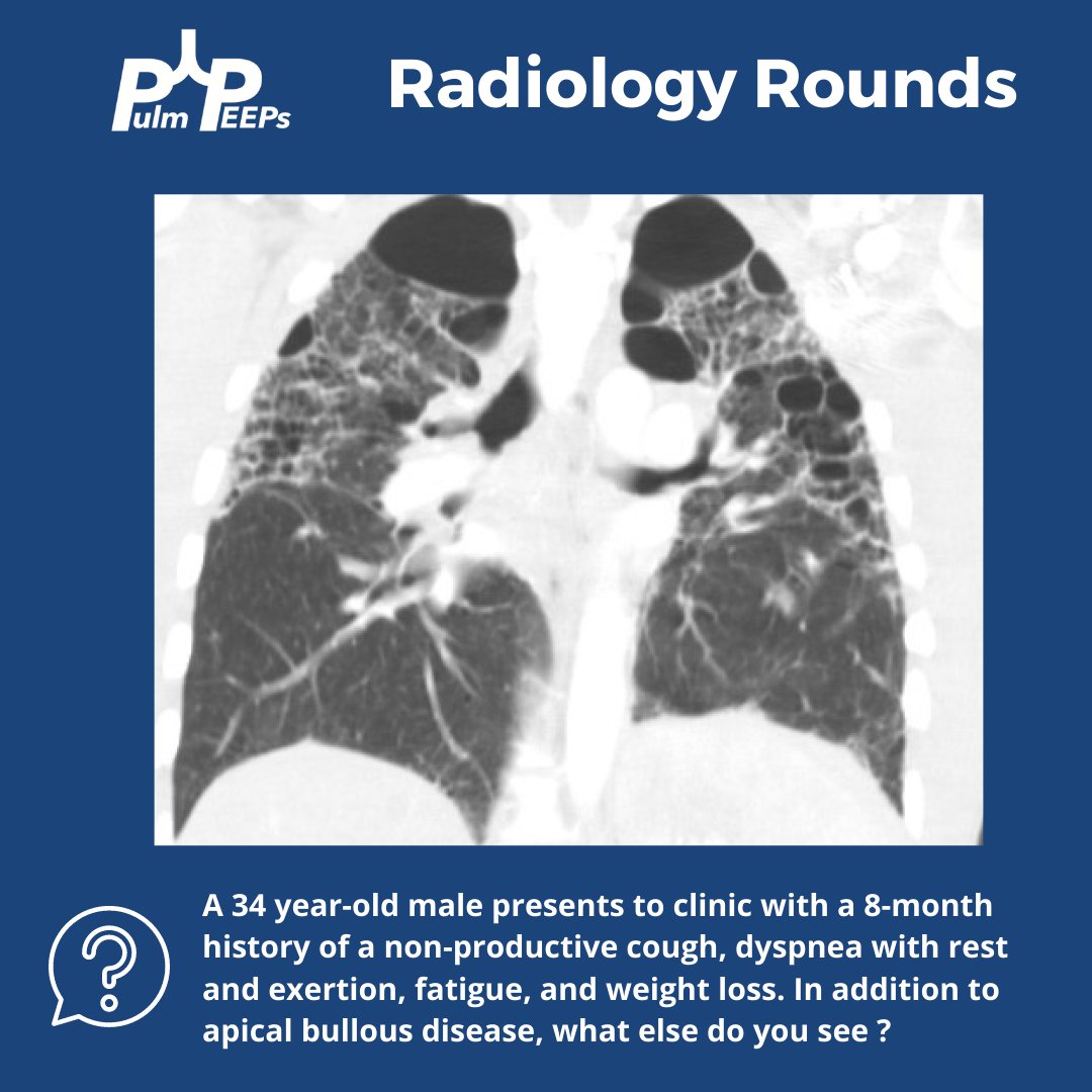
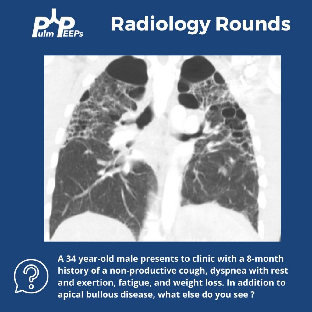
We have another #RadiologyRounds for you today! You are seeing a new patient in the clinic with dyspnea who brings in prior CT chest imaging. A representative coronal image is shown.

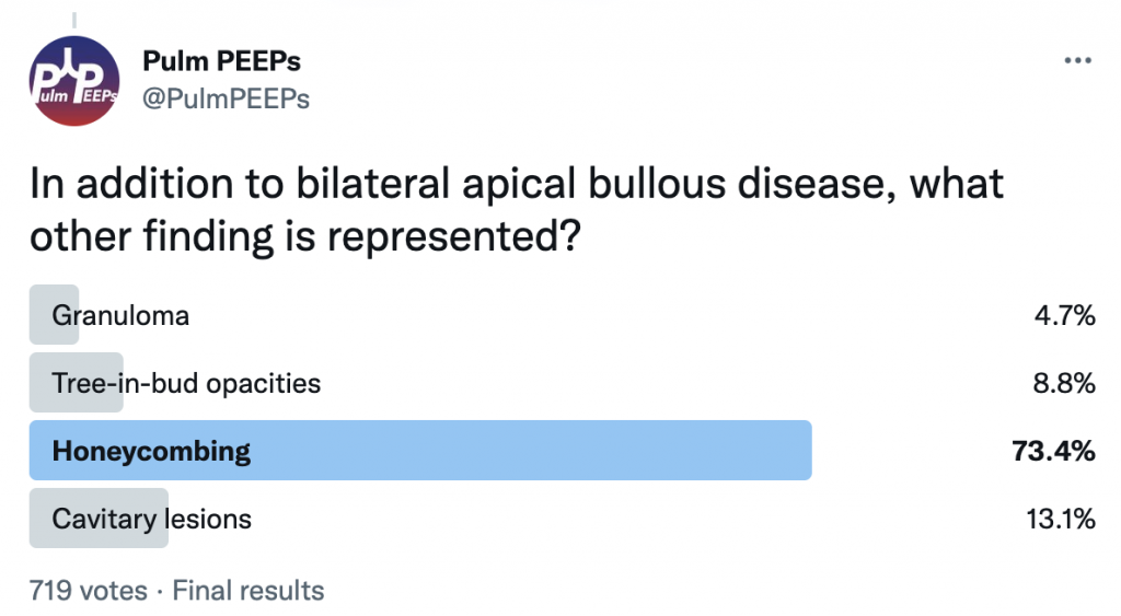
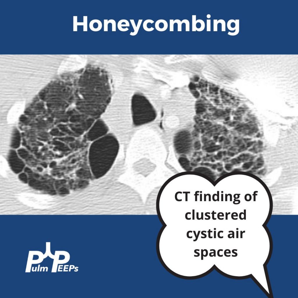
In addition to bullous disease, you see bilateral honeycombing with evidence of fibrosis primarily in the upper lung fields.

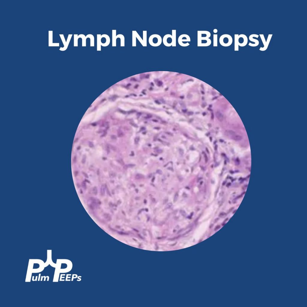
As part of your evaluation, an EBUS is performed showing the following representative lymph node tissue pathology.
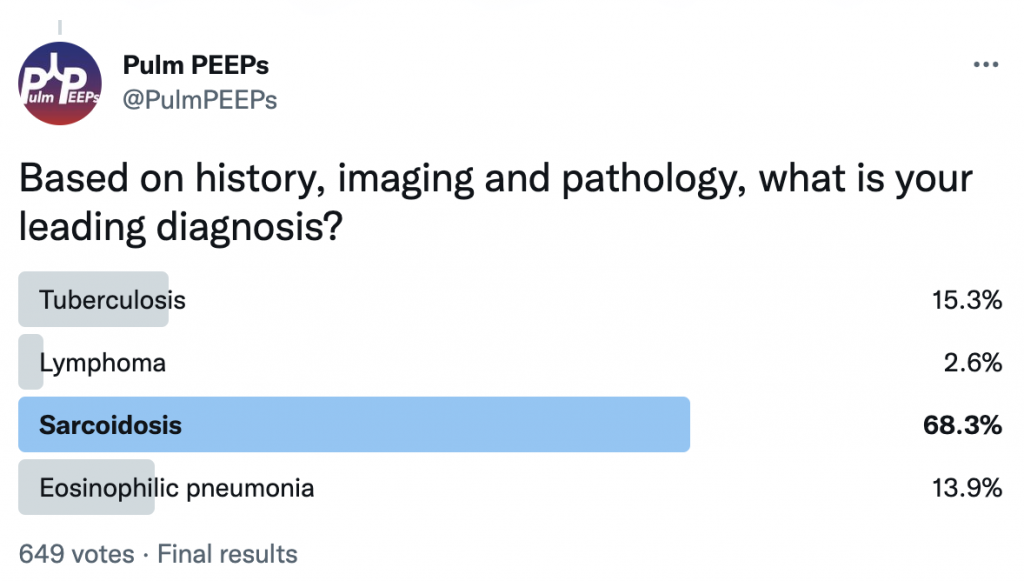
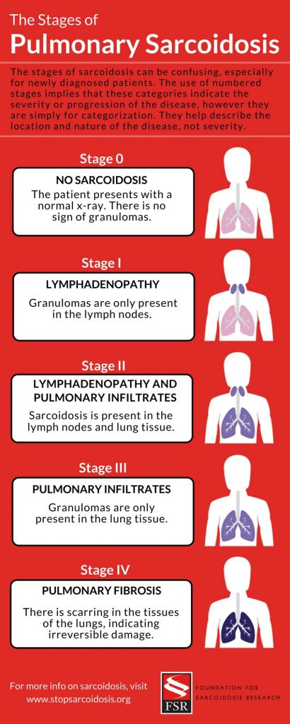
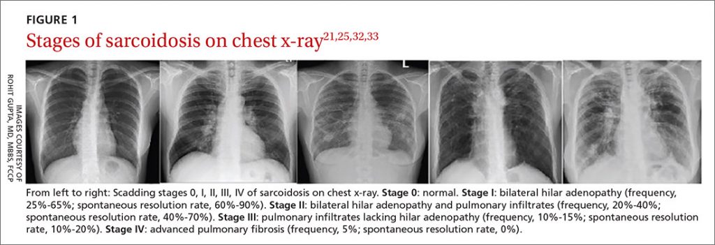
We had evidence of noncaseating granulomas, evidence of fibrocystic changes on chest imaging, and we excluded other causes of granulomatous disease. Given his symptoms and clinical context, we were concerned about Stage IV pulmonary sarcoidosis which can be categorized below.

Image source: https://www.stopsarcoidosis.org/stages-of-sarcoidosis/

Radiology Rounds 10: Pulmonary Arterial Hypertension – CT Signs

1

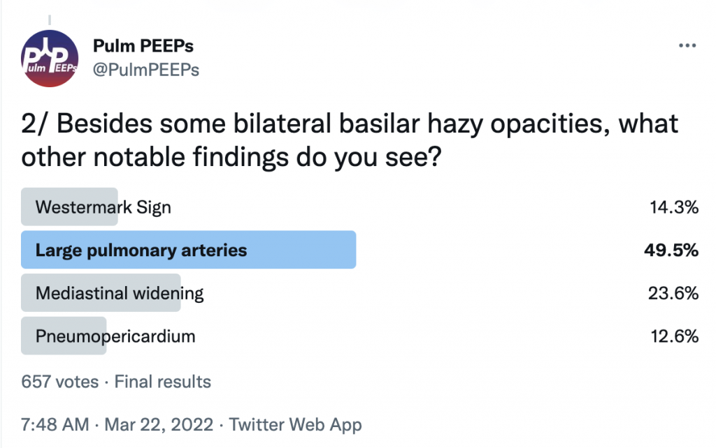
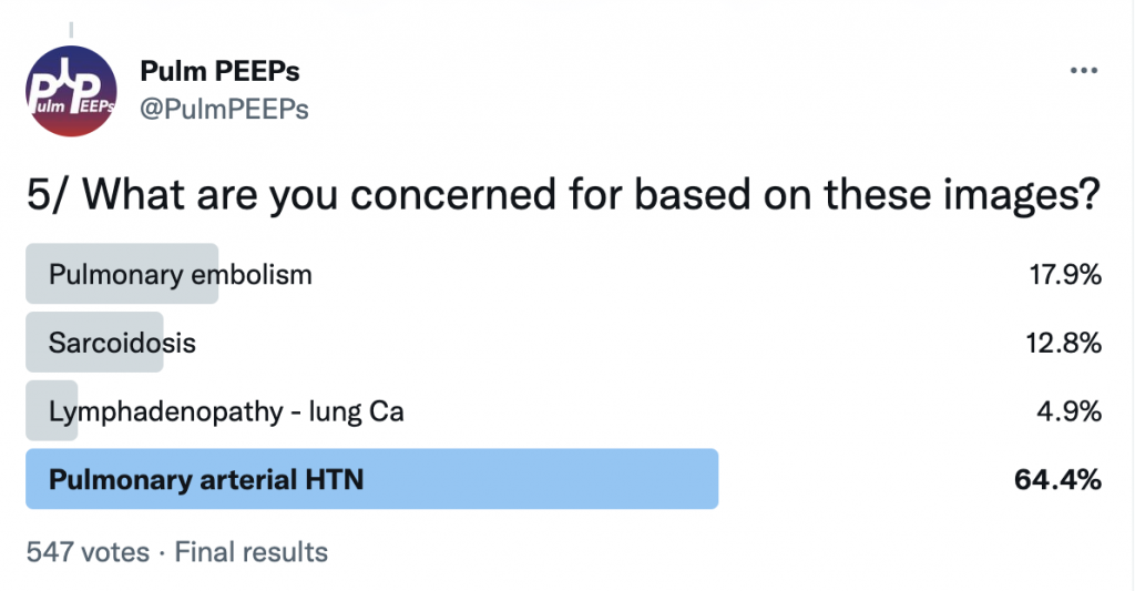
For Radiology Rounds this week we’ll be looking at multiple imaging modalities to help solve the case. Please follow along on Twitter for our live polls and for ongoing discussion of the case.

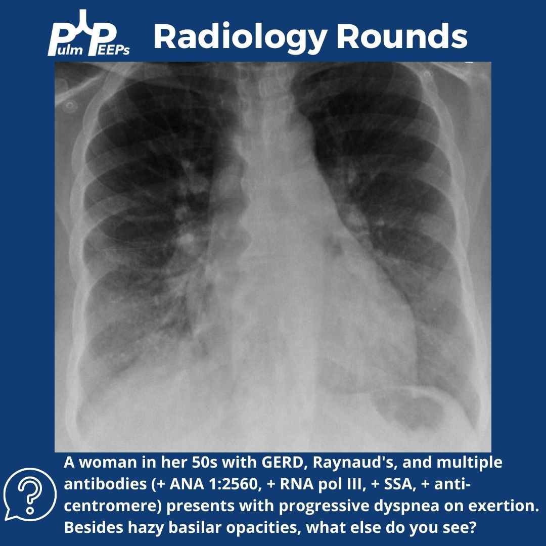
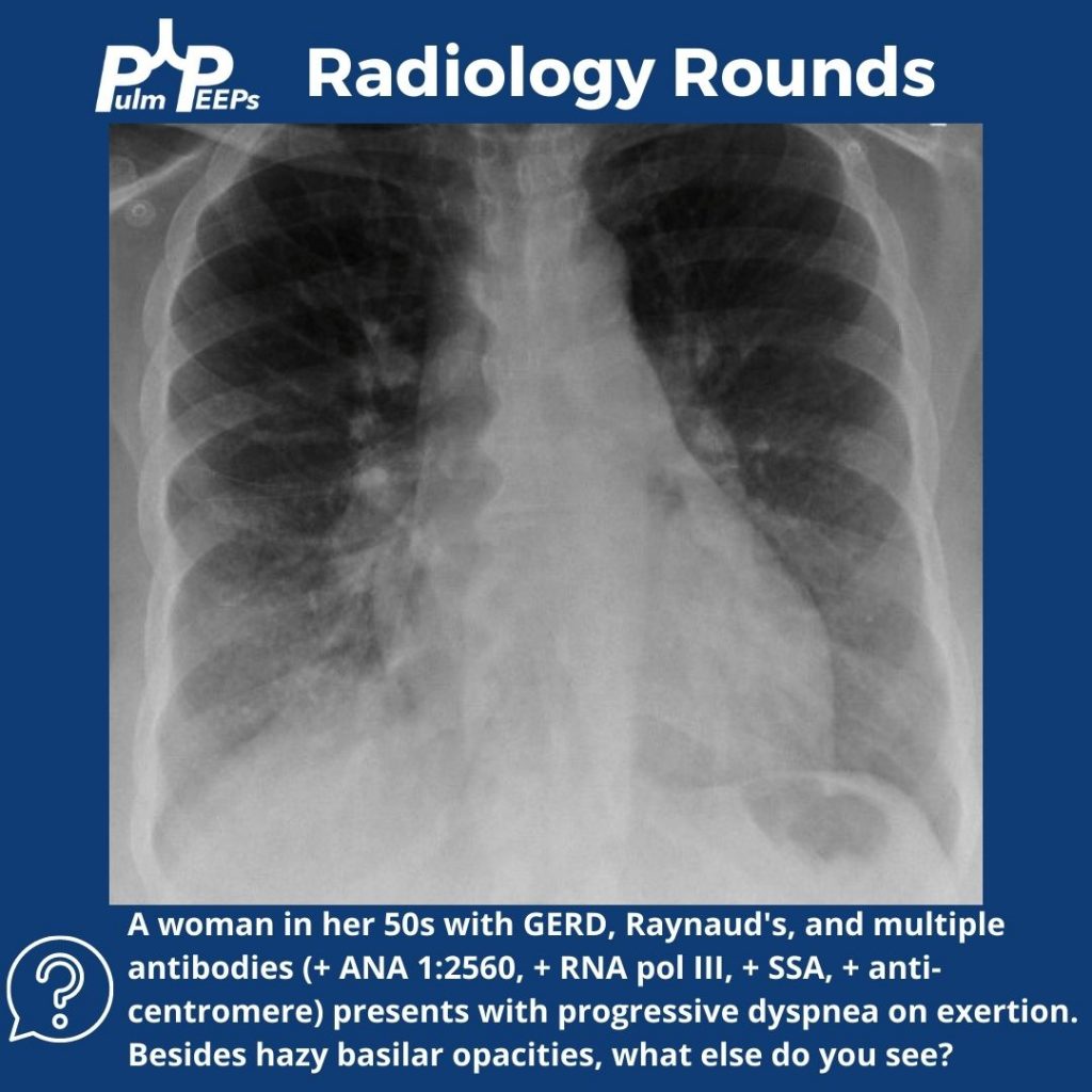
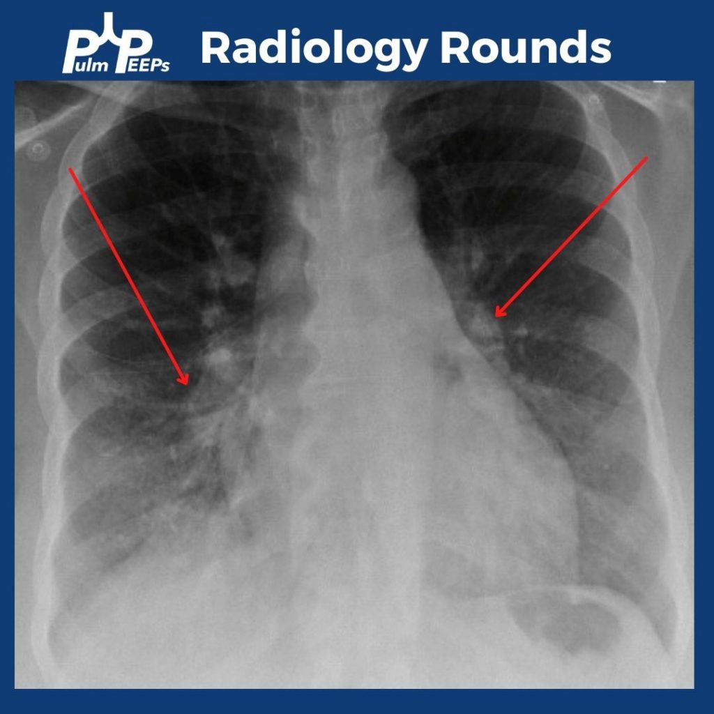
In addition to bibasilar opacities, the CXR shows bilateral enlarged pulmonary arteries.

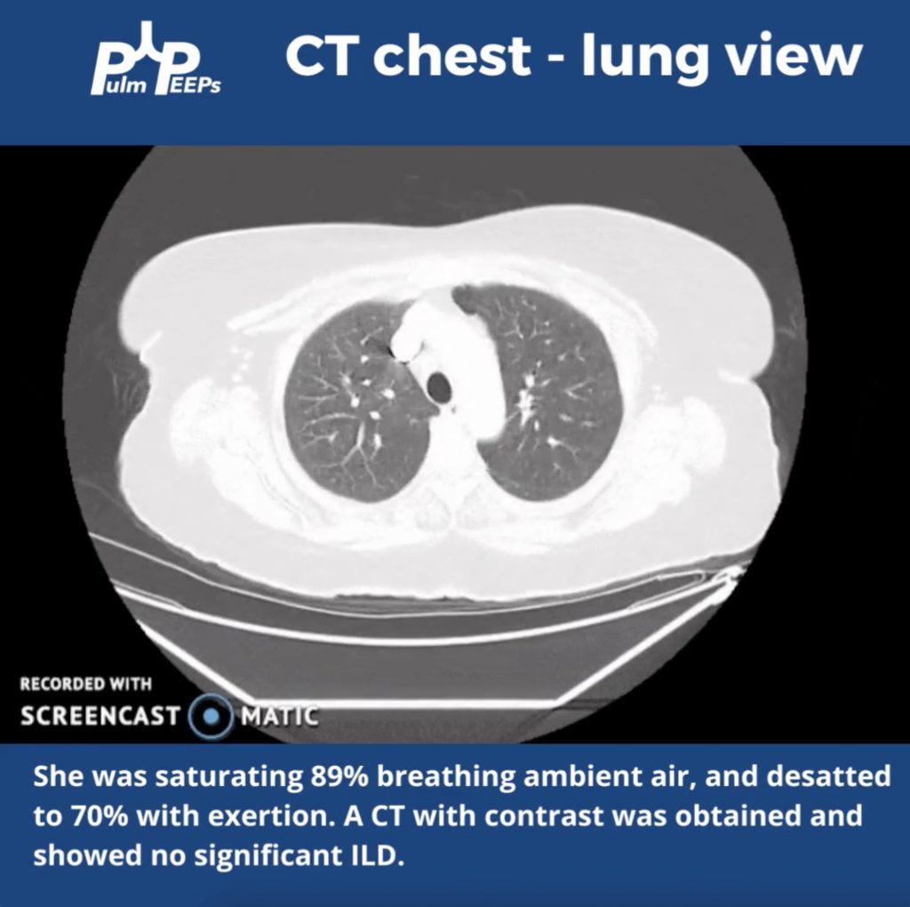
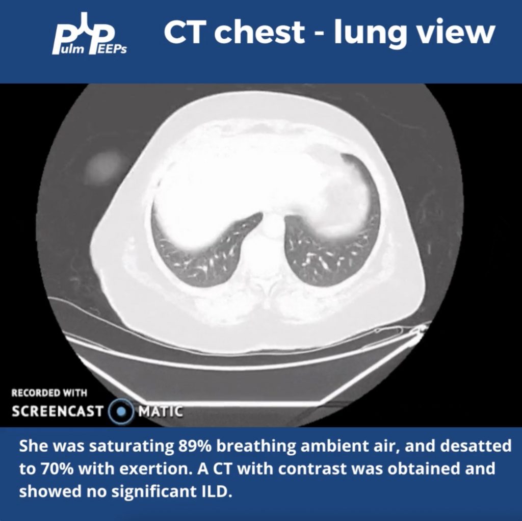
The patient was hypoxemic on room air and desatted into the 70s with ambulation. Given her possible connective tissue disease a CT chest was obtained.

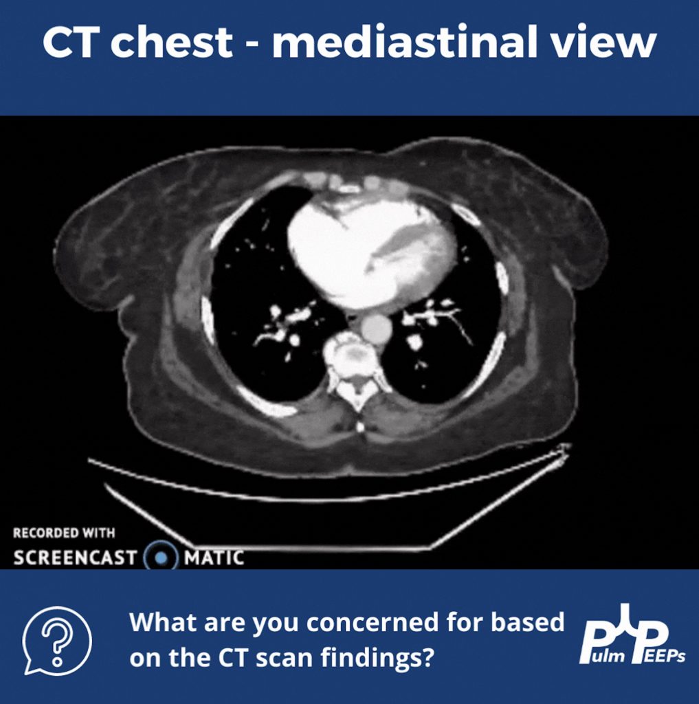
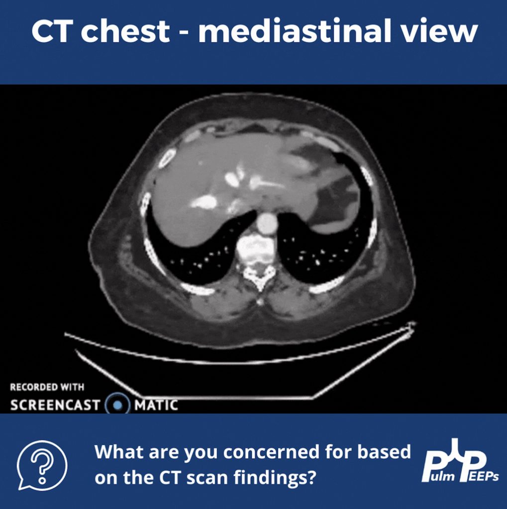
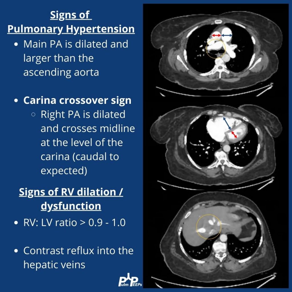
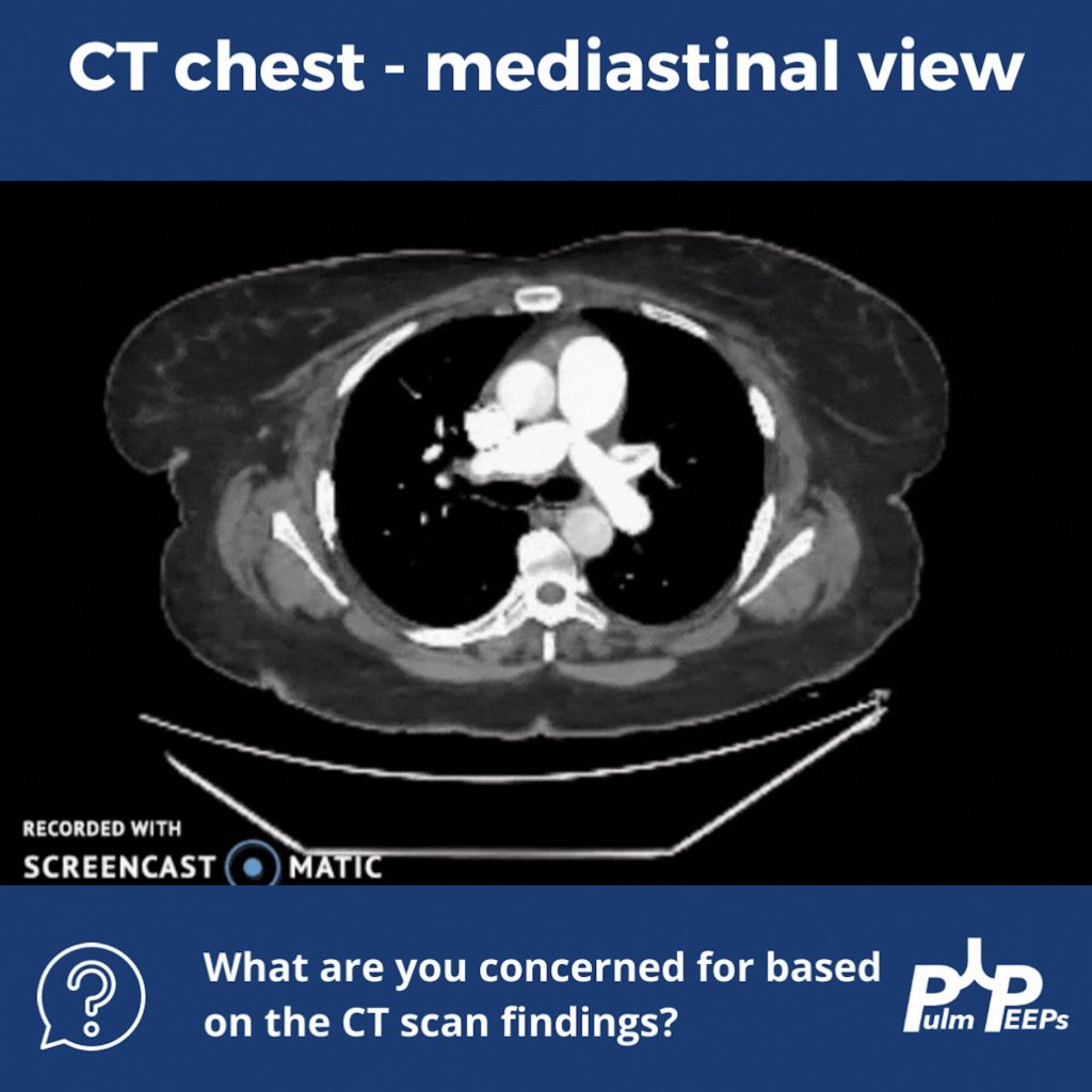
Although no ILD was revealed on the CT scan, let’s take a look at the mediastinal windows:

There are multiple signs of pulmonary hypertension, and right ventricular dilation and strain on this CT scan. Let’s review them:

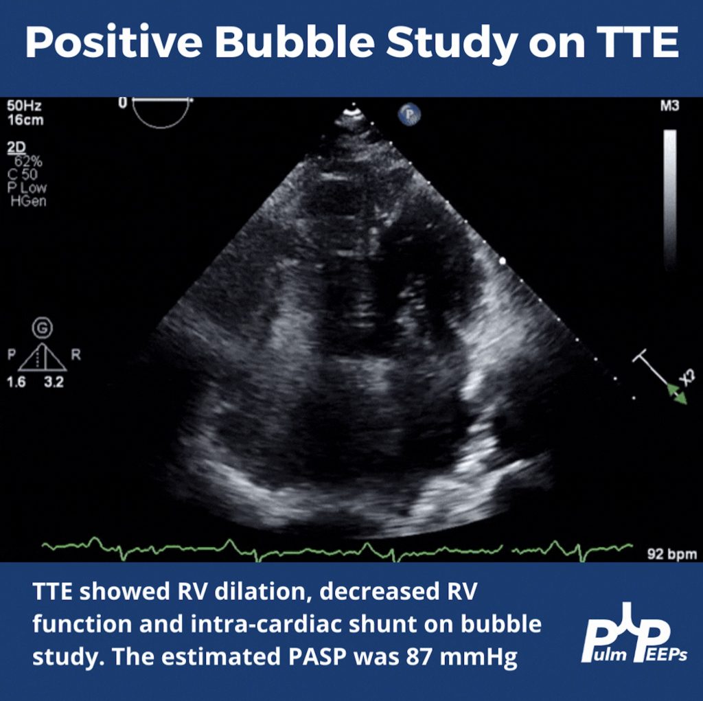
To help explain the patient’s hypoxemia, an ECHO with bubble study was ordered next

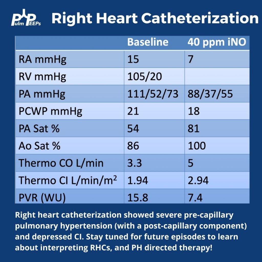
Since pulmonary hypertension can only truly be diagnosed on right heart catheterization, this was performed next and revealed severe pre- and post- (mostly pre)-capillary pulmonary hypertension

9. Top Consults: Interstitial Lung Disease Diagnosis

Top Consults ILD

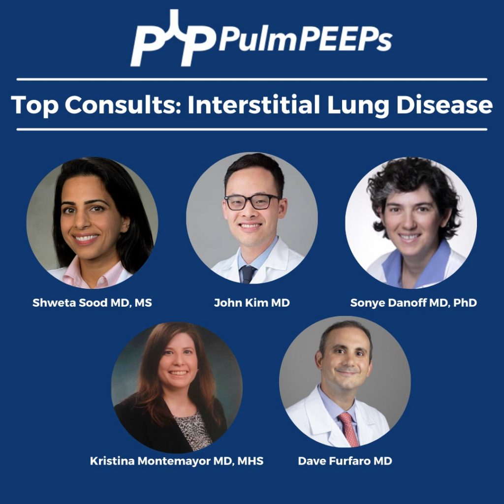
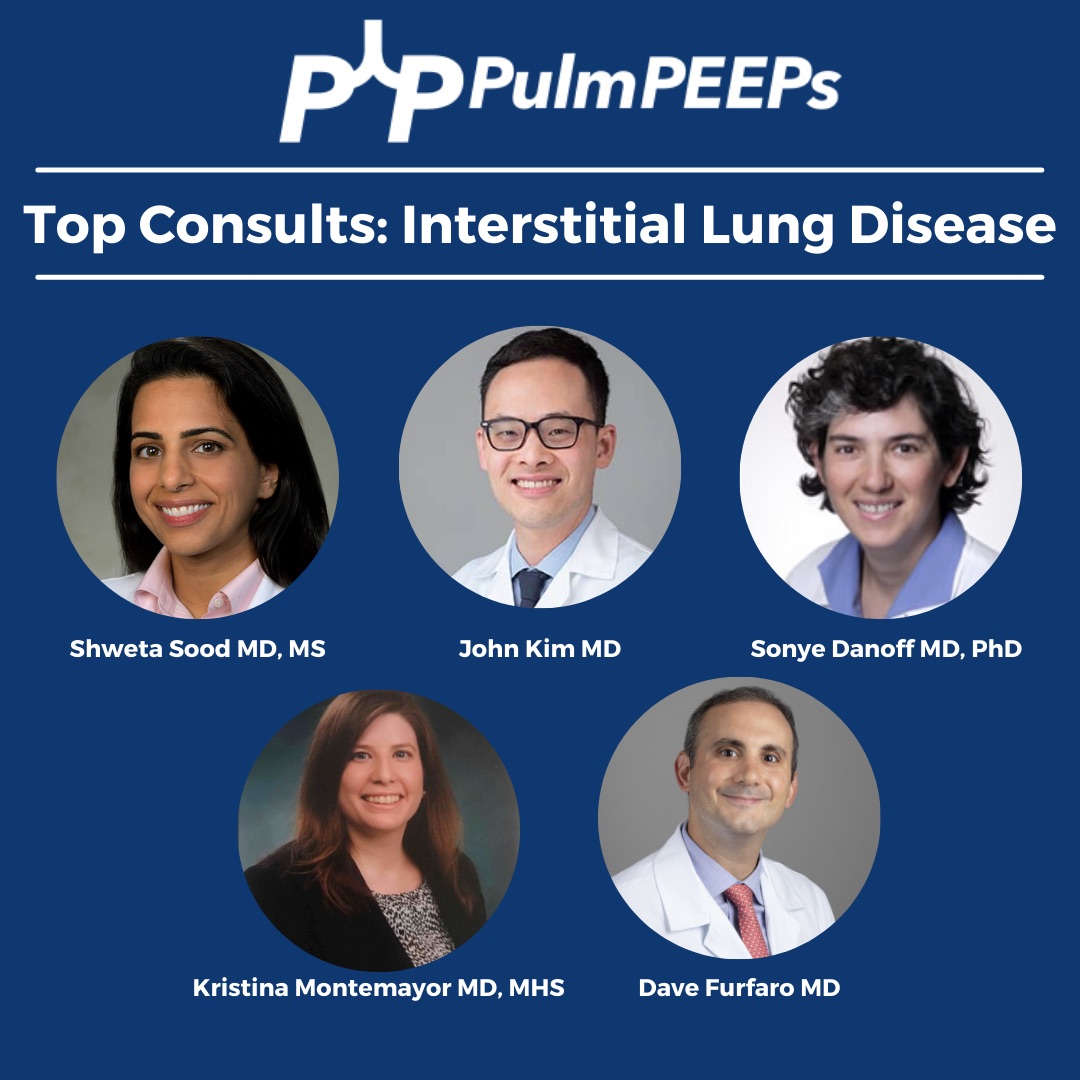
This week we are absolutely thrilled to be joined by three Interstitial Lung Disease experts to discuss the workup and differential for a patient with a new presentation of suspected ILD. This is also our first episode in a collaboration between the Pulm PEEPs and the American Thoracic Society Clinical Problems Assembly. In a series of episodes, we will be joined by pulmonary experts from around the country who are leaders in the ATS CP Assembly to provide content on common and cutting-edge topics in PCCM.

Meet Our Guests

Sonye Danoff is an Associate Professor of Medicine at Johns Hopkins and is Co-Director of the John Hopkins Interstitial Lung Disease and Pulmonary Fibrosis program. She also serves as the Assembly Chair of the Clinical Problems Assembly for the American Thoracic Society.

John Kim is an Assistant Professor of Medicine at UVA and has both clinical and research expertise in interstitial lung disease with a focus on pulmonary fibrosis.

Shweta Sood is an Assistant Professor of Medicine at Penn Medicine whose expertise is in Interstitial Lung Disease. She is an integral part of fellowship training where she leads the monthly ILD conference for fellows as well as provides didactics for ILD cases.

Consult Patient

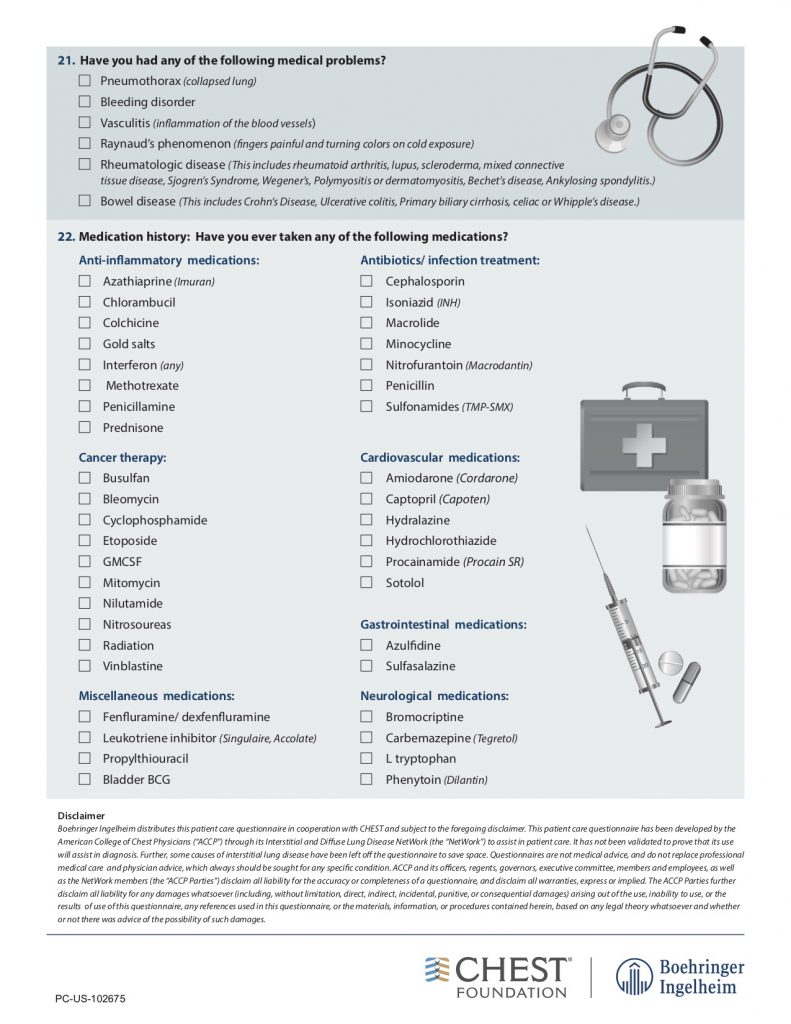
A 66-year-old man who is a never smoker with a past medical history of hypertension and osteoarthritis was admitted to the hospital after presenting with progressive dyspnea on exertion to dyspnea at rest and was found to be hypoxemic. He reports 4 months of progressive dyspnea on exertion but on further questioning, thinks he was last normal about 1.5 years ago when he could walk 2 miles at a time. Currently, he can only walk 0.25 to 0.5 miles before needing to stop. He reports an intermittent, dry cough throughout the day that is not associated with eating, position, or sleeping. A full ROS is negative including for rashes or joint pains. His family history is notable only for hypertension and hyperlipidemia. He is a never smoker, drinks in moderation 1-2 x a week, and lives in the suburbs with his wife. His house has central heating and air conditioning, they have no pets, and they have carpeted floors. He is a retired police detective.

His physical exam is notable for fine crackles at the bilateral bases on pulmonary auscultation, and he is breathing comfortably on nasal cannula although mildly tachypneic to 18 breaths per minute. He has no signs of volume overload, no peripheral clubbing, no rashes, and joint exam does not reveal swelling or synovitis.

Key Learning Points

Take away points from our guests:

— ILD is a symptom, not a diagnosis

— The first time a patient is evaluated for interstitial lung disease is the best chance for making the diagnosis so take the time to evaluate them thoughtfully

— Start the physical exam with the hands first. The hands can reveal a lot about the patient (clubbing, cyanosis, joint, skin, and nailbed findings) and it establishes a personal connection

— When doing the pulmonary exam, percuss first from top to bottom to learn the size of the lungs, and then listen from bottom to top

— When reading a CT scan the simplest approach is “Is it a UIP pattern or not?”. This can be your first diagnostic divide. UIP is consistent with IPF, and in select circumstances connective tissue disease, occupational lung disease, or advanced hypersensitivity pneumonitis. Non-UIP patterns have a broader differential

— A multi-disciplinary interstitial lung disease conference is the gold standard for establishing an ILD diagnosis

Gathering the history:

— Ask about onset: acute or chronic. “When was the last time your breathing was entirely normal?”

— Symptoms can be shortness of breath, a lingering cough, or often fatigue and decreased energy. Occurrence is important! Do symptoms occur only with exertion or at rest too?

— “Have you ever had chest imaging before?”

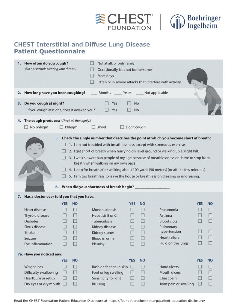
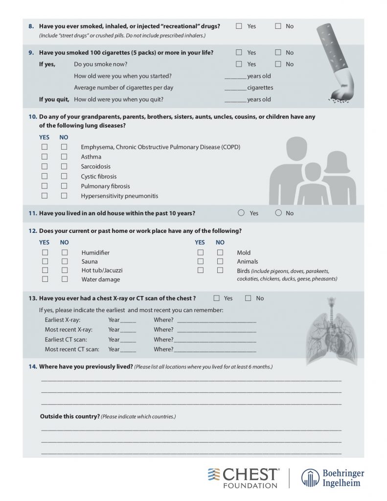
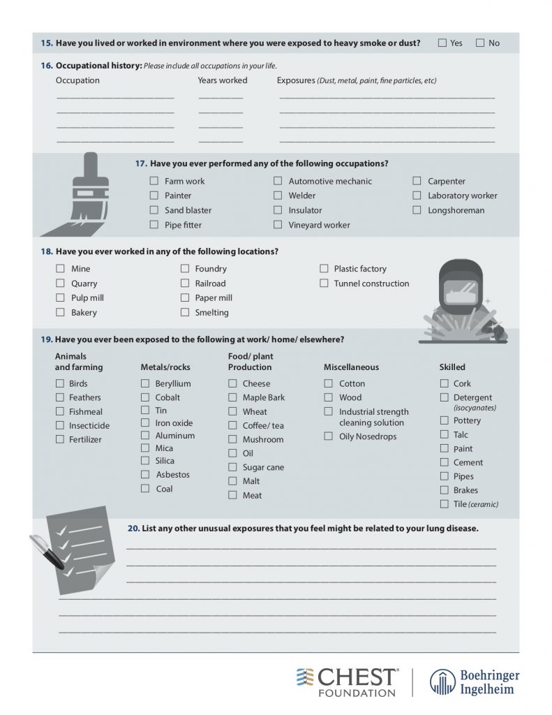
— Take a thorough exposure history, and the weird questions are all necessary! Ask about birds and feathers (pet birds, bird feeders, down pillows or blankets, hunting, taxidermy), mold or water damage, organic or inorganic compounds from work (landscaping, ship yards, coal mines)

American College of Chest Physicians – Interstitial Lung Disease Patient Questionnaire

Physical Exam:

— Assess stability first and foremost

— Lung findings: crackles, inspiratory squeaks (often in hypersensitivity pneumonitis)

— Look for evidence of alternative diagnoses: volume overload, liver disease, signs of infection

— Evaluate for signs of connective tissue disease: examine the skin around the forehead and mouth for signs of scleroderma, look for rashes, perform a thorough joint examination and look at their hands, and assess muscle strength

Imaging:

— Order a high-resolution CT scan without contrast. High resolution means thin slices that are 1-2 mm thick. Contrast should be avoided if possible because it makes looking for subtle reticulations or ground-glass opacities harder

— Inspiratory and expiratory films help evaluate for gas trapping. If present, this may indicate hypersensitivity pneumonitis

— Prone films allow you to distinguish reticular changes in the dependent portions of the lungs from atelectasis

Reading the CT scan:

— Look at the distribution first. Is it uniform from top to bottom or not? Is it subpleural, peripheral predominant, or central?

— Identify key features: reticulation, traction bronchiectasis, honeycombing, ground-glass opacities, cysts, and nodules

Laboratory evaluation:

— ANA, Scl-70, DS DNA, anti-RNP, anti-centromere, RF, CCP, SSA, SSB, RNA pol 3, HIV, myositis panel, Hypersensitivity pneumonitis panel

Pulmonary function tests:

— A restrictive ventialtory defect is the classic pattern and can tell you about severity. In ILD, TLC, VC, FRC, and RV are generally all reduced in proportion

— Identify if there is obstruction or not, because if present this may indicate coexisting emphysema or hypersensitivity pneumonitis

–The DLCO can be helpful for disease severity, and for raising concern about other co-existing diagnoses such as pulmonary vascular disease, or emphysema

— A DLCO < 50% predicted may predict that the patient will need oxygen with exertion and the patient should be walked

6 Minute Walk Test:

— This should be performed for all new patients because it is important for prognosis

— In the first year after diagnosis, a 6MWD should be performed every 3 – 4 months to assess disease trajectory. It can be done every 6 – 12 months after that.

Differential Diagnosis:

Silo ILDs into two large buckets

1) An exposure, trigger, or underlying cause is present: hypersensitivity pneumonitis, medication-induced, occupational lung disease, connective tissue disease, granulomatous disorder

2) Idiopathic interstitial pneumonia

References and links for further reading

  1. Raghu G, Collard HR, Egan JJ, et al. An official ATS/ERS/JRS/ALAT statement: idiopathic pulmonary fibrosis: evidence-based guidelines for diagnosis and management. Am J Respir Crit Care Med. 2011;183(6):788-824. doi:10.1164/rccm.2009-040GL
  2. Raghu G, Brown KK. Interstitial lung disease: clinical evaluation and keys to an accurate diagnosis. Clinics in Chest Medicine. 2004;25(3):409-419. doi:10.1016/j.ccm.2004.05.007
  3. Bradley B, Branley HM, Egan JJ, et al. Interstitial lung disease guideline: the British Thoracic Society in collaboration with the Thoracic Society of Australia and New Zealand and the Irish Thoracic Society. Thorax. 2008;63 Suppl 5:v1-58. doi:10.1136/thx.2008.101691
  4. American Thoracic Society, European Respiratory Society. American Thoracic Society/European Respiratory Society International Multidisciplinary Consensus Classification of the Idiopathic Interstitial Pneumonias. This joint statement of the American Thoracic Society (ATS), and the European Respiratory Society (ERS) was adopted by the ATS board of directors, June 2001 and by the ERS Executive Committee, June 2001. Am J Respir Crit Care Med. 2002;165(2):277-304. doi:10.1164/ajrccm.165.2.ats01
  5. Lederer DJ, Martinez FJ. Idiopathic Pulmonary Fibrosis. New England Journal of Medicine. 2018;378(19):1811-1823. doi:10.1056/NEJMra1705751
  6. Travis WD, Hunninghake G, King TE, et al. Idiopathic nonspecific interstitial pneumonia: report of an American Thoracic Society project. Am J Respir Crit Care Med. 2008;177(12):1338-1347. doi:10.1164/rccm.200611-1685OC
  7. Wijsenbeek M, Cottin V. Spectrum of Fibrotic Lung Diseases. New England Journal of Medicine. 2020;383(10):958-968. doi:10.1056/NEJMra2005230
  8. Hariri LP, Roden AC, Chung JH, et al. The Role of Surgical Lung Biopsy in the Diagnosis of Fibrotic Interstitial Lung Disease: Perspective from the Pulmonary Fibrosis Foundation. Annals ATS. 2021;18(10):1601-1609. doi:10.1513/AnnalsATS.202009-1179FR
  9. Exposures. hpLung. Accessed February 12, 2022. https://www.hplung.com/

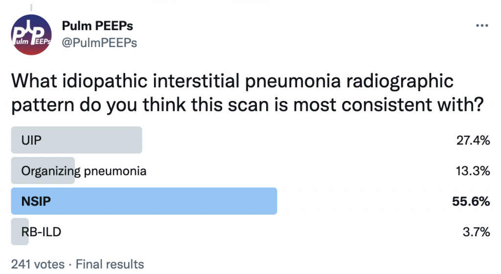
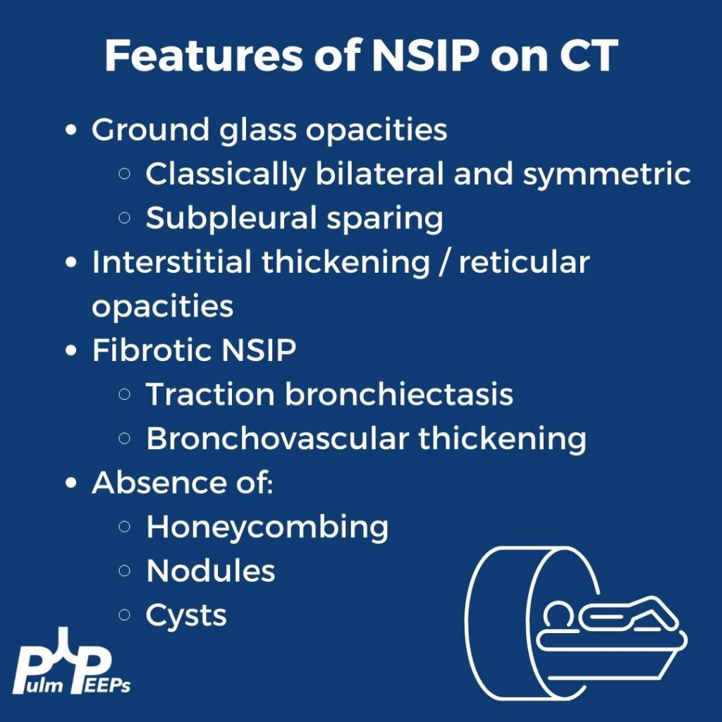
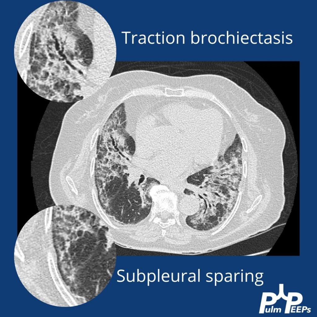
Radiology Rounds 7: Fibrotic NSIP

Fibrotic NSIP GIF

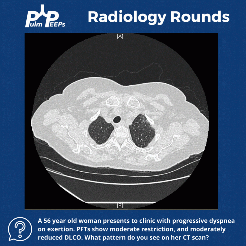
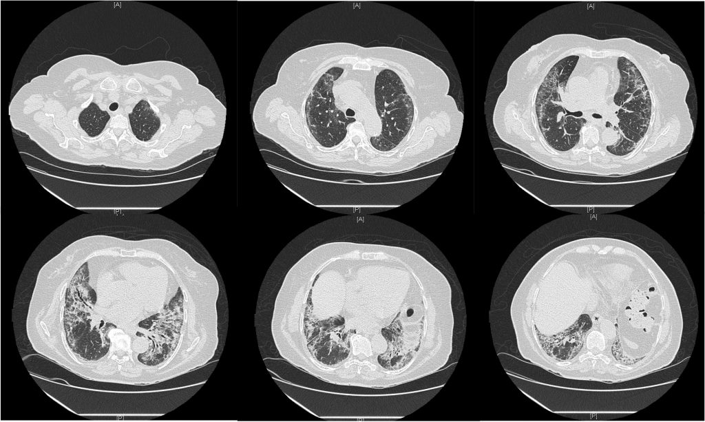
Evaluating a patient with suspected interstitial lung disease is a challenge for all pulmonary clinicians from medical students to attending physicians. This week’s Radiology Rounds covers some findings seen in one classic pattern. Make sure to listen to our show next week for more about ILD diagnosis!

8. A Case of Dyspnea and Lymphadenopathy

Case Dyspnea and Lymphadenopathy Face Page

Dave Furfaro, Kristina Montemayor, and Ansa Razzaq are back to tackle another pulmonary case! Listen in and solve the case yourself, and we’ll share some diagnostic pearls along the way. Let us know any additional thoughts on Twitter.

Patient Presentation

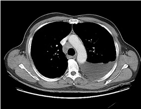
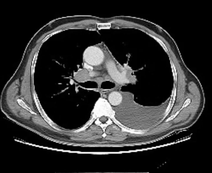
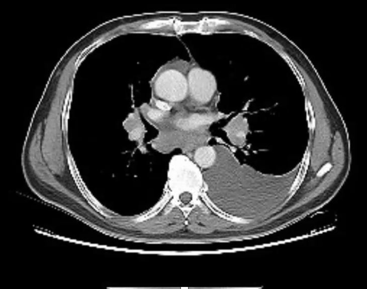
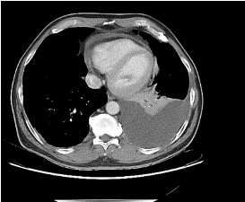
The is patient is 57-year-old man with hypertension and asthma who presents with dyspnea and left-sided pleuritic chest pain for 3 weeks. He was in his usual state of health until 3 weeks prior to admission, when he developed dyspnea and sharp left-sided chest pain that worsens with deep breathing. His symptoms are accompanied by unintentional 30-pound weight loss over the past several months as well as an intermittent cough that is nonproductive.

On physical exam, he is mildly tachypneic and saturating well on room air with otherwise normal vital signs. He has decreased breath sounds at the right lung base.

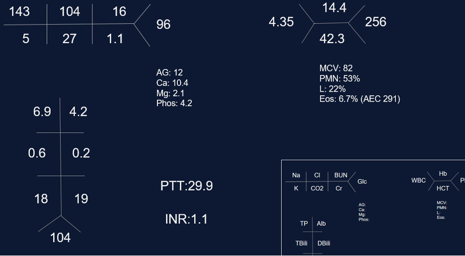
Initial labs

Key Learning Points

**Spoilers Ahead** If you want to think through the case on your own we advise listening to the episode first before looking at the infographics below

Physical Exam Pearls

Reasons for decreased breath sounds on physical exam

1. Increased thickness of chest wall

2. Reduced airflow to part of the lung

3. Overinflation to part of the lung

4. Something between the lung and chest wall — air or fluid

Determine bradypnea and tachypnea quickly by matching your breahting rate to the patient’s respiratory rate

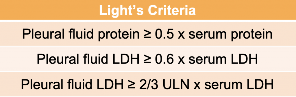
Pleural Effusions

Transudative effusion partial differential: heart failure, nephrotic syndrome, hepatohydrothorax, fluid overload, hypoalbuminemia, urinothorax, amyloidosis*, chlothorax*, hypothyrodism*, malignancy*, sarcoidosis*

*these effusions often present as exudates but can be transudates

Exudative effusion partial differential: Infection, hemothorax, malignancy, connective tissue disease, chylothorax, pancreatitis, esophageal perforation.

Sarcoidosis

Key Elements of Diagnosis:

1. Is there a compatible presentation (imaging, physical exam)

2. Detection of non-necrotizing granulomatous inflammation in one or more tissue samples

3. Exclusion of other disease that may present similarily

Pulmonary stages of sarcoidosis:

Key to remember that patients don’t always progress through these stages. The system is useful for prognosticating and determining treatment based on the risk for disease progression.

Image source: Gupta Et Al. J Fam Pract. 2021 April;70(3):E4-E15 | 10.12788/jfp.0177

Treatment for pulmonary sarcoidosis

Patients with stage 1, and even many with stage 2, often don’t require treatment

The first-line agent is oral glucocorticoids and the typical starting dose is prednisone 20 – 40 mg by mouth daily. The patient should be evaluated closely, and ideally, this dose can be tapered starting at about 4 – 6 weeks. Following this, the prednisone dose is tapered slowly over 6 months – 1 year while monitoring for symptom recurrence.

Second-line steroid-sparing agents are methotrexate, azathioprine, or mycophenolate. These are often used if the patient relapses, or is on more then 10mg daily for 3 months after the initial taper with intolerance of steroids

Third-line agents: tumor necrosis factor (TNF)- alpha antagonists (small molecule or monoclonal antibody therapy)

References and links for further reading

  1. Light RW. Pleural Effusion. New England Journal of Medicine. 2002;346(25):1971-1977. doi:10.1056/NEJMcp010731
  2. Feller-Kopman D, Light R. Pleural Disease. New England Journal of Medicine. 2018;378(8):740-751. doi:10.1056/NEJMra1403503
  3. Sarcoidosis: An FP’s primer on an enigmatic disease. MDedge Family Medicine. 2021;70(3). doi:10.12788/jfp.0177
  4. Iannuzzi MC, Rybicki BA, Teirstein AS. Sarcoidosis. New England Journal of Medicine. 2007;357(21):2153-2165. doi:10.1056/NEJMra071714
  5. Drent M, Crouser ED, Grunewald J. Challenges of Sarcoidosis and Its Management. New England Journal of Medicine. 2021;385(11):1018-1032. doi:10.1056/NEJMra2101555