This week on #RadiologyRounds we are extremely excited to share a case brought to you by one of our new Associate Editors, Leon Mirson! Enjoy, and follow us on Twitter and Instagram for content delivered to you weekly!
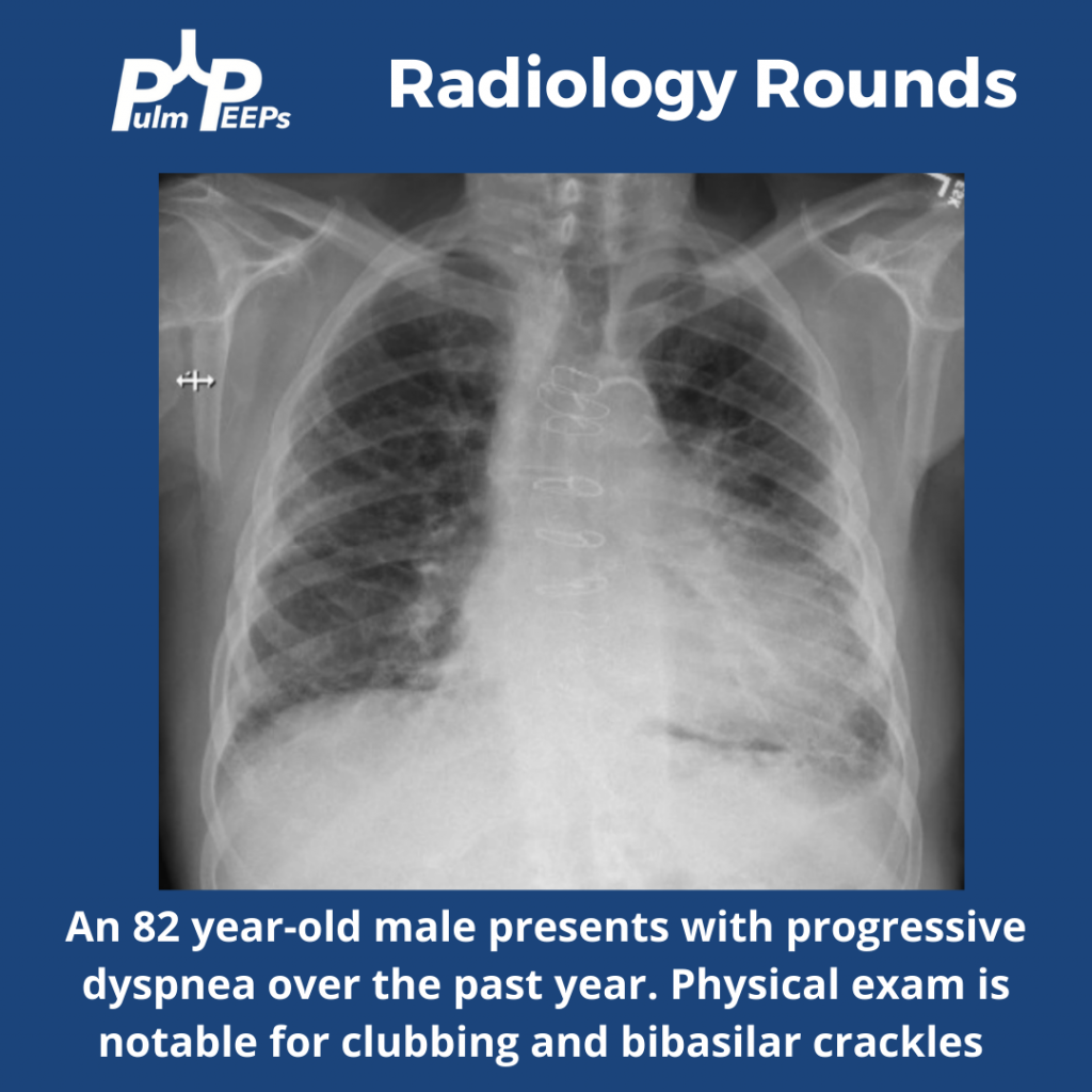
What abnormalities do you see on this CXR to help explain the patient’s presentation?
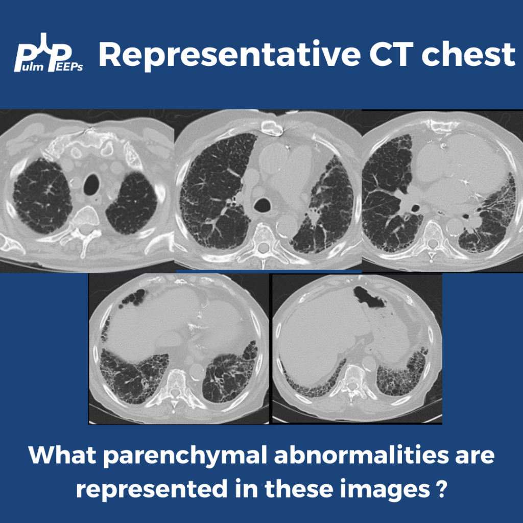
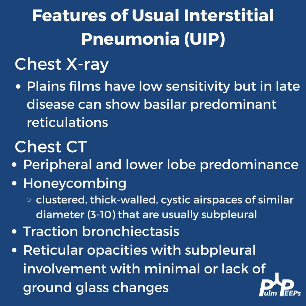
The CT scan has reticular changes consistent with interstitial lung disease and there are multiple features that help us define the pattern of the ILD. His CT notably has very few ground-glass opacities, there is traction bronchiectasis, and honeycombing with a basilar and peripheral / sub-pleural predominance.
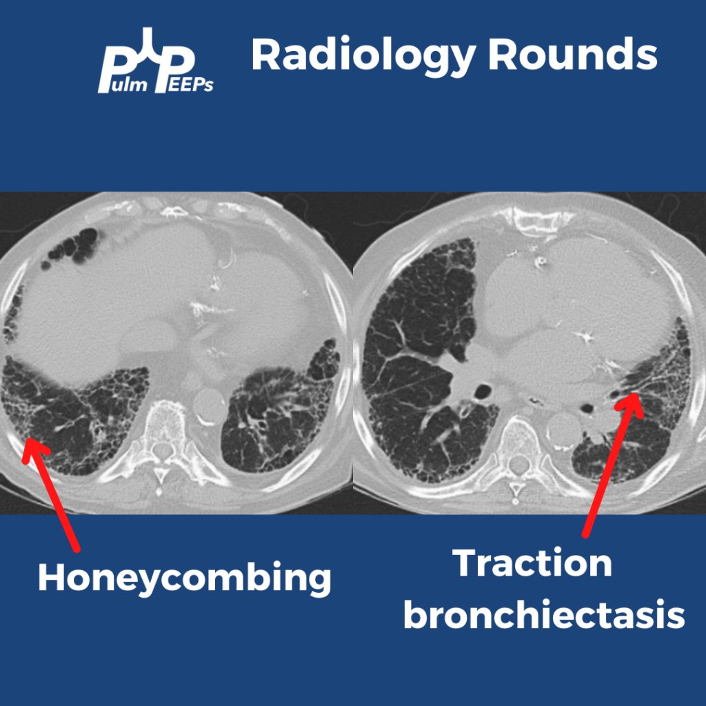
Taking all these features together, the patient’s radiographic presentation is consistent with Usual Interstitial Pneumonia (UIP)
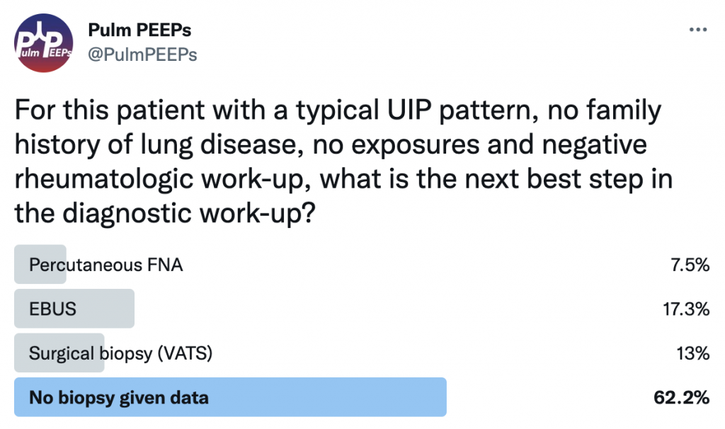
This patient had a thorough history taken and he had no prior smoking and no occupational or environmental exposures of significance. He had no family history of interstitial lung disease. A broad history was taken regarding symptoms of connective tissue disease and a broad serologic workup was sent, all of which were unremarkable. What would you want to do next diagnostically?
If you want to learn more about diagnosing interstitial lung disease, listen to our prior Top Consults episode on diagnosing ILD with experts in the field and see these prior #RadiologyRounds on Fibrotic NSIP and Sarcoidosis.