This week’s Radiology Rounds comes from Pulm PEEPs Associate Editor Tess Litchman!

Pulmonary and Critical Care content for learners and practitioners of all levels
We’re excited to share this Radiology Rounds prepared in collaboration with Nick Ghionni!
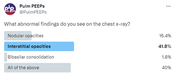
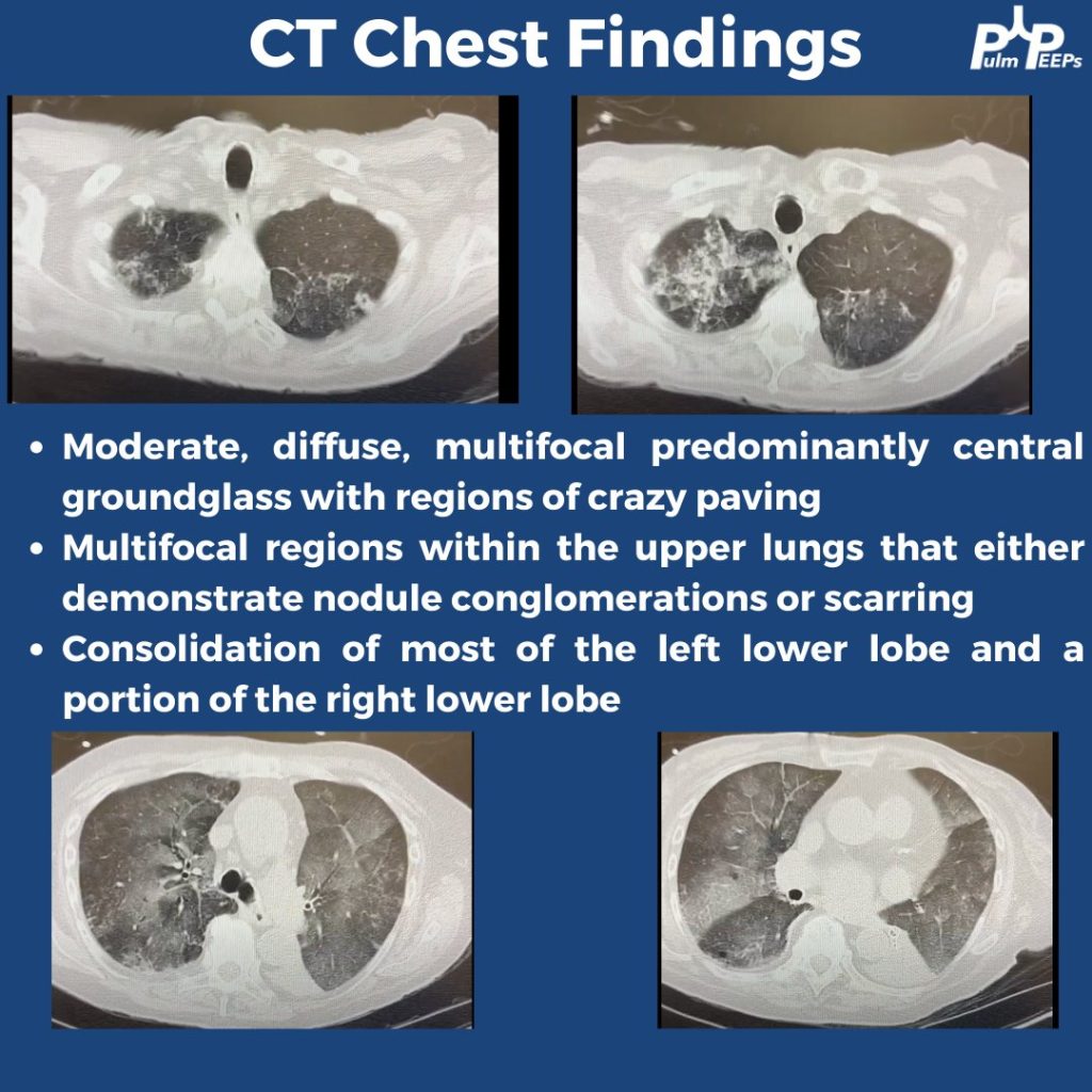
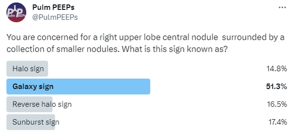
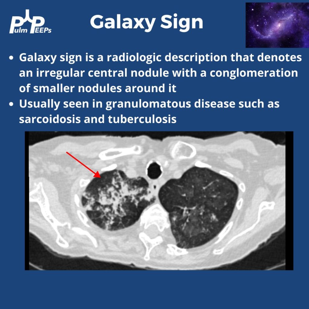
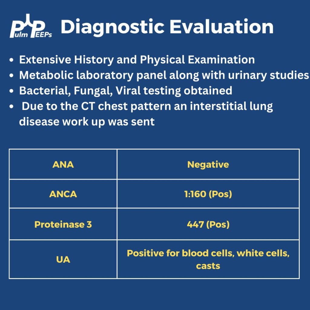
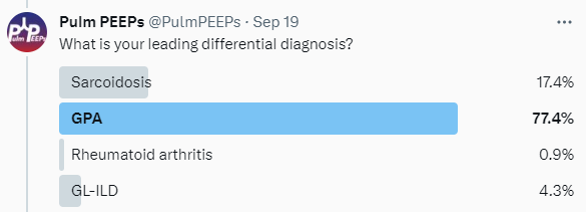
The patient was ultimately diagnosed with GPA.

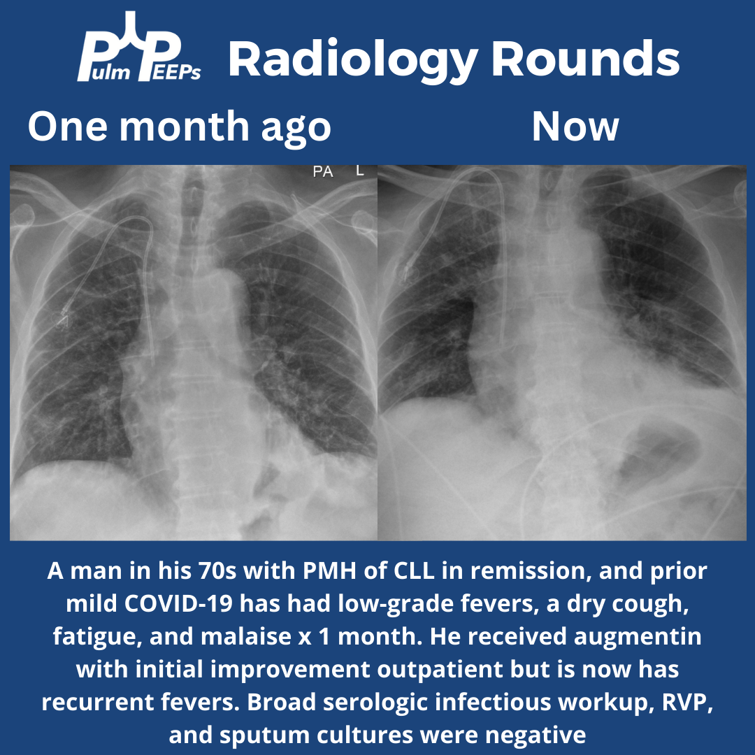
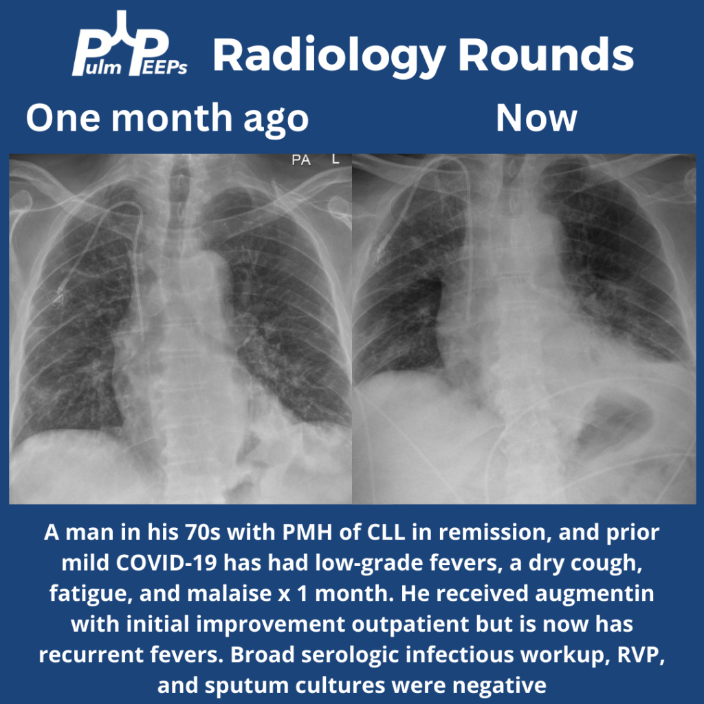
Time for #RadiologyRounds! This week we have a man in his 70s with CLL s/p treatment now in remission. He had fevers, a dry cough, and malaise that initially improved outpatient with antibiotics but have now recurred.
Check out the video of his CT scan here
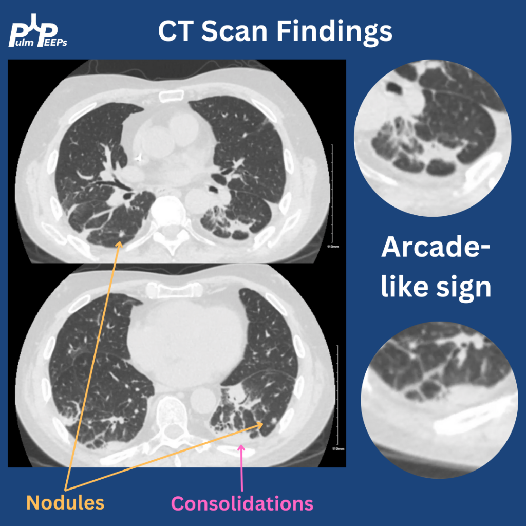
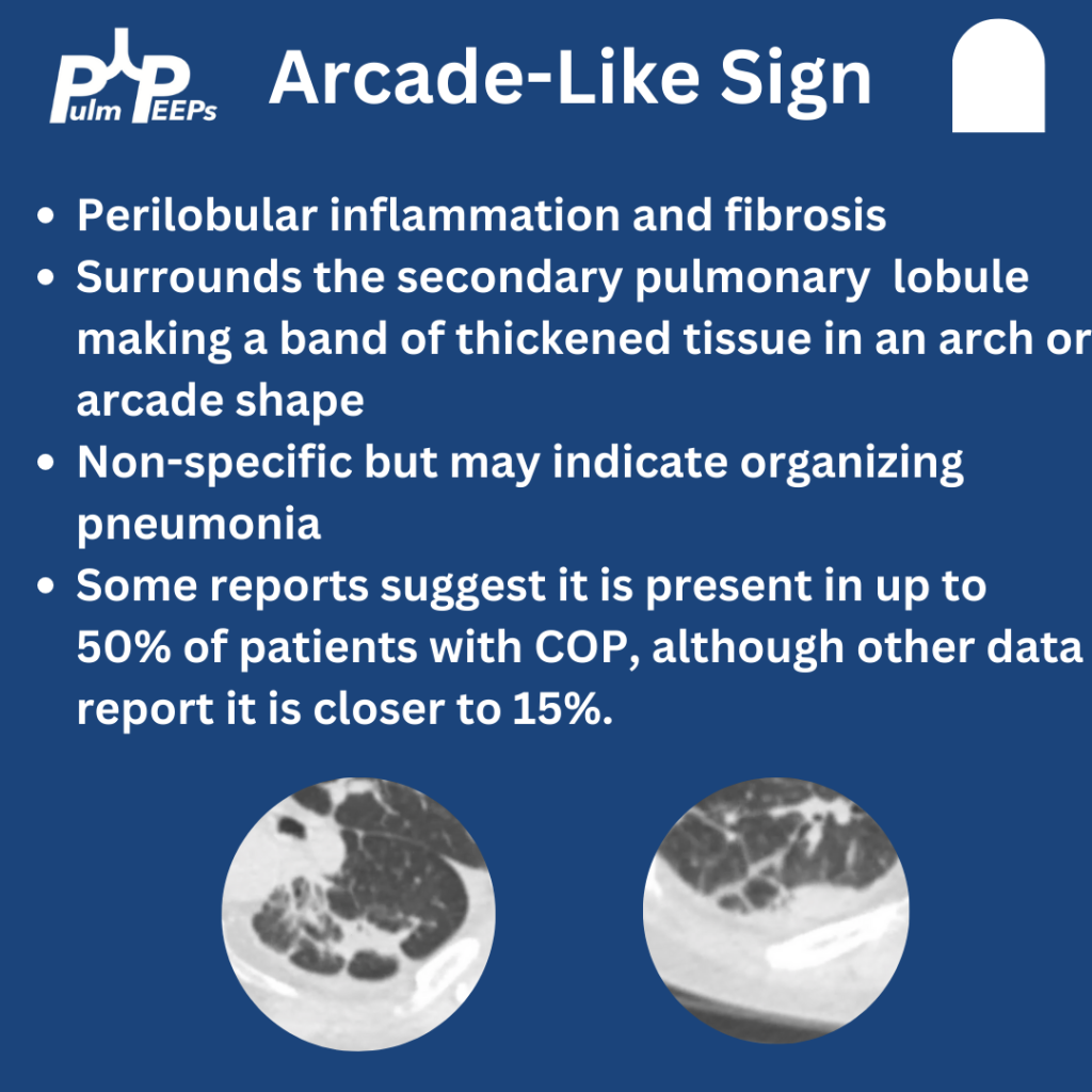
The CT had multiple findings including small nodules, consolidations, and bands of fibrosis around the secondary pulmonary nodules. This is known as Arcade-Like Sign and has classically been described with COP. While it can make you think COP, it has a low sensitivity
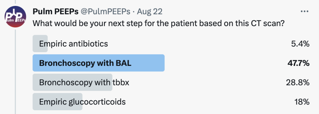
The patient underwent bronchoscopy with Tbbx given the suspicion for OP. The BAL was negative for bacterial, viral, fungal, or mycobacterial infection. The histopathology revealed organizing pneumonia. The patient was started on glucocorticoids and all symptoms improved
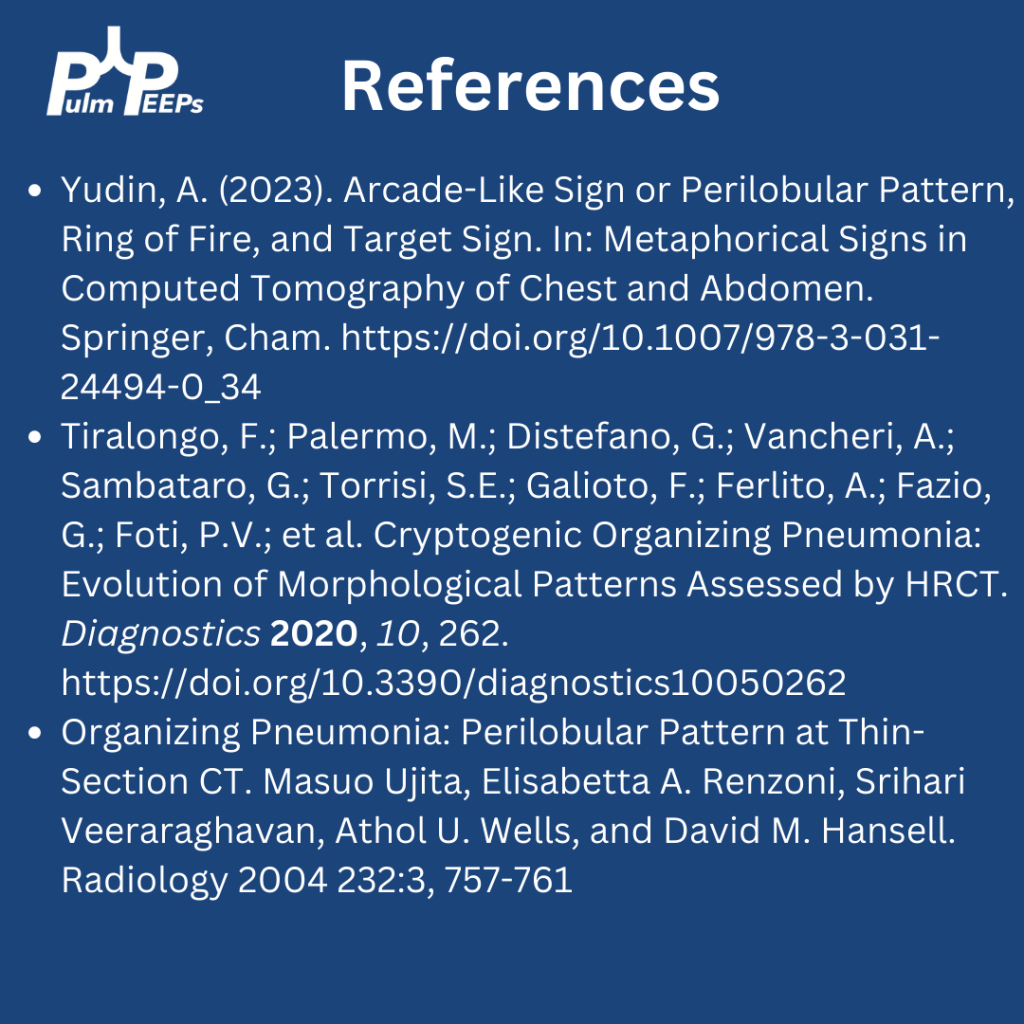
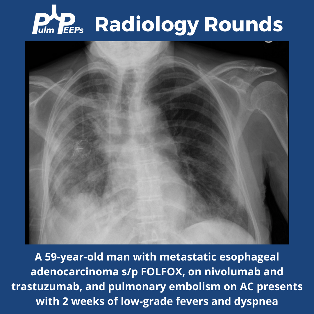
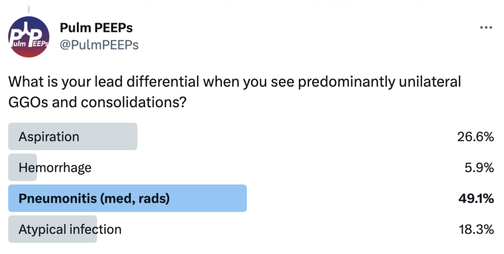
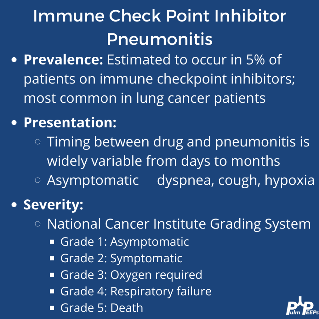
A middle-aged man with metastatic esophageal cancer presents with 2 weeks of low-grade fevers and more recently progressive dyspnea. His exam is notable for late inspiratory crackles throughout the right lung.
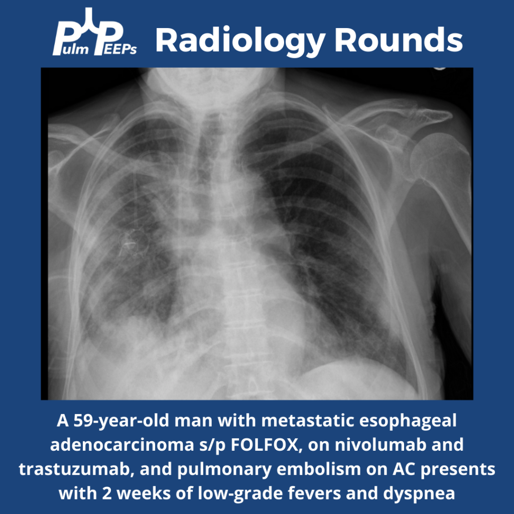
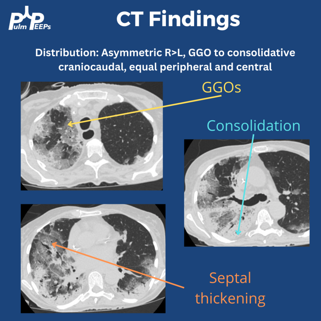
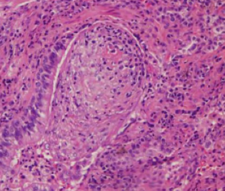
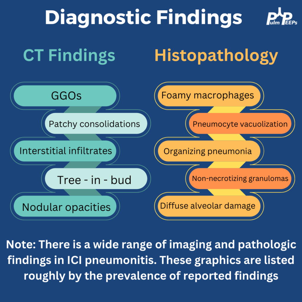
A bronchoscopy was performed and an expanded infectious panel was sent and all were negative, as were serum infectious studies (e.g. histoplasmosis ag, urine legionella etc). A trans bronchial biopsy showed plugs of granulation tissue filling the alveolar spaces
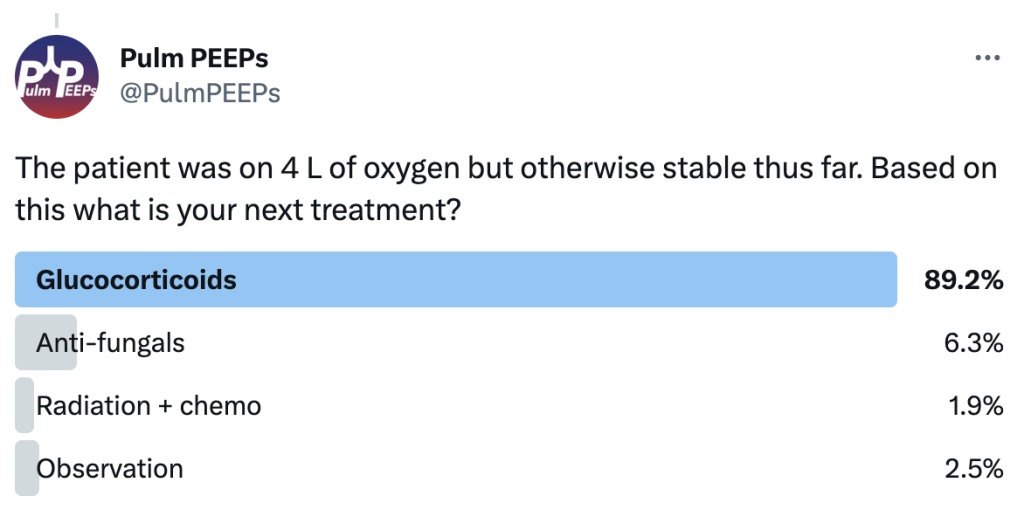
He was treated with steroids but her hypoxemia progressed and she was ultimately intubated. What is your treatment of choice for refractory ICI pneumonitis?
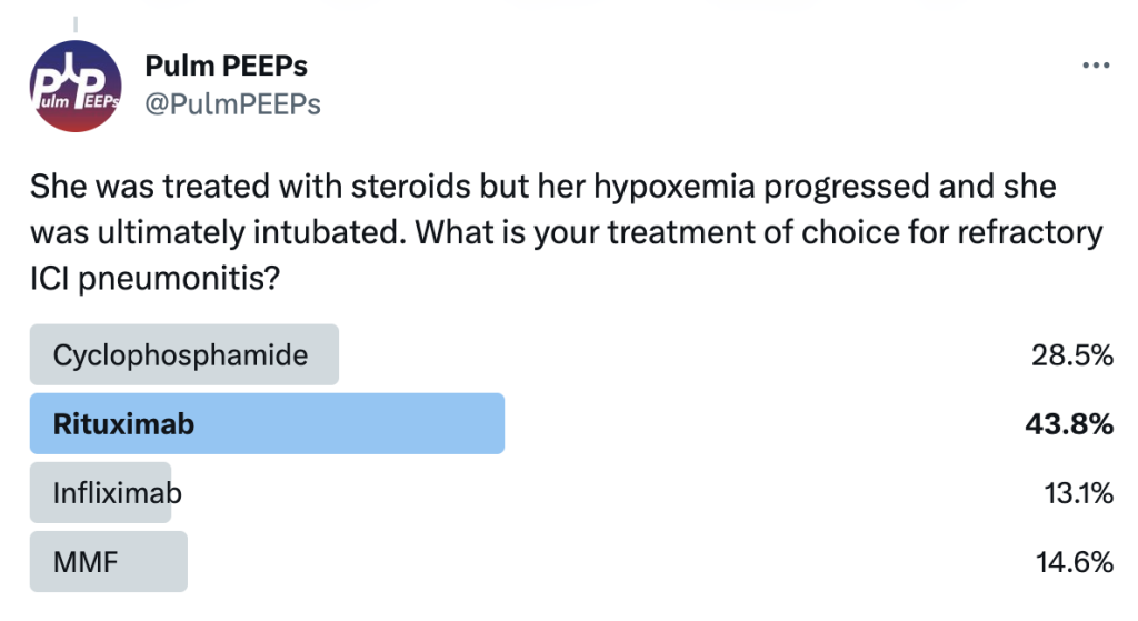
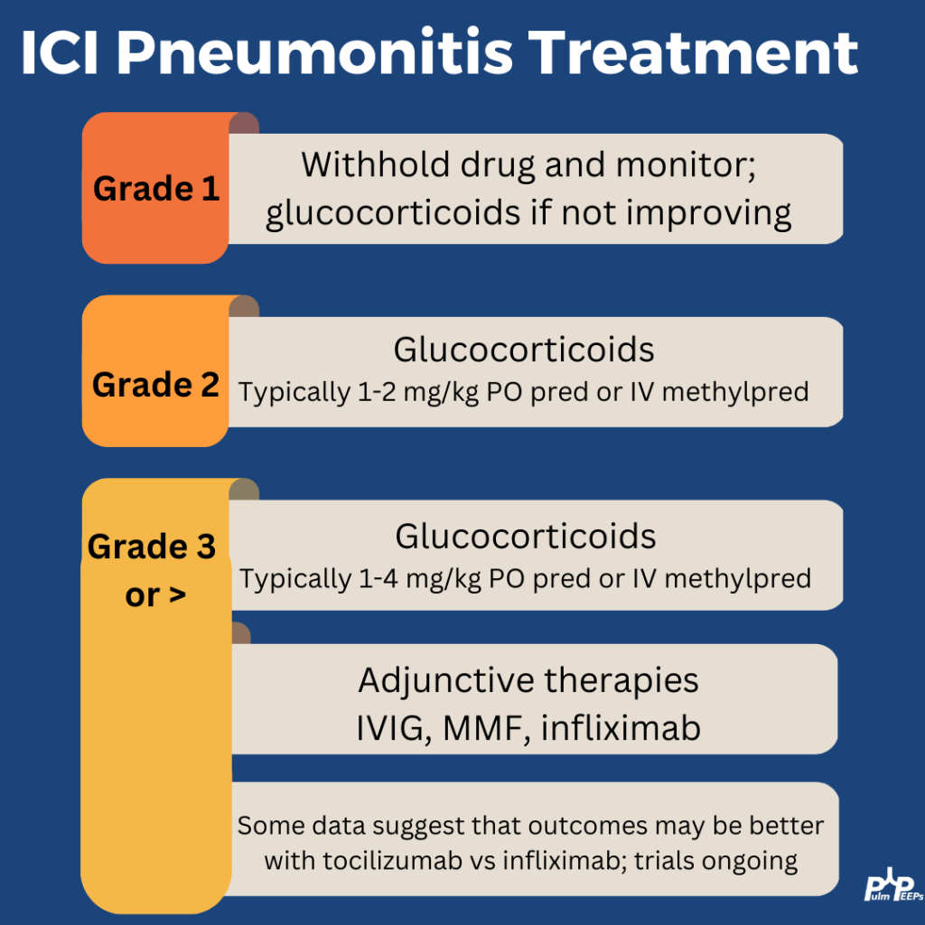
Severe ICI is rare and the treatment for steroid refractory disease is still based on small series and case reports This patient was treated with high-dose steroids (2mg/kg), IVIG, MMF, and tocilizumab. With that, he improved after 10 days intubated and was weaned to room air.
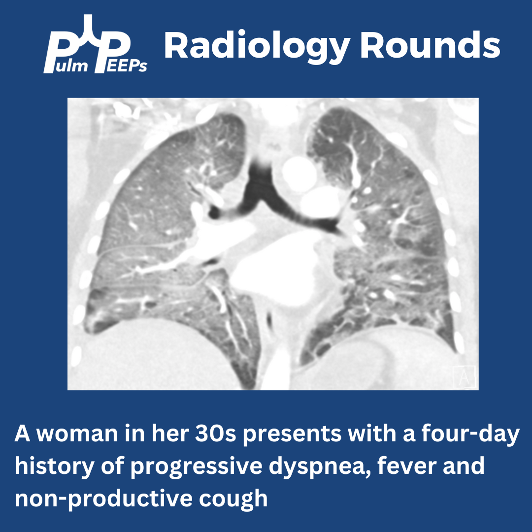
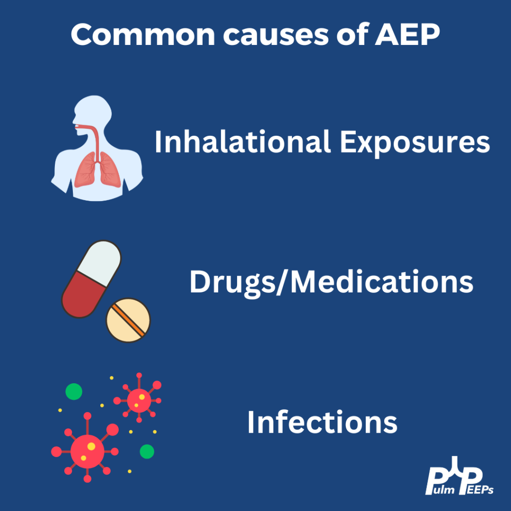
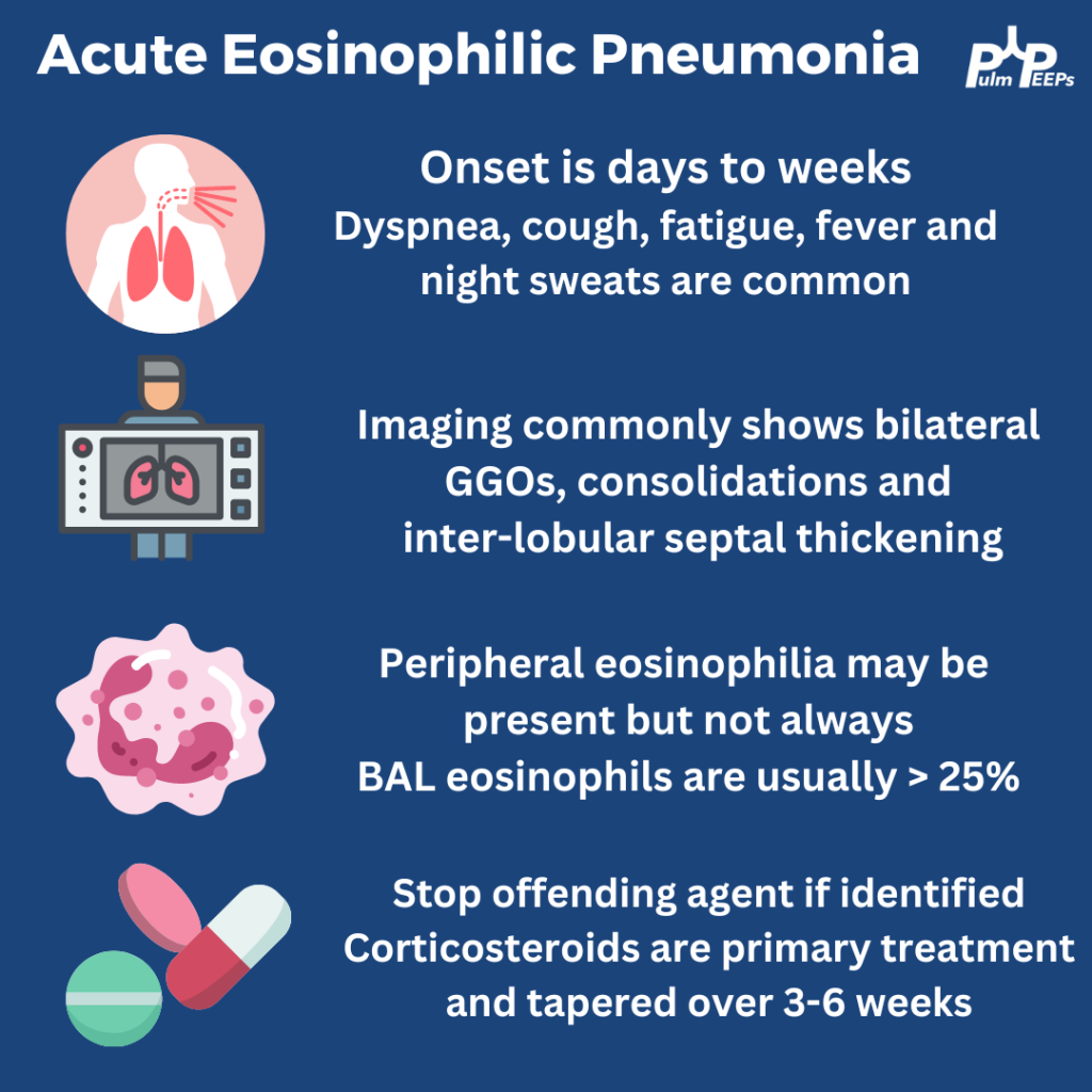
A woman in her 30s with no past medical history presents with acute dyspnea, fever and a non-productive cough. She has no sick contacts or recent travel. She is not on home medications.. She smokes 1/2 cigarettes per day and and occasionally vapes.
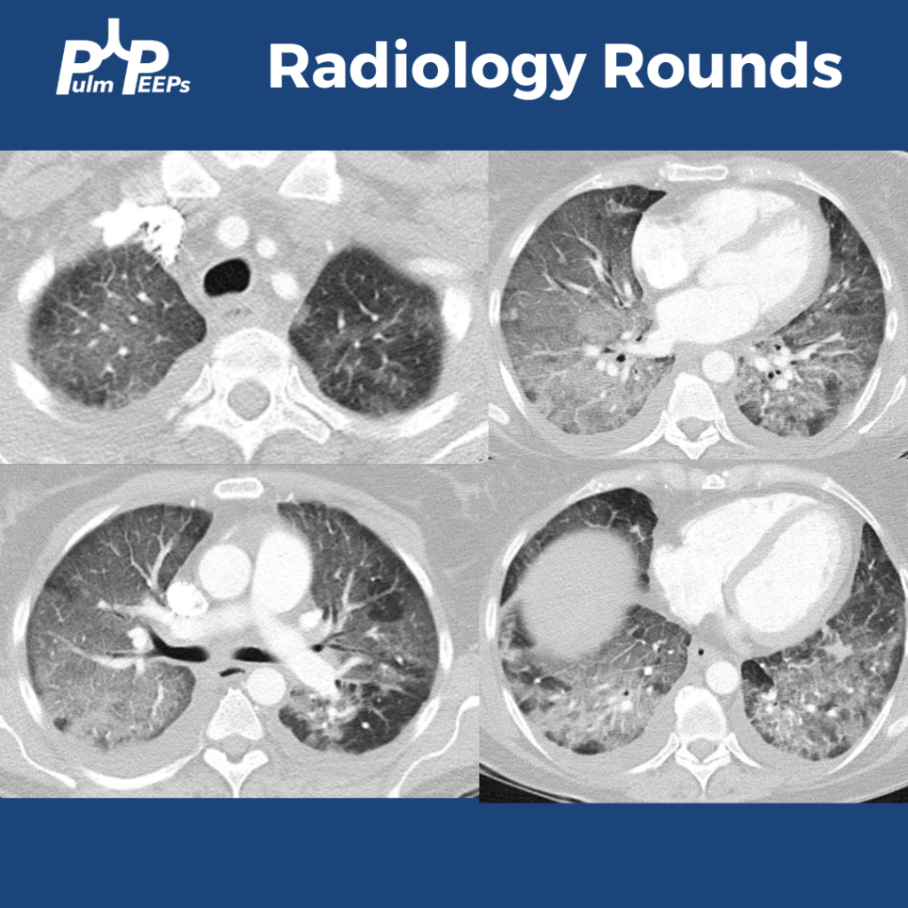
She is found to have diffuse bilateral patchy ground-glass opacities with some interlobular septal thickening. She has escalating oxygen requirements and was initially started on broad-spectrum empiric antimicrobial therapies.
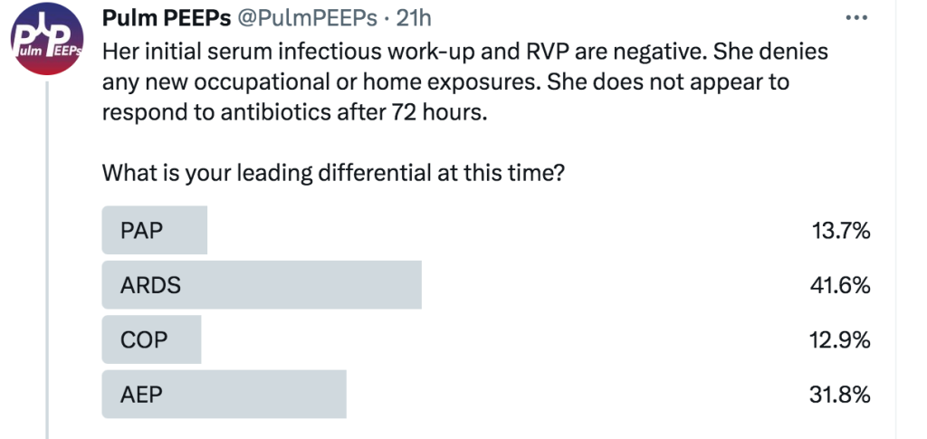
Her initial serum infectious work-up and RVP are negative. She denies any new occupational or home exposures. She does not appear to respond to antibiotics after 72 hours.
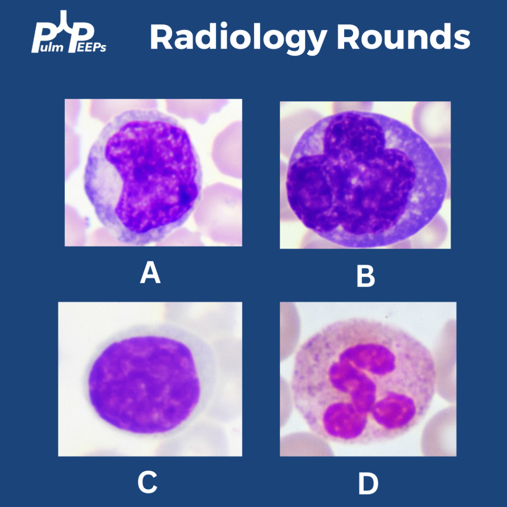
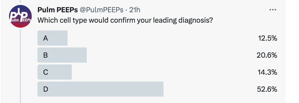
You start empiric steroids (prednisone 60 mg daily) given concern for acute eosinophilic pneumonia. Within 48 hours she is weaned off HFNC and was discharged home on D7 on room air with close pulmonary follow-up.
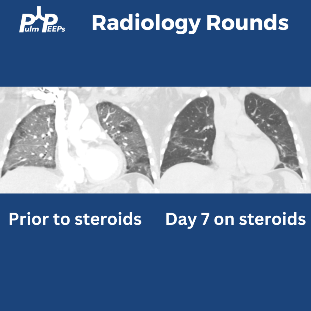
She was amenable to tobacco cessation therapies and was followed closely outpatient with tapering of her steroids over 4 weeks.
This week on Pulm PEEPs we are thrilled to share a collaboration with the American Thoracic Society Assembly on Respiratory Cell & Molecular Biology. We are joined by two expert members of the ATS RCMB Assembly who have done basic and translational research in respiratory biology and lung disease. We explore the topic of Short Telomeres and their role in lung disease. With the annual ATS Conference just around the corner, this is a great intro episode for everyone from aspiring researchers and clinical pulmonologists.
Meet The Guests
Mark Snyder is an Assistant Professor of Medicine at the University of Pittsburgh Medical Center, and a member of the Graduate Program in Microbiology and Immunology there. He does research on the role of the adaptive immune system’s role in chronic rejection after lung transplantation and has received both a Parker B Francis Foundation award and an NIH K23 grant for this work.
Jonathan Alder is an Assistant Professor of Medicine at the University of Pittsburgh. His research focuses on telomeres and their role in human health and disease. He is an accomplished researcher, was a Parker B Francis fellow, and now has an NIH RO1 studying Telomere-mediated Lung disease.
Further Reading and References
Podcast: Play in new window | Download
Subscribe: Apple Podcasts | Spotify | Amazon Music | Android | iHeartRadio | Podcast Index | RSS
Today’s #RadiologyRounds is a reminder that pulmonary care extends beyond the lungs! Although we’ll have a CT chest too
A 70-year-old woman with a diagnosis of seronegative RA presents with progressive dyspnea. PFTs show a moderate restrictive defect and moderately reduced DLCO. Here is the CT scan:
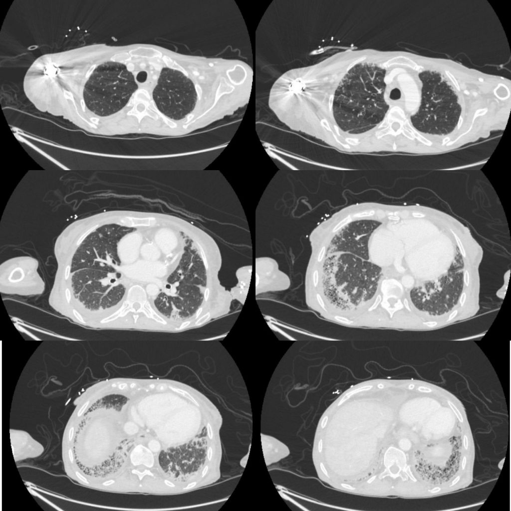
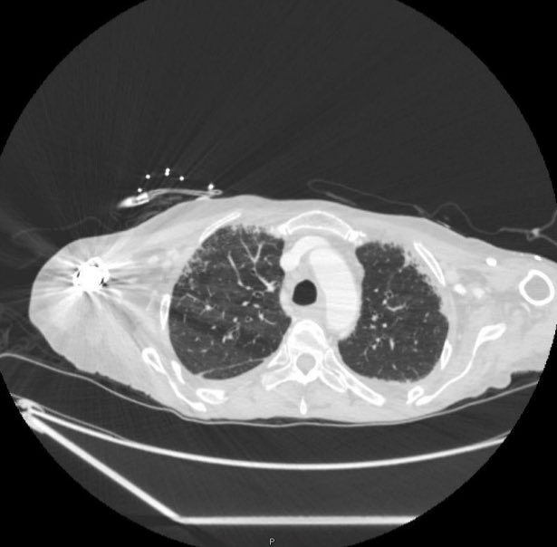
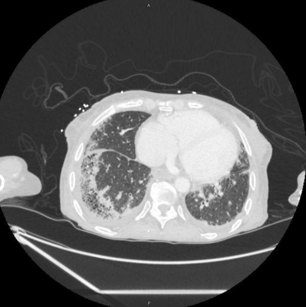
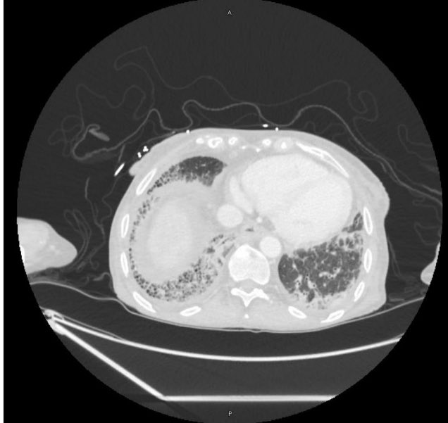
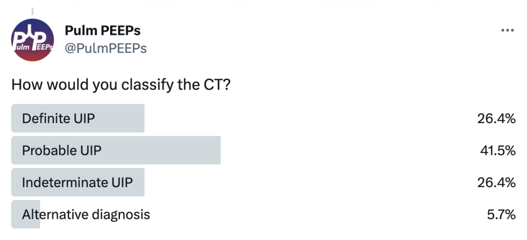
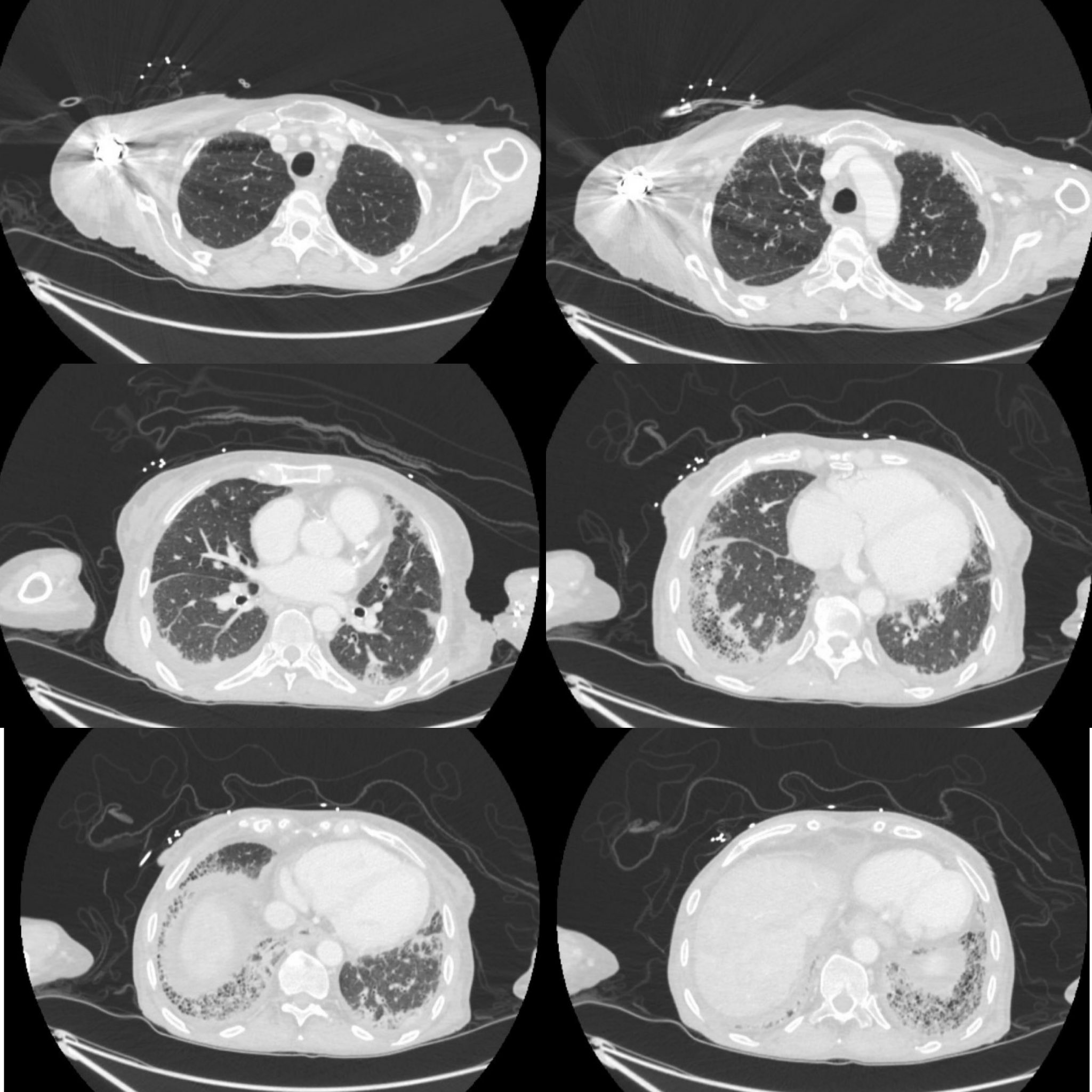
The CT was interpreted as indeterminate for UIP / fibrosing NSIP. The basilar and subpleural fibrosis and honeycombing are consistent with UIP, but with reticular nodular infiltrates and pleural effusions that are less consistent. In reviewing her history: RF, CCP, and anti-SCL70 were all negative. On hand exam, she had MCP enlargement, some PIP synovitis, and slight R hand ulnar deviation. She did not have clear telangiectasias. She had a history of GERD. She denied Raynauds. She had significant skin thickening over her legs and limited ankle mobility. X-rays were obtained:
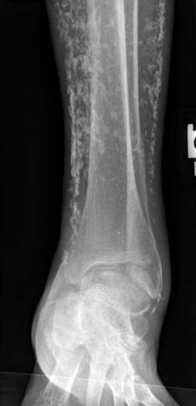
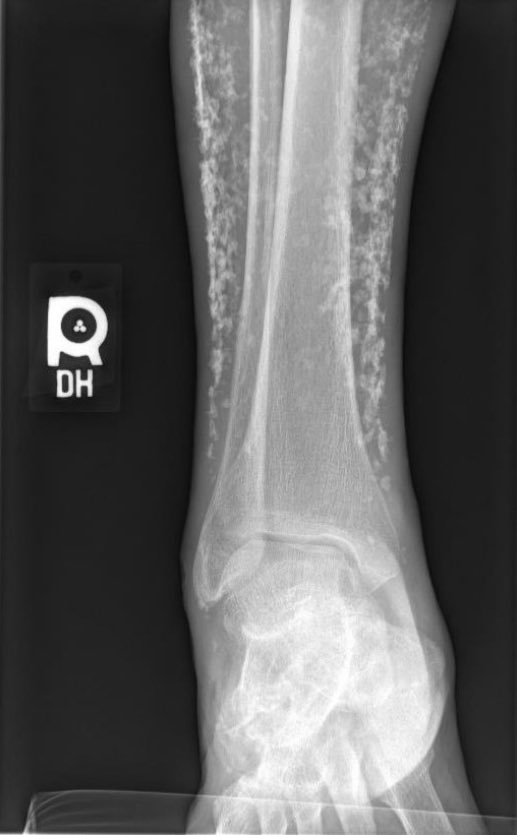
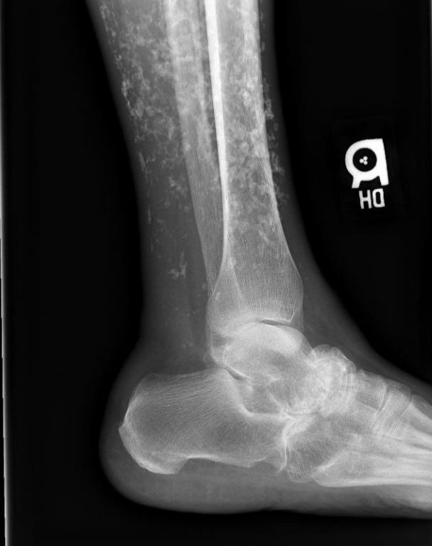
This showed severe dystrophic calcifications and no bony erosions. This degree of extensive dystrophic calcifications is consistent with scleroderma and the patient was felt to have limited scleroderma, and possibly an overlap syndrome
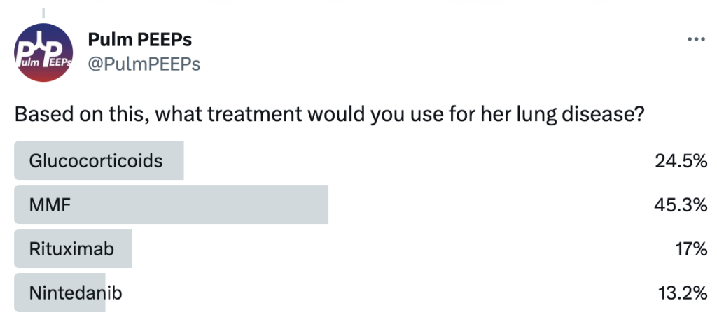
The patient was started on MMF as the initial agent based on the Scleroderma Lung Study II (SLSII): https://pubmed.ncbi.nlm.nih.gov/27469583/ Glucocorticoids were avoided given the risk of scleroderma renal crisis with steroids. An anti-fibrotic was also considered but not started as 1st line.
Join us as we head to the Texas Medical Center and are joined by Dr. Bitar and Dr. Gotur from Houston Methodist Pulmonary and Critical Care Medicine.
Meet our Guests
Dr. Mohamad Bitar is a Pulmonary and Critical Care Fellow at the Houston Methodist Pulmonary and Critical Care Program in Houston, Texas. Dr. Bitar completed his medical school at Misr University for Science and Technology and his Internal Medicine residency at Good Samaritan Hospital.
Dr. Deepa Gotur is an Associate Professor of Clinical Medicine at Weill Cornell Medical College and an Adjunct Associate professor at Texas A&M. She is also the Pulmonary and Critical Care Program Director at Houston Methodist. She is a sepsis and ARDS researcher, as well as a dedicated educator.
Case Presentation
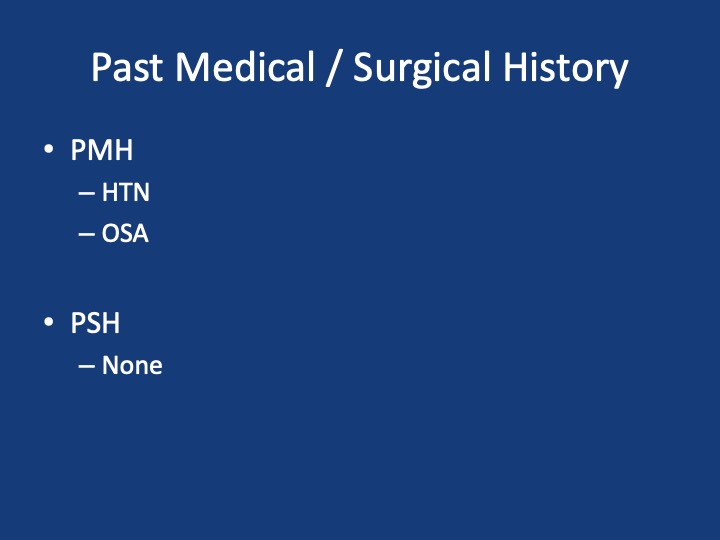
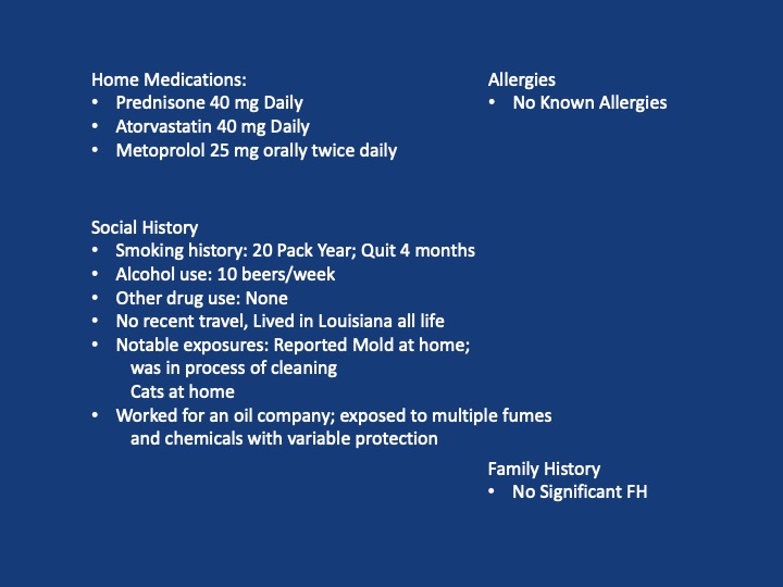
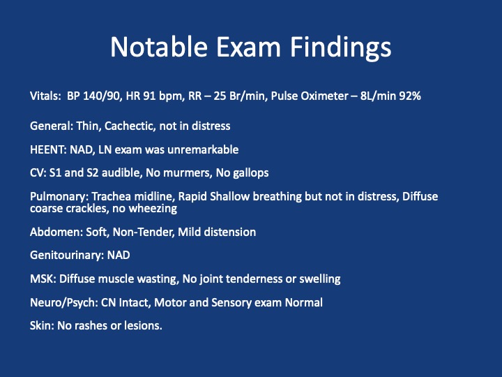
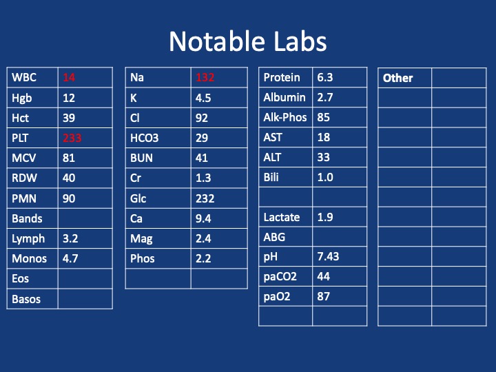
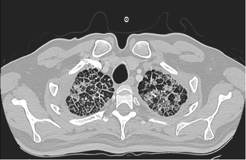
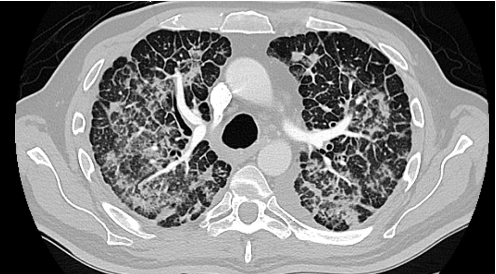
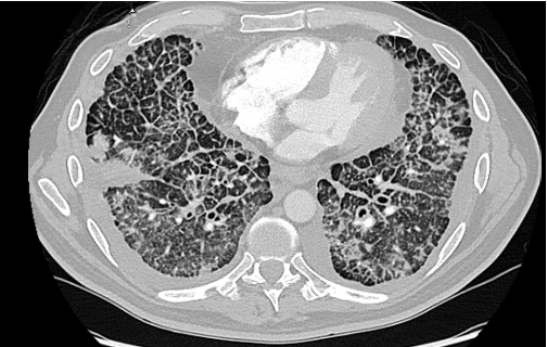
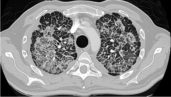
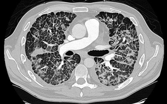
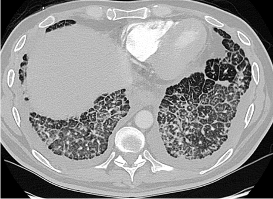
A 40s-year-old male presents with 3 months of progressive shortness of breath. He was diagnosed with COVID and given treatment with steroids, and antibiotics with no response.
He presented to the hospital 3 months later after not being able to carry out daily activities and was found to have low oxygen saturation
Admitted, initial CT Chest – showed “Non-specific patchy ground-glass bilateral pulmonary infiltrates with Coarsened interstitial markings that could be related to COVID-19/atypical infection”
References and further reading
1. Silva, C. Isabela S. MD, PhD; Müller, Nestor L. MD, PhD. Idiopathic Interstitial Pneumonias. Journal of Thoracic Imaging 24(4):p 260-273, November 2009.
2.Gruden JF, Naidich DP, Machnicki SC, Cohen SL, Girvin F, Raoof S. An Algorithmic Approach to the Interpretation of Diffuse Lung Disease on Chest CT Imaging: A Theory of Almost Everything. Chest. 2020 Mar;157(3):612-635.
3. Ryu JH, Daniels CE, Hartman TE, Yi ES. Diagnosis of interstitial lung diseases. Mayo Clin Proc. 2007 Aug;82(8):976-86.
Podcast: Play in new window | Download
Subscribe: Apple Podcasts | Spotify | Amazon Music | Android | iHeartRadio | Podcast Index | RSS
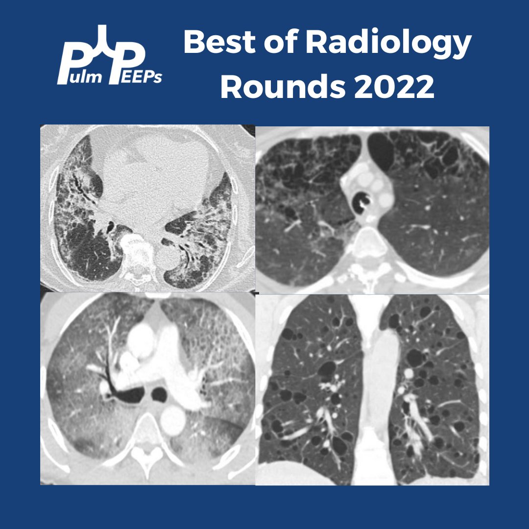
For our last #RadiologyRounds of 2022, we present four high-yield cases of the past year for you to review
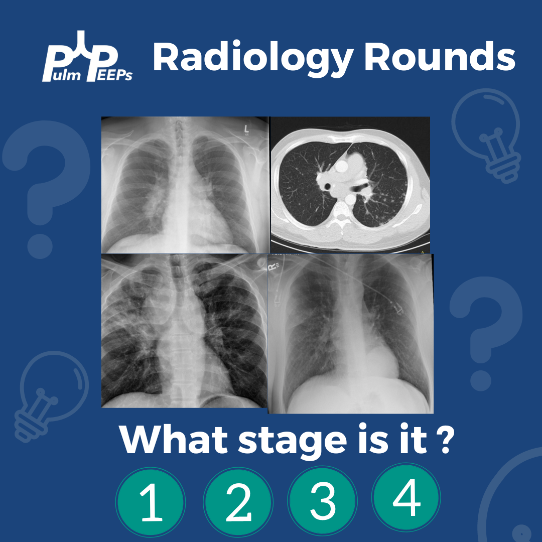
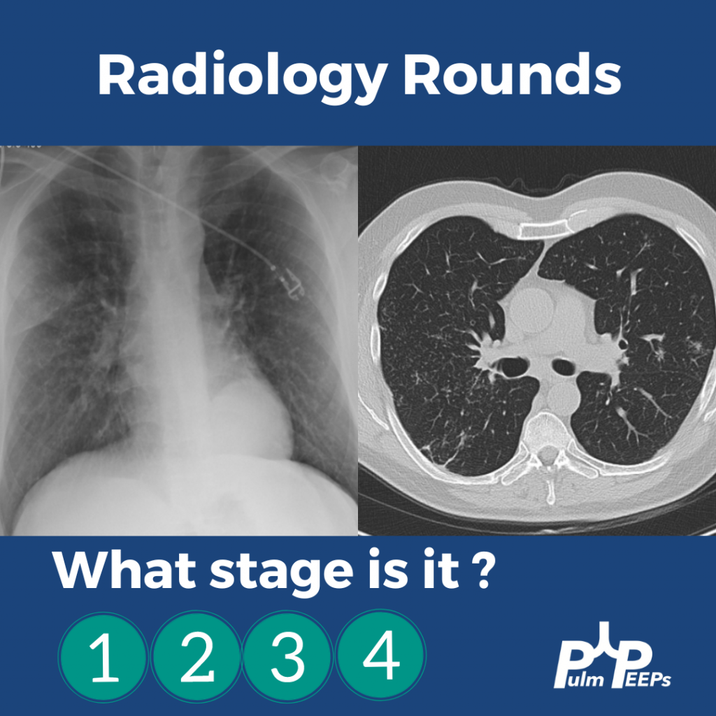
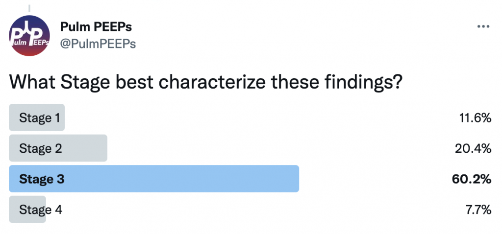
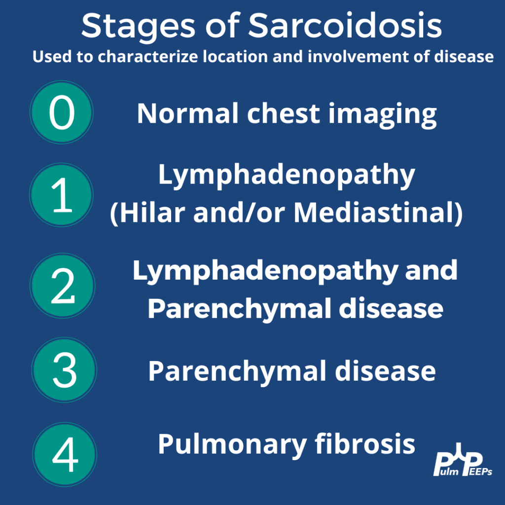
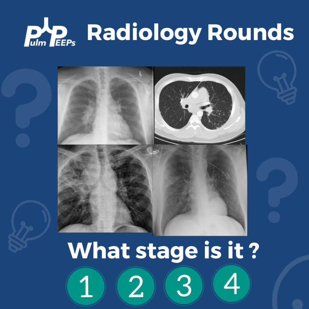
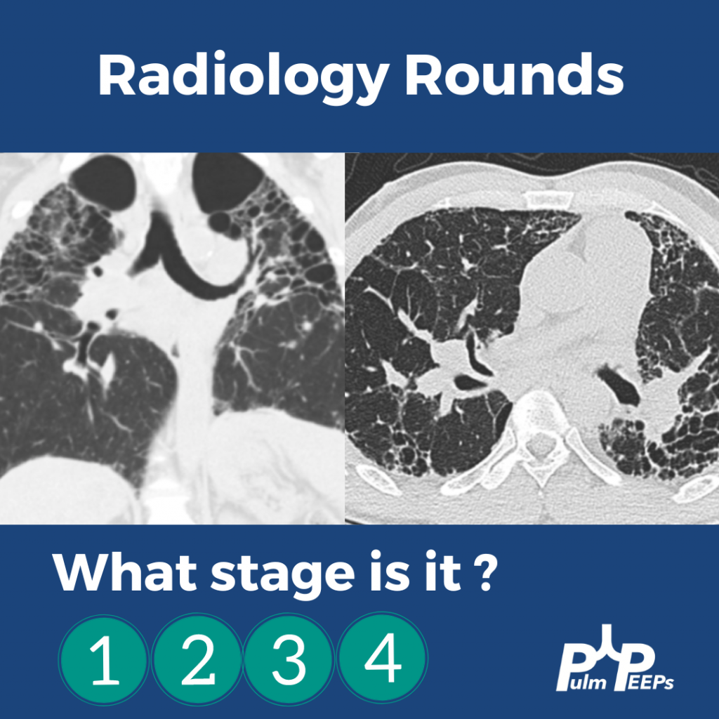
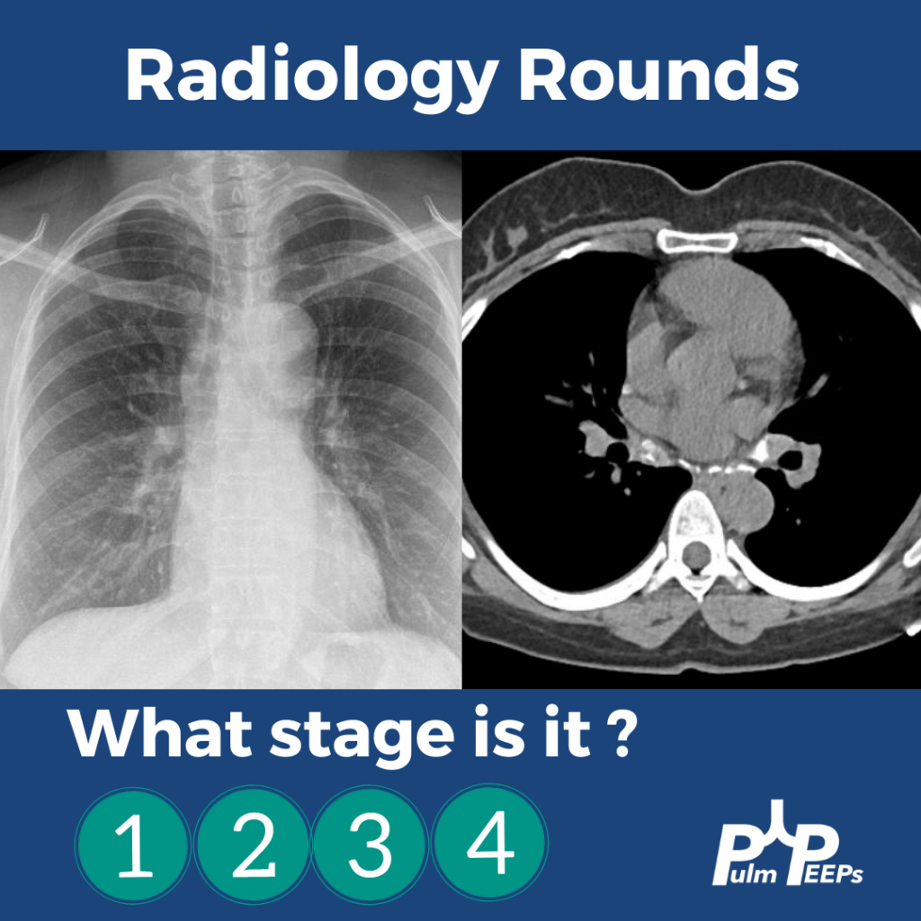
It is Tuesday and we have another Radiology Rounds we can’t wait to share with you. Follow along and see if you select the right answer as we go through different presentations of sarcoidosis and pick your answer! What stage is it?!
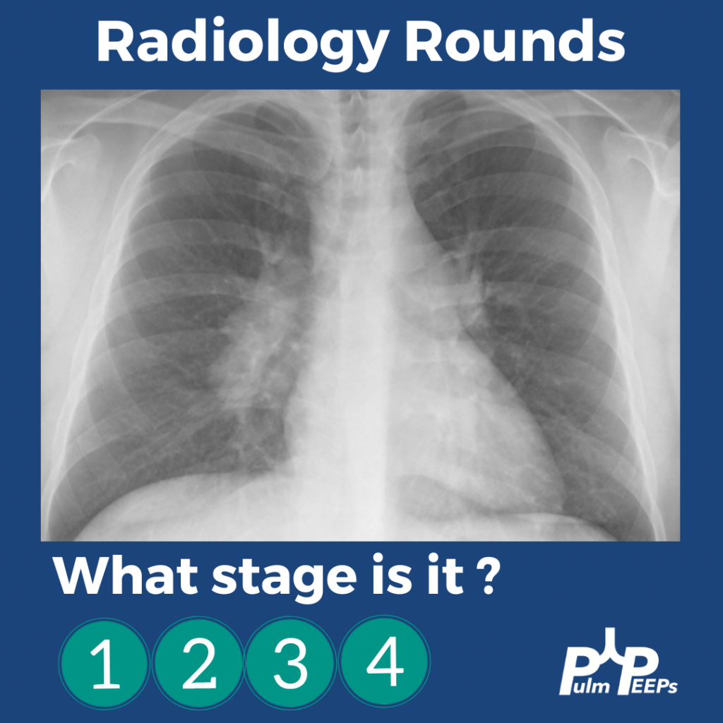
A middle-aged man presents to you after he was found to have hilar adenopathy on a routine chest x-ray.
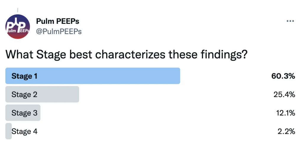
A middle-age man presents with dyspnea on exertion, night sweats and weight loss. You see evidence of bilateral apical disease, and fibrosis with evidence of honeycombing on chest CT.
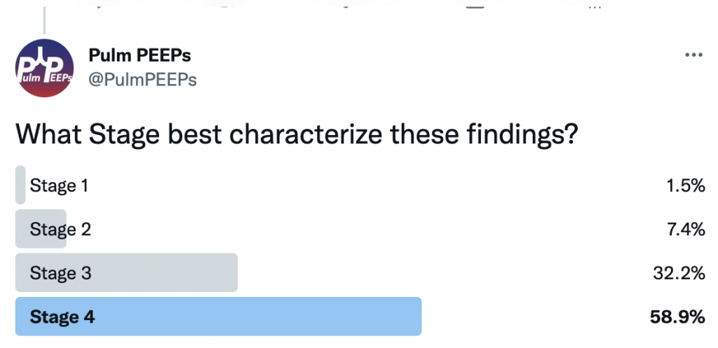
A young woman presents with dyspnea on exertion and was found to have hilar adenopathy with parenchymal disease.
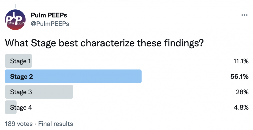
An elderly man presents with dyspnea on exertion and was found to have nodular parenchymal disease without extensive lymphadenopathy.