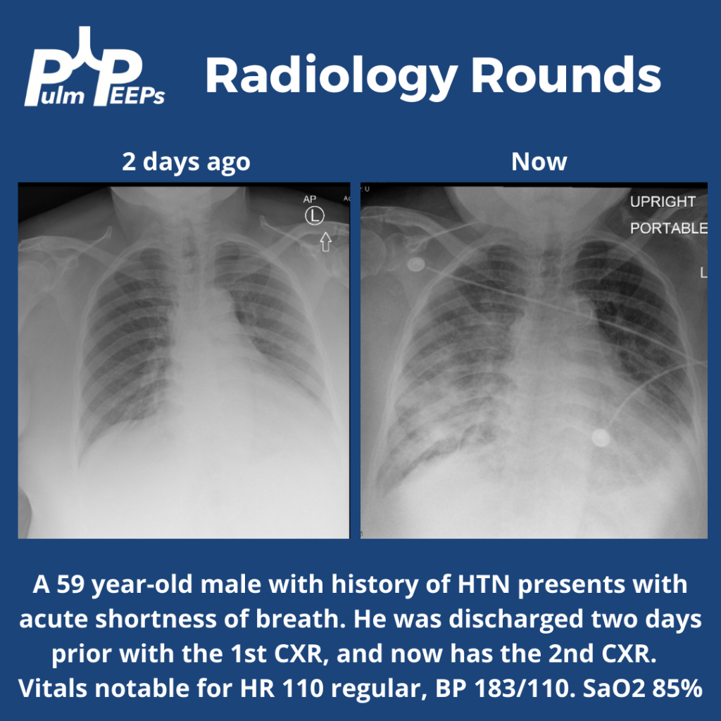
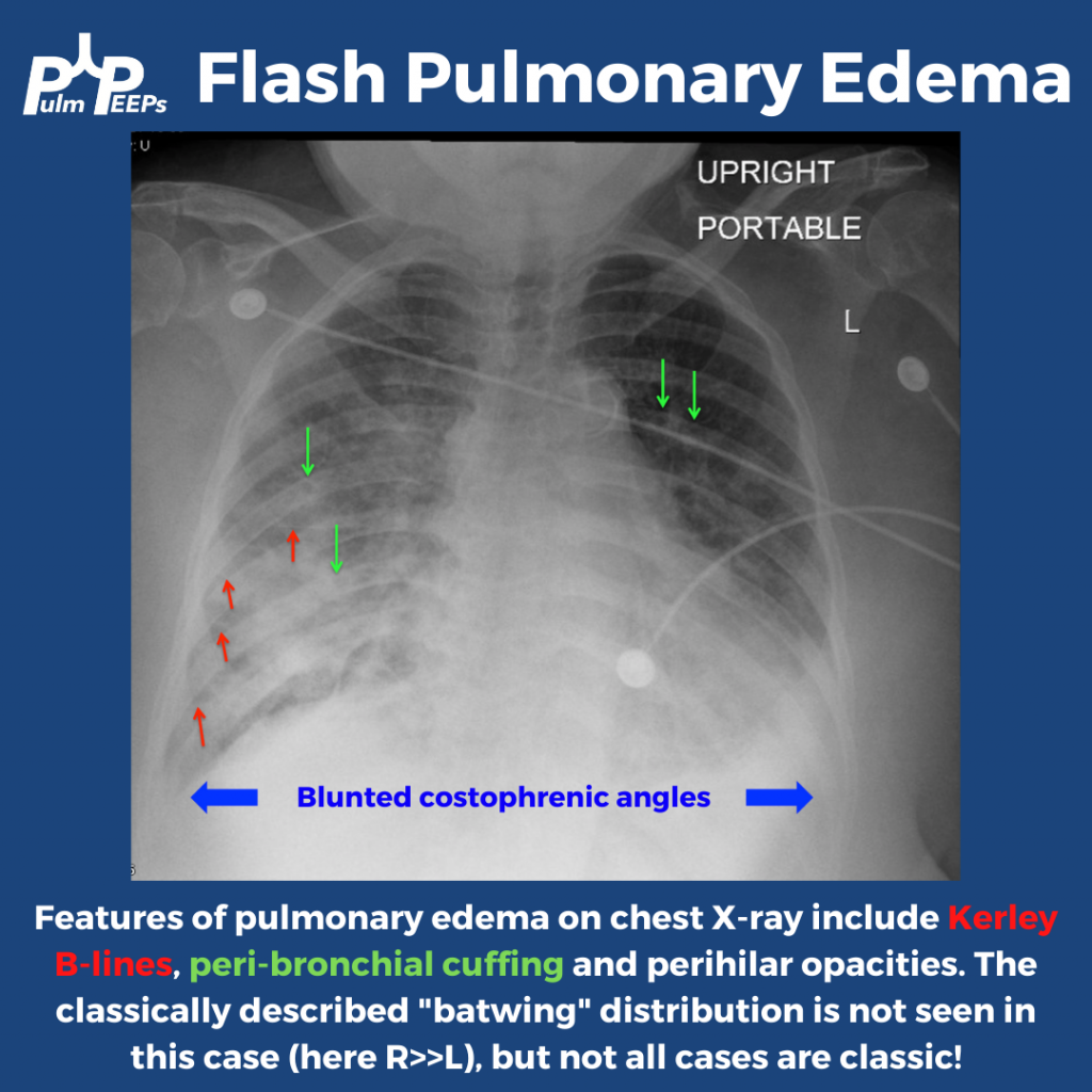
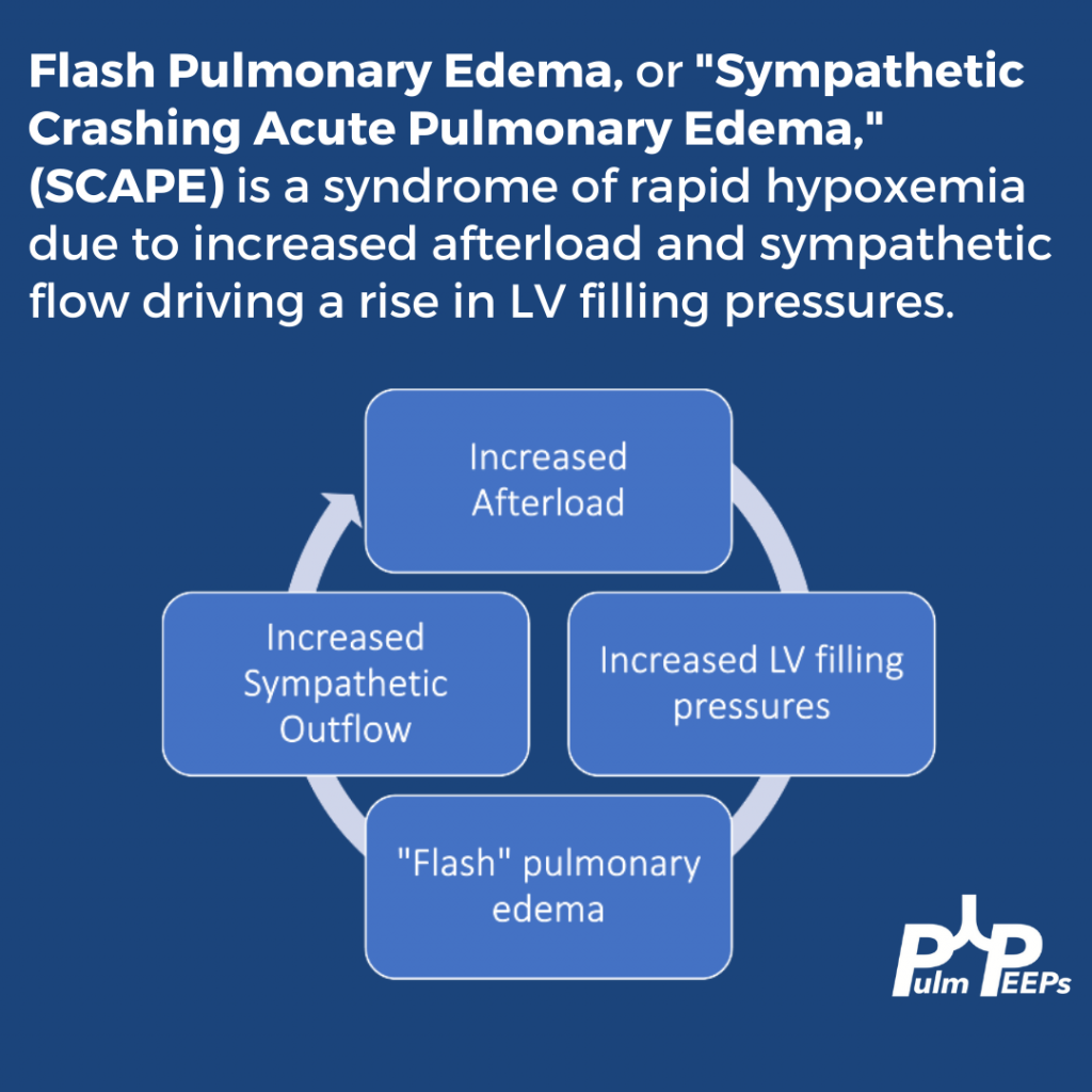
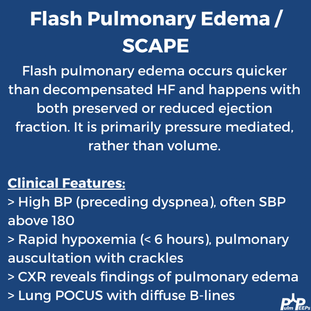
28. Fellows’ Case Files: Harvard – MGH & BIDMC

Welcome back to our Pulm PEEPs Fellows’ Case Files series! We are joined this week by a fellow and the program director from the Harvard combined PCCM fellowship at Massachusettes General Hospital and Beth Israel Deaconess Medical Center. Listen in for a great learning case and let us know on Twitter, if you have a great case to share!

Meet our Guests

Brian Rosenberg is a third year fellow at the Harvard MGH/BI program. He completed his undergraduate degree at Harvard, received his MD  from Yale where he also got a PhD in cell biology, and then did his internal medicine residency at Columbia University Medical Center in NYC.

Asha is an Assistant Professor Medicine at Beth Israel Deaconess Medical Center and Harvard Medical School, and is the Program Director of the Harvard MGH/BI combined fellowship. She is also the Director of the Pulmonary Consult Service at BIDMC, was a Rabkin Fellow in Medical Education and has received multiple leadership and teaching awards

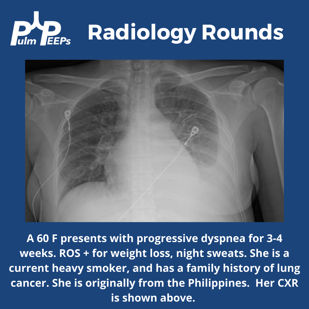
Radiology Rounds – 10/11/22

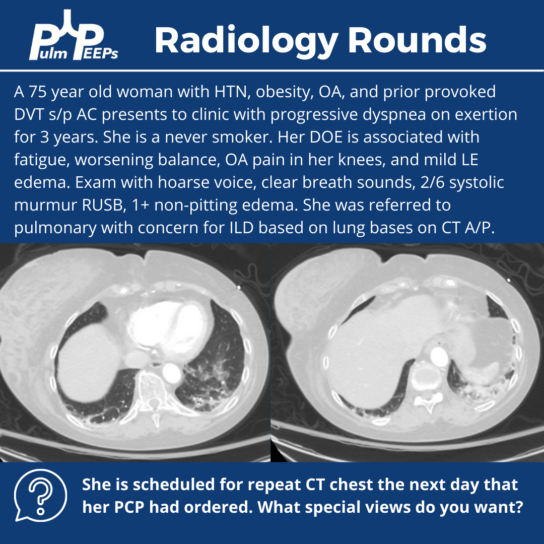
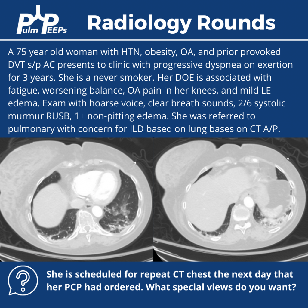
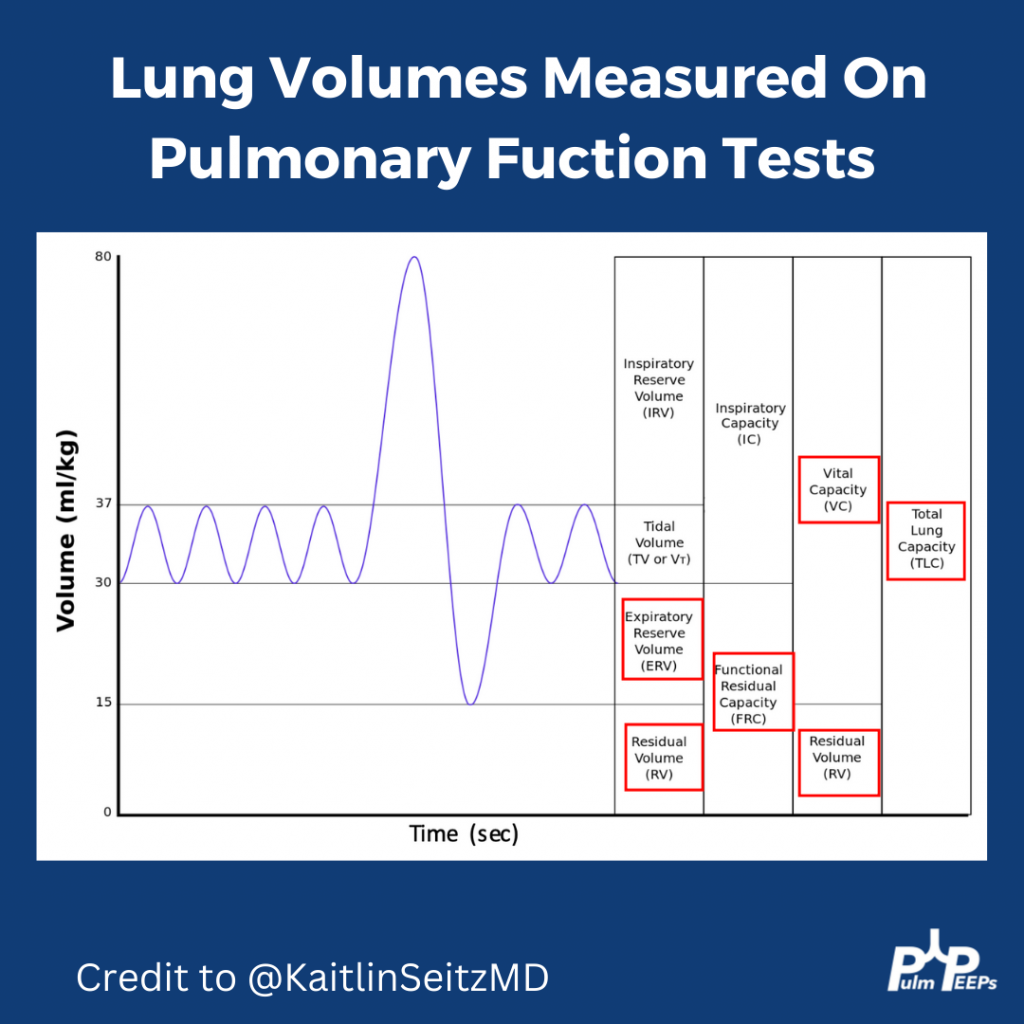
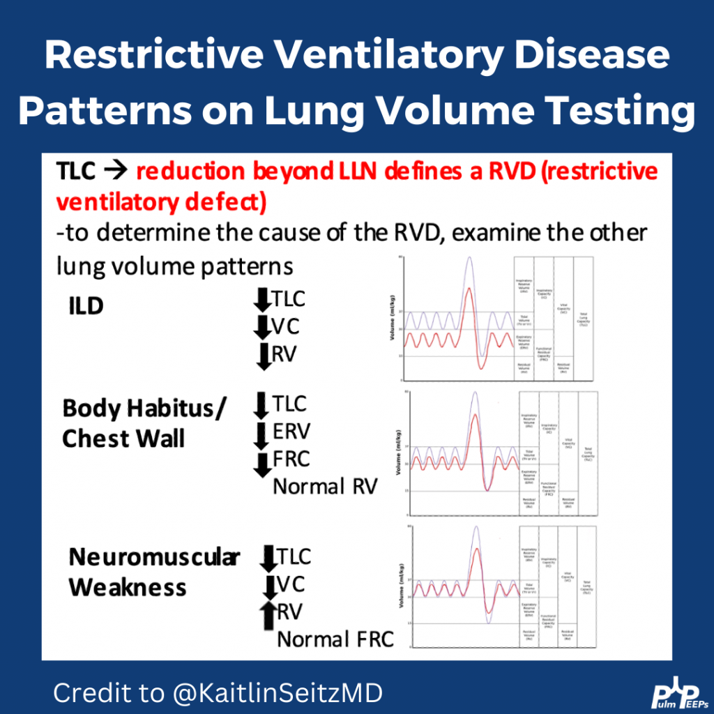
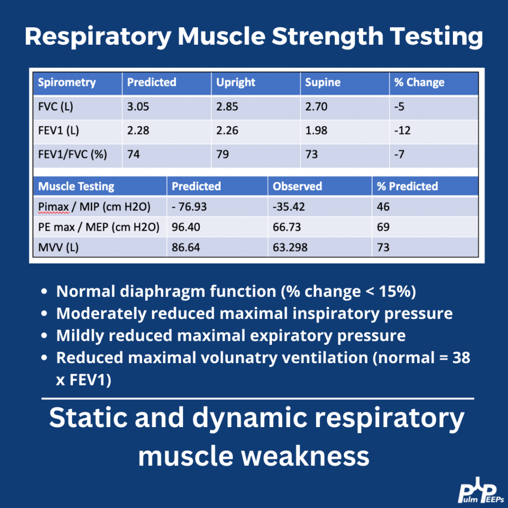
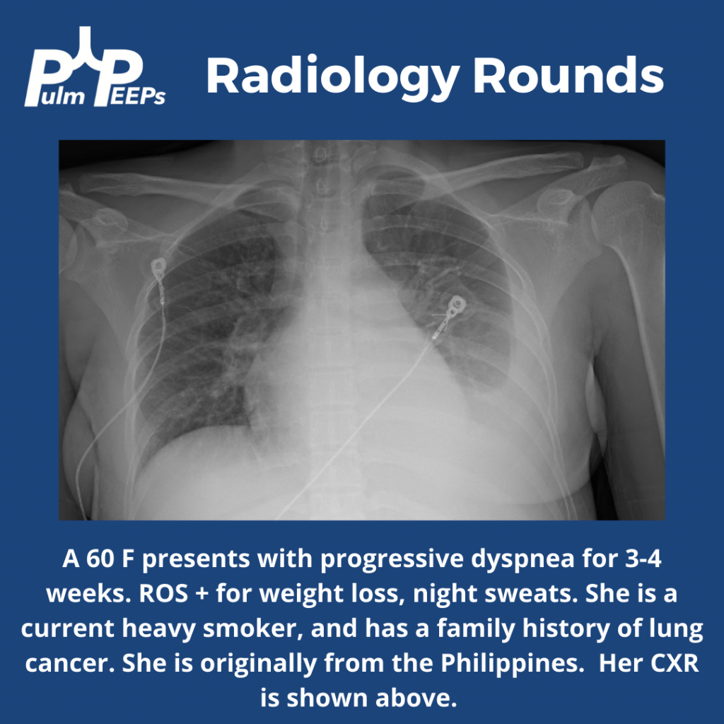
For #RadiologyRounds this week we have a mystery case from the pulmonary clinic complete with imaging and exploration of PFTs. Follow along for some great clinical pearls and teaching points about lung function tests. Graphics made with the help of outstanding educator Kaitlin Seitz.

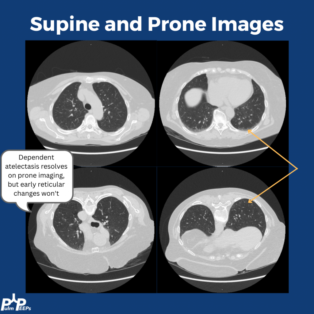
What imaging views would you get next?

A) Supine and prone

B) Inspiratory and expiratory

C) High resolution

D) With contrast

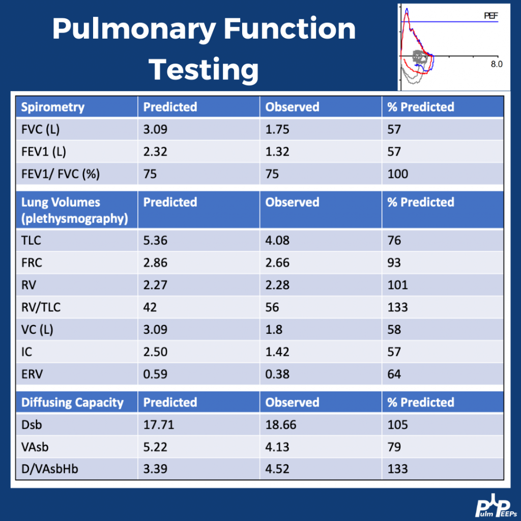
What test would you get next?

A) Bronchoscopy

B) ECHO

C) PFTs

D) Lung US

What do these PFTs show?

A) Restriction concerning for ILD

B) Restriction concerning for obesity

C) Restriction concerning for weakness

D) Mixed obstruction and restriction

The patient was referred to neurology and ultimately diagnosed with severe mixed sensory and motor chronic axonal polyneuropathy.

25. ARDS Precision Medicine & Phenotypes Roundtable

We’re very excited this week on Pulm PEEPs to be resuming our Roundtable series. We are joined by two outstanding critical care doctors to discuss precision medicine in the ICU, specifically ARDS phenotypes. This is a topic of increasing clinical and research interest, and personalized medicine in the ICU will certainly change the landscape of how care is delivered in the coming years and decades. We are honing in on ARDS today and how phenotyping can influence future research and clinical care.

Meet Our Guests

Carolyn Calfee is a Professor of Medicine and Anesthesia at the University of California, San Francisco. She is a leader in the field of ARDS research and a pioneer in the field of ARDS phenotyping research. She has received numerous NIH grants and has literally 100s of publications on ARDS and other topics. She is also a previous ATS CC Assembly chair, and in 2022 received the ATS Recognition Award for Scientific Accomplishments.

Annette Esper is an Associate Professor of Medicine at Emory University School of Medicine. She works clinically in critical care and is the Medical Director of the stepdown Intensive Care Unit at Grady Memorial Hospital. In addition to her clinical activities, Annette does both clinical and translational research in ARDS, and was the Assembly Chair for the ATS Critical Care Assembly from 2021 – 2022.

Key Learning Points

Berlin Criteria of ARDS:

— Acute symptoms developing within 7 days of a known insult

— Bilateral airspace opacites on chest imaging

— Hypoxemia not fully explained by cardiogenic pulmonary edema

— P:F ratio < 300 on a PEEP of 5

Heterogeneity in ARDS

— ARDS has a broad definition so it is comprised of people with a wide range of disease characteristics and severity

— There is heterogeneity in clinical characteristics, but also underlying biological drivers of disease

— Heterogeneity stymies research efforts to identify effective therapies in ARDS

Phenotyping in ARDS

— There are many ways of phenotyping for critical illness and ARDS

1. Etiology. Examples: COVID vs non-COVID, pulmonary vs non-pulmonary, bacterial vs viral

2. Physiologic phenotypes: Severity (Berlin criteria P:F ratio); Compliance, Ventilatory ratio

3. Biological phenotypes: Different underlying drivers of disease

— The motivation for phenotyping is to find treatment-responsive subgroups within the broader heterogeneous subgroups

— Phenotyping embodies more than risk factors, because it includes information about the host response, not just predictors of outcome

Biomarkers in ARDS

— There is probably a role for biomarkers in ARDS clinically and in research

> Prognostication

> Identify who will be responsive to specific therapies

> May not be one biomarker, will likely be a panel

— What is the perfect ARDS biomarker?

> Specific: identify a group of patients that are at risk, or respond to therapies differently

> Easily measurable at the bedside

> Reliable

> Reproducible

— Challengers in identifying useful biomarkers

> Heterogeneity of disease

> Real world applicability. For example, can you get IL-6 back in real-time? Can you apply it consistently when labs have different testing techniques and scales?

> Temporal stability – how do biomarkers change over the time course of ARDS?

— Biomarkers of interest

> Inflammatory markers (IL-6, IL-8, TNF)

> sRAGE – Soluble receptor for advanced glycation end products

> Highest levels on type 1 alveolar epithelial cells

> Seems to be a marker of alveolar epithelial injuries

> Meta-genomic sequencing of patients in a real-time environment

Latent class analysis

— Clustering technique that, agnostic to outcomes, looks for existing groups within the data

— Ideally, identifies biologically distinct phenotypes that may have different prognoses or response to therapy

Omics in ARDS

— Existing risk scores are quite limited, so using biological data to distinguish patients seems promising.

— Unbiased approach to identifying subgroups to identify patients that behave similarly biologically

— Omics is really thinking about endotyping patients and identifying the biological processes that are driving phenotypes

Hypo and hyperinflammatory phenotypes in ARDS

— Described by LCA incorporating demographics, clinical data, labs, vital signs, 6-8 plasma protein biomarkers

— Importantly, the groups were identified agnostically to outcomes.

— Distinguished by:

> Inflammatory biomarkers (IL-6, IL-8, TNF 1)

> Acidosis

> Shock, vasopressor requirement, and multi-system organ failure

— Consistently across 8 different data sets

— Both RCTs and observational cohorts

— Hyperinflammatory phenotype has dramatically worse clinically outcomes (higher mortality, fewer VFD)

— The different phenotypes respond differently to therapies retrospectively in RCTs

— The phenotypes did respond differently to PEEP, fluids conservative therapy, and simvastatin.

— This was not seen universally (rosuvastatin did not have differential treatment response)

— Note: We don’t really know that inflammation is at the heart of the pathogenesis of what distinguishes these two groups. The “hypoinflammatory” phenotype still has elevated levels of inflammatory biomarkers compared to controls.

What is next?

— This is all just subgroup analysis.

— These hypotheses still need to be tested prospectively

— Need to be able to easily identify the phenotypes quickly and easily

— Working on biomarker-based and non-biomarker-based clinical classifications

Key Quote:

Dr. Calfee “My takeaway point would be, there is no one best or one right way to phenotype these patients. I think there are numerous different approaches that we’re probably going to be using over the years. But I would say that what we want to focus on is what has the potential to change outcomes for our patients and to really identify individual patients or groups of patients that respond differently to therapies. And I think if we can keep that goal in mind and start testing some of these hypotheses prospectively we’re going to make progress.”

References and links for further reading

  1. Sinha P, Calfee CS. Phenotypes in ARDS: Moving Towards Precision Medicine. Curr Opin Crit Care. 2019;25(1):12-20. doi:10.1097/MCC.0000000000000571
  2. Calfee CS, Delucchi KL, Sinha P, et al. Acute respiratory distress syndrome subphenotypes and differential response to simvastatin: secondary analysis of a randomised controlled trial. Lancet Respir Med. 2018;6(9):691-698. doi:10.1016/S2213-2600(18)30177-2
  3. Matthay MA, Arabi YM, Siegel ER, et al. Phenotypes and personalized medicine in the acute respiratory distress syndrome. Intensive Care Med. 2020;46(12):2136-2152. doi:10.1007/s00134-020-06296-9
  4. Wilson JG, Calfee CS. ARDS Subphenotypes: Understanding a Heterogeneous Syndrome. Crit Care. 2020;24(1):102. doi:10.1186/s13054-020-2778-x
  5. Yang P, Esper AM, Martin GS. The Future of ARDS Biomarkers: Where Are the Gaps in Implementation of Precision Medicine? In: Vincent JL, ed. Annual Update in Intensive Care and Emergency Medicine 2020. Annual Update in Intensive Care and Emergency Medicine. Springer International Publishing; 2020:91-100. doi:10.1007/978-3-030-37323-8_7

22. Fellows’ Case Files: University of Maryland

This week we are absolutely thrilled to be launching a new series here at Pulm PEEPs. This is the first episode in our new Fellows’ Case Files series. The purpose of this series is to highlight the incredible clinical work that is done by pulmonary and critical care fellows everywhere, share fascinating cases from across the world, and assemble a diverse network of pulmonary and critical care educators. For each episode, we will visit a different institution, and be joined by a current fellow and the Pulmonary and Critical Care Fellowship Program Director. Our aim is to learn from them, amplify some incredible teaching points, and hear about their program. We hope you enjoy it, and if you have a case you want to bring on the series reach out to us on Twitter or at our email pulmpeeps@gmail.com.

Meet Our Guests

Fahid Alghanim is a senior pulmonary and critical care fellow at the University of Maryland. He attended medical school at the Lebanese American University Gilbert and Rose-Marie Chagoury School of Medicine and completed his internal medicine residency at Johns Hopkins Bayview. He has published on topics ranging from lung transplants to patient navigators in the ICU.

Dr. Van Holden is an Associate Professor of  Medicine at the University of Maryland School of Medicine and the Pulmonary and Critical Care Fellowship Program director. Clinically, she specializes in interventional pulmonology. She is also an accomplished educator and is very active with the American Thoracic Society. She helped write the 2021 Critical Care Core Curriculum and helped coordinate the 2022 Resident Boot Camp.

Patient Presentation

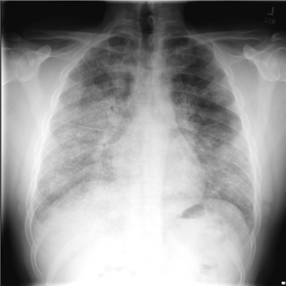
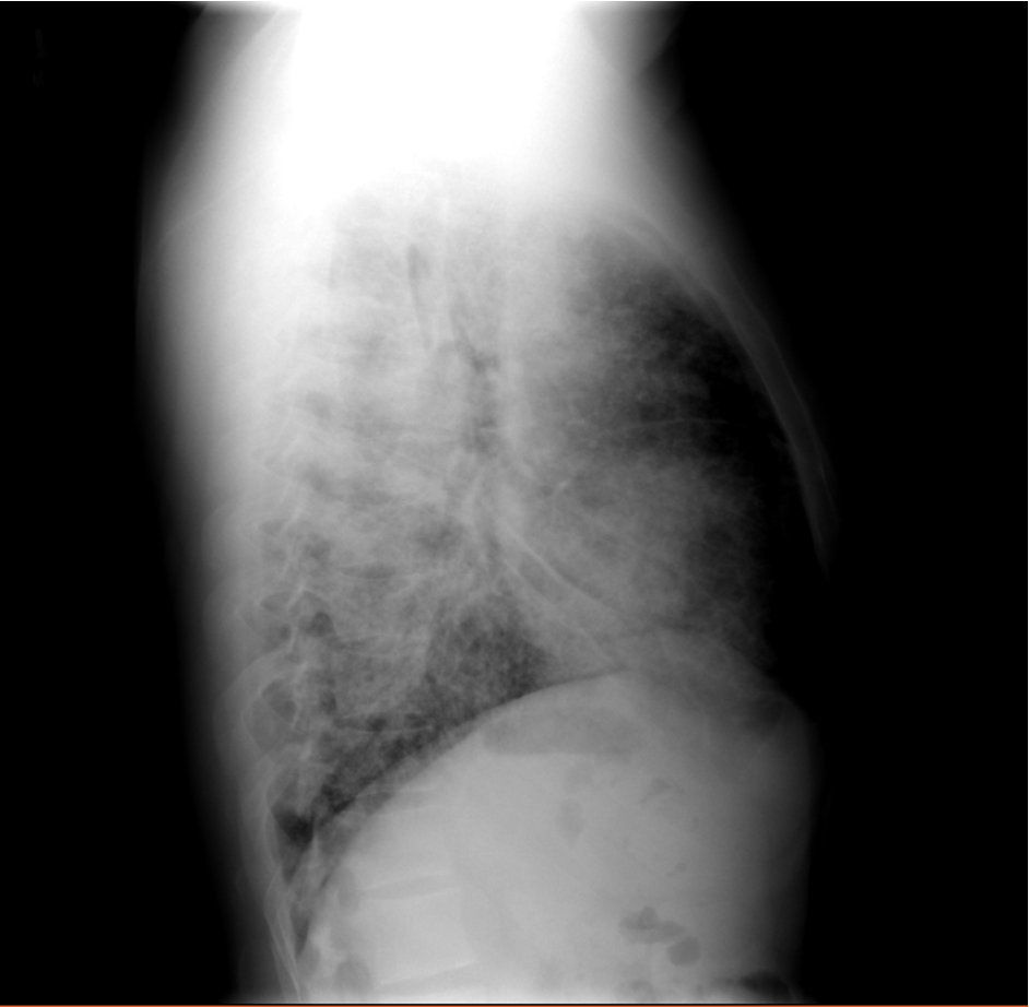
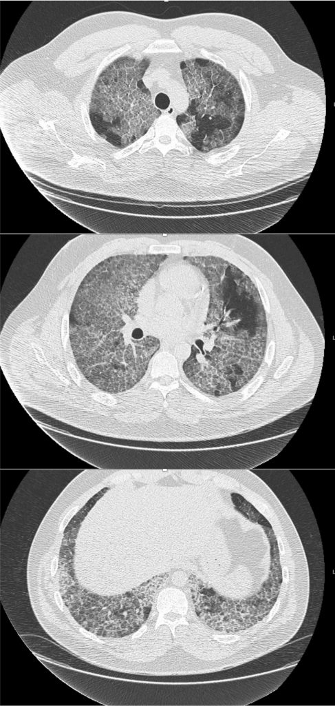
A 26-year-old man presents to his primary care doctor with 1.5 months of intermittent dyspnea, cough, chest tightness, and fatigue. His dyspnea was initially exertional, and he noticed he could do less at the gym. However, in the past 3-4 weeks it has progressed to being even with mild movement. His brother was recently diagnosed and treated for acute bronchitis so he thought this could be similar. In the office, he is noted to be tachypneic with an oxygen saturation of 83% breathing ambient air. A chest X-ray is obtained and he is sent urgently to the emergency department.

Key Learning Points

**Spoilers Ahead** If you want to think through the case on your own we advise listening to the episode first before looking at the infographics below

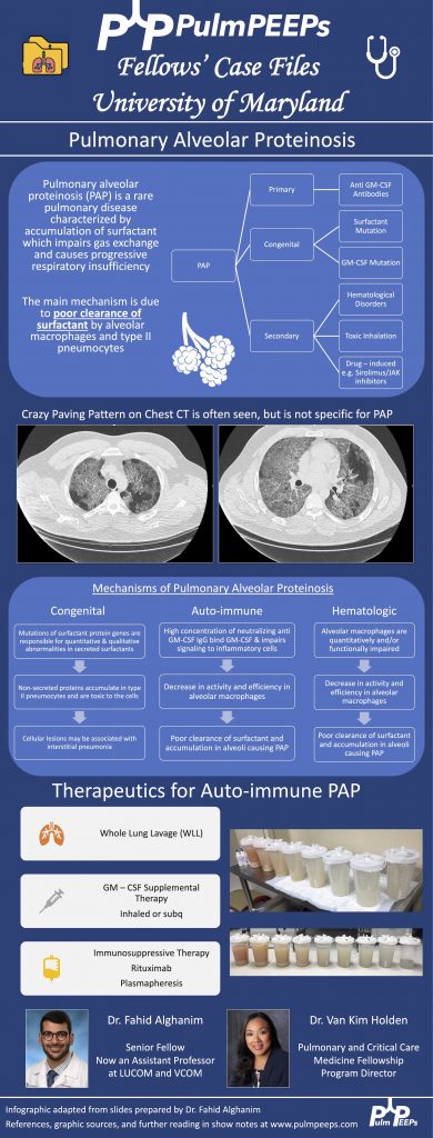
  1. Crazy Paving is a radiological term describing ground glass opacities with superimposed interlobular septal thickening. The differential diagnosis is broad and includes infectious, neoplastic, and autoimmune processes. It is not limited to just Pulmonary alveolar proteinosis (PAP) but is suggestive in an appropriate clinical setting.
  2. PAP is a disorder of surfactant production or clearance and its etiology is divided into three major subgroups. Primary or autoimmune; Secondary such as from toxic inhalations, hematological disorders, or medications; and Congenital
  3. PAP is diagnosed by positive Periodic acid-Schiff (PAS) staining of lipo-proteinaceous material in the distal bronchioles and alveoli on lung biopsy. The diagnosis can be made with PAS-positive BAL staining, but this has limited sensitivity and lung biopsy is necessary for the diagnosis in up to 30 – 35% of cases.
  4. It is important not to anchor on a diagnosis when a patient presents to you for re-evaluation even if seen by a prior expert. This was pivotal in this case!
  5. Please don’t put anything in your lung. Any toxic inhalation exposure could result in significant damage to lung parenchyma and morbidity as a result.

References and Further Reading

  1. Borie R, Danel C, Debray MP, et al. Pulmonary alveolar proteinosis. Eur Respir Rev. 2011;20(120):98-107. doi:10.1183/09059180.00001311
  2. Carey B, Trapnell BC. The molecular basis of pulmonary alveolar proteinosis. Clin Immunol. 2010;135(2):223-235. doi:10.1016/j.clim.2010.02.017
  3. Inoue Y, Trapnell BC, Tazawa R, et al. Characteristics of a large cohort of patients with autoimmune pulmonary alveolar proteinosis in Japan. Am J Respir Crit Care Med. 2008;177(7):752-762. doi:10.1164/rccm.200708-1271OC
  4. Kavuru MS, Malur A, Marshall I, et al. An open-label trial of rituximab therapy in pulmonary alveolar proteinosis. Eur Respir J. 2011;38(6):1361-1367. doi:10.1183/09031936.00197710
  5. Michaud G, Reddy C, Ernst A. Whole-lung lavage for pulmonary alveolar proteinosis. Chest. 2009;136(6):1678-1681. doi:10.1378/chest.09-2295
  6. Smith BB, Torres NE, Hyder JA, et al. Whole-lung Lavage and Pulmonary Alveolar Proteinosis: Review of Clinical and Patient-centered Outcomes. J Cardiothorac Vasc Anesth. 2019;33(9):2453-2461. doi:10.1053/j.jvca.2019.03.047
  7. Tazawa R, Ueda T, Abe M, et al. Inhaled GM-CSF for Pulmonary Alveolar Proteinosis. New England Journal of Medicine. 2019;381(10):923-932. doi:10.1056/NEJMoa1816216
  8. Tung AH, Grace J, O’Kane GM, Kumar K. Transbronchial lung biopsy (TBLB) in diagnosing pulmonary alveolar proteinosis (PAP): forgotten role in Australia? Respirology Case Reports. 2015;3(4):145-147. doi:10.1002/rcr2.129
  9. Werner AK, Koumans EH, Chatham-Stephens K, et al. Hospitalizations and Deaths Associated with EVALI. New England Journal of Medicine. 2020;382(17):1589-1598. doi:10.1056/NEJMoa1915314

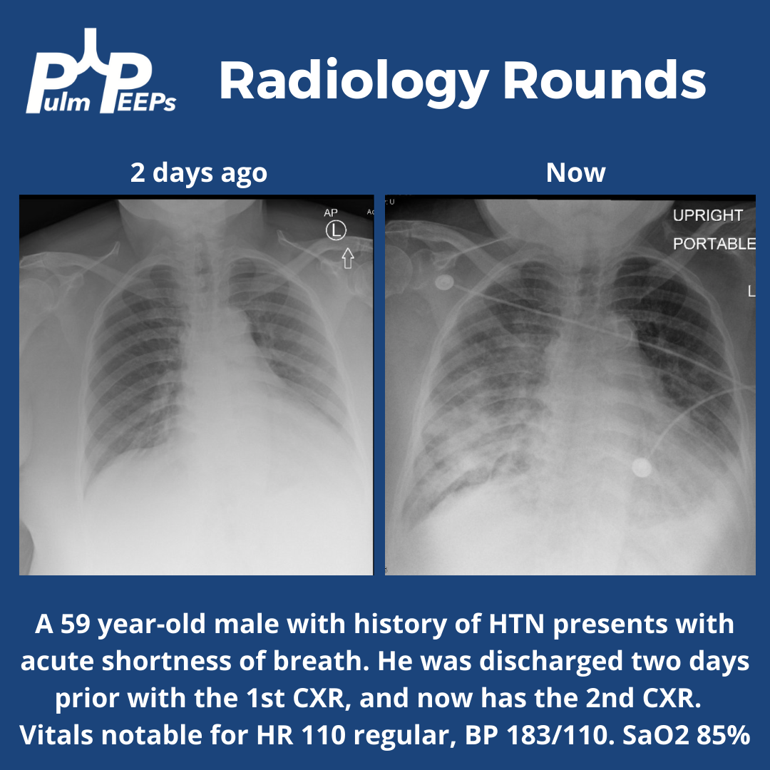
Radiology Rounds – 6/28/22

This week’s #RadiologyRounds is brought to you by our newest Contributor, Matthew Tsai! Matt will be continuing to work with us to bring you great cases and images and we are thrilled to have him on the team! Follow us on Twitter and Instagram for our Radiology Rounds, podcast episode releases, and more!

7. Top Consults: Severe Asthma Exacerbation

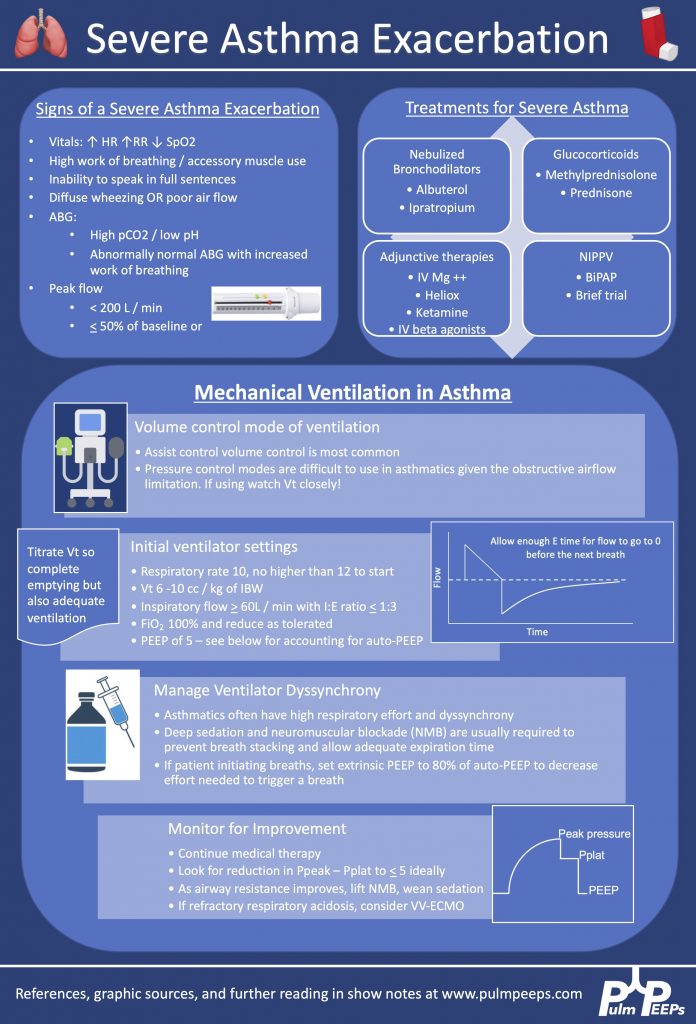
We are excited to bring you another episode in our Pulm PEEPs Top Consults series! Kristina Montemayor and David Furfaro, are joined by Sandy Zaeh to discuss the assessment and management of a patient with a severe asthma exacerbation. We’ll follow a consult patient from the emergency department to the ICU, and cover everything from the physiology of pulsus paradoxus in asthma to how to manage the ventilator in status asthmaticus. Listen today and please send any questions our way on Twitter @pulmPEEPS.

Meet Our Guests

Sandy Zaeh is an Instructor of Medicine and Pulmonary & Critical Care Medicine physician at Yale School of Medicine.

Key Learning Points

References and links for further reading

  1. Chung KF, Wenzel SE, Brozek JL, et al. International ERS/ATS guidelines on definition, evaluation and treatment of severe asthma. European Respiratory Journal. 2014;43(2):343-373. doi:10.1183/09031936.00202013
  2. Rodrigo GJ, Rodrigo C, Hall JB. Acute asthma in adults: a review. Chest. 2004;125(3):1081-1102. doi:10.1378/chest.125.3.1081
  3. Godwin HT, Fix ML, Baker O, Madsen T, Walls RM, Brown CA. Emergency Department Airway Management for Status Asthmaticus With Respiratory Failure. Respir Care. 2020;65(12):1904-1907. doi:10.4187/respcare.07723
  4. Althoff MD, Holguin F, Yang F, et al. Noninvasive Ventilation Use in Critically Ill Patients with Acute Asthma Exacerbations. Am J Respir Crit Care Med. 2020;202(11):1520-1530. doi:10.1164/rccm.201910-2021OC
  5. Brenner B, Corbridge T, Kazzi A. Intubation and Mechanical Ventilation of the Asthmatic Patient in Respiratory Failure. Proc Am Thorac Soc. 2009;6(4):371-379. doi:10.1513/pats.P09ST4
  6. Laher AE, Buchanan SK. Mechanically Ventilating the Severe Asthmatic. J Intensive Care Med. 2018;33(9):491-501. doi:10.1177/0885066617740079
  7. Leatherman J. Mechanical ventilation for severe asthma. Chest. 2015;147(6):1671-1680. doi:10.1378/chest.14-1733

6. PEEP in ARDS Roundtable

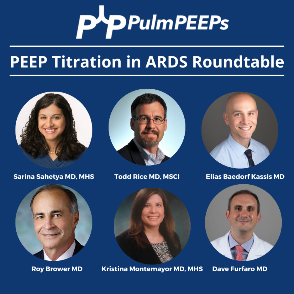
This week on Pulm PEEPs, Dave Furfaro and Kristina Montemayor are joined by experts in the field of critical care medicine and ARDS to discuss all things PEEP! Drs. Roy Brower, Sarina Sahetya, Todd Rice, and Elias Baedorf-Kassis discuss everything ranging from PEEP basics to their approach to optimizing PEEP in patients with ARDS.

Meet Our Guests

Roy Brower is a Professor of Medicine at Johns Hopkins where he served as the MICU director for over 33 years, and he has been one of the pioneers for lung-protective ventilation for patients with ARDS.

Elias Baedorf-Kassis is an Assistant Professor of Medicine at Beth Israel Deaconess Medical Center and Harvard Medical School. He is the Medical Director of Respiratory Care at BIDMC, and helps lead the VV-ECMO program.

Todd Rice is an Associate Profess of Medicine in the Division of Allergy, Pulmonary, and Critical Care Medicine at Vanderbilt University and Vice President for Clinical Trial Innovation and Operations in the Vanderbilt Institute for Clinical and Translational Research.

Sarina Sahetya is an Assistant Professor of Medicine at Johns Hopkins Hospital and does research in the diagnosis and treatment of ARDS.


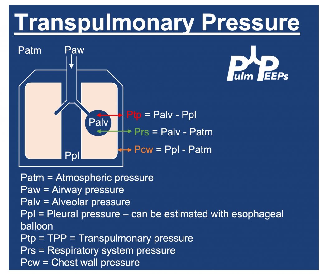
Key Learning Points

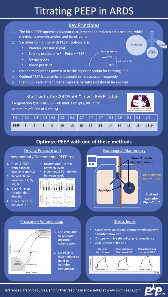
Driving Pressure figure from Amato et al. 2015. Stress index figure from Hess 2014.
  • The plateau pressure can be measured on the ventilator with an inspiratory hold maneuver
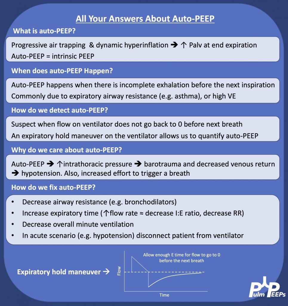
  • Extrinsic PEEP is applied by the ventiilator, while intrinsic PEEP, or auto-PEEP, occurs when there is incomplete emptying of the lungs due to inadequate time for exhalation. This often happens with obstructive lung disease. Intrinsic PEEP can be measured on the ventilator with an end-expiratory hold maneuver
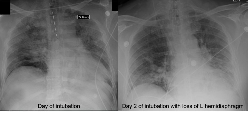
  • We utilize PEEP in all intubated patients to minimize atelectasis. When patients are supine, the heart moves back 2 cm and the diaphragm raises by 2 cm, so often the left lower lobe of the lung is compressed and there is atelectasis there. This is often seen on CXR:

References, Image Sources, and Further Reading

  1. Higher versus Lower Positive End-Expiratory Pressures in Patients with the Acute Respiratory Distress Syndrome. New England Journal of Medicine. 2004;351(4):327-336. doi:10.1056/NEJMoa032193
  2. Amato MBP, Meade MO, Slutsky AS, et al. Driving Pressure and Survival in the Acute Respiratory Distress Syndrome. New England Journal of Medicine. 2015;372(8):747-755. doi:10.1056/NEJMsa1410639
  3. Writing Group for the Alveolar Recruitment for Acute Respiratory Distress Syndrome Trial (ART) Investigators. Effect of Lung Recruitment and Titrated Positive End-Expiratory Pressure (PEEP) vs Low PEEP on Mortality in Patients With Acute Respiratory Distress Syndrome: A Randomized Clinical Trial. JAMA. 2017;318(14):1335-1345. doi:10.1001/jama.2017.14171
  4. Beitler JR, Sarge T, Banner-Goodspeed VM, et al. Effect of Titrating Positive End-Expiratory Pressure (PEEP) With an Esophageal Pressure-Guided Strategy vs an Empirical High PEEP-Fio2 Strategy on Death and Days Free From Mechanical Ventilation Among Patients With Acute Respiratory Distress Syndrome: A Randomized Clinical Trial. JAMA. 2019;321(9):846-857. doi:10.1001/jama.2019.0555
  5. LaFollette R, Hojnowski K, Norton J, DiRocco J, Carney D, Nieman G. Using pressure–volume curves to set proper PEEP in acute lung injury. Nursing in Critical Care. 2007;12(5):231-241. doi:10.1111/j.1478-5153.2007.00224.x
  6. Hess DR. Respiratory mechanics in mechanically ventilated patients. Respir Care. 2014;59(11):1773-1794. doi:10.4187/respcare.03410
  7. Sahetya SK, Hager DN, Stephens RS, Needham DM, Brower RG. PEEP Titration to Minimize Driving Pressure in Subjects With ARDS: A Prospective Physiological Study. Respir Care. 2020;65(5):583-589. doi:10.4187/respcare.07102
  8. Umbrello M, Chiumello D. Interpretation of the transpulmonary pressure in the critically ill patient. Ann Transl Med. 2018;6(19):383. doi:10.21037/atm.2018.05.31
  9. Kenny JES. ICU Physiology in 1000 Words: Driving Pressure & Stress Index. PulmCCM. Published February 13, 2016. Accessed January 1, 2022. https://pulmccm.org/review-articles/icu-physiology-in-1000-words-driving-pressure-stress-index/

Radiology Rounds – 12/28/21

Today we’re bringing you a special edition of Radiology Rounds complete with classic imaging, and some key critical care and ventilator physiology. This case is a perfect lead-in for next week’s Pulm PEEPs Roundtable on PEEP titration, so make sure to tune in!

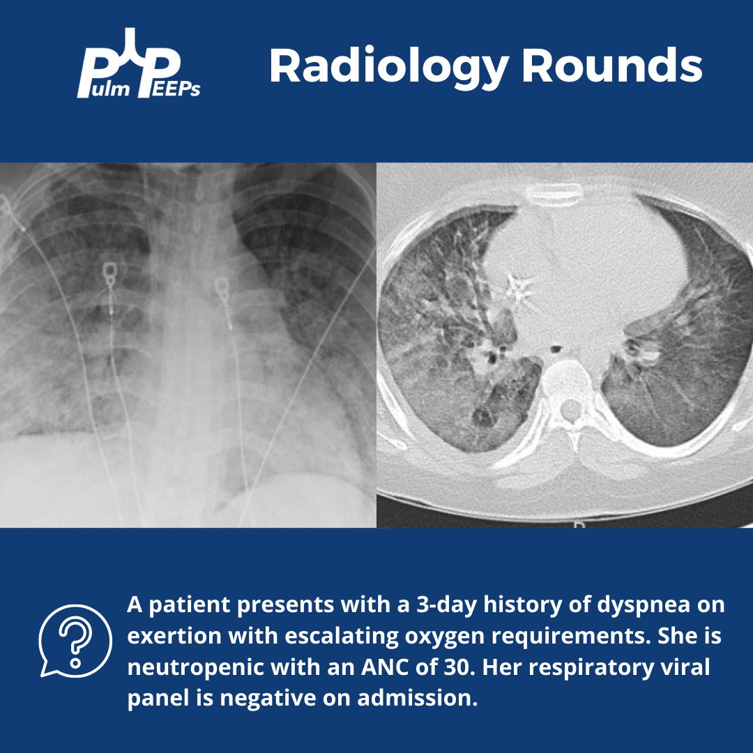
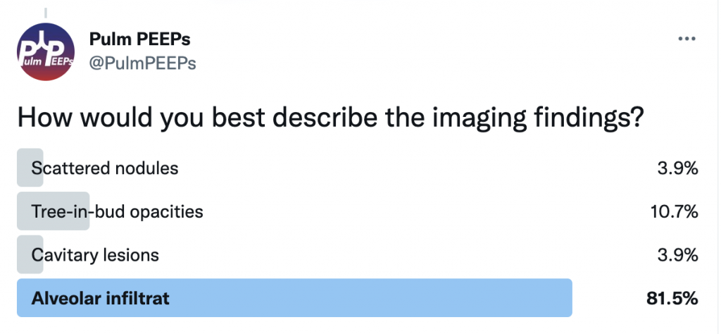
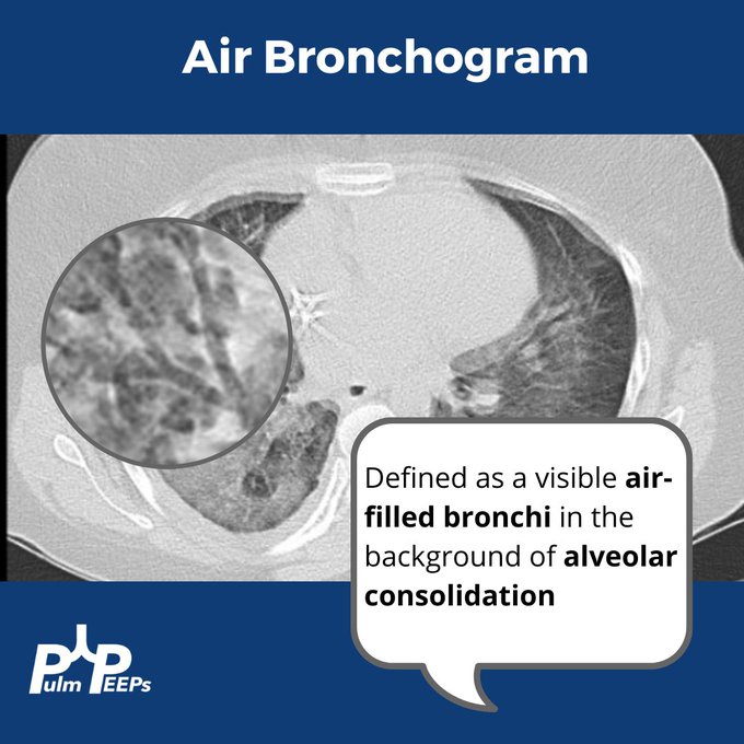
How would you best describe the imaging findings?


There are bilateral, diffuse alveolar infiltrates noted on imaging with evidence of an air bronchogram on the CT image.

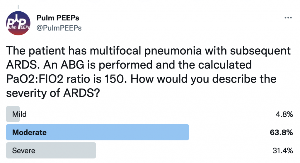
The patient develops worsening hypoxemia requiring mechanical intubation. The patient has multifocal pneumonia and requires intubation. ABG is performed and the calculated PaO2:FIO2 ratio is 150. How would you describe the severity of ARDS?


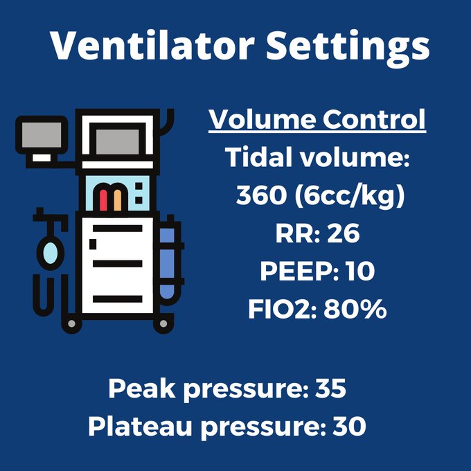
This patient has moderate ARDS based on a PaO2:FIO2 ratio that is between 100 and 200. The patient’s initial ventilator settings on volume control are:

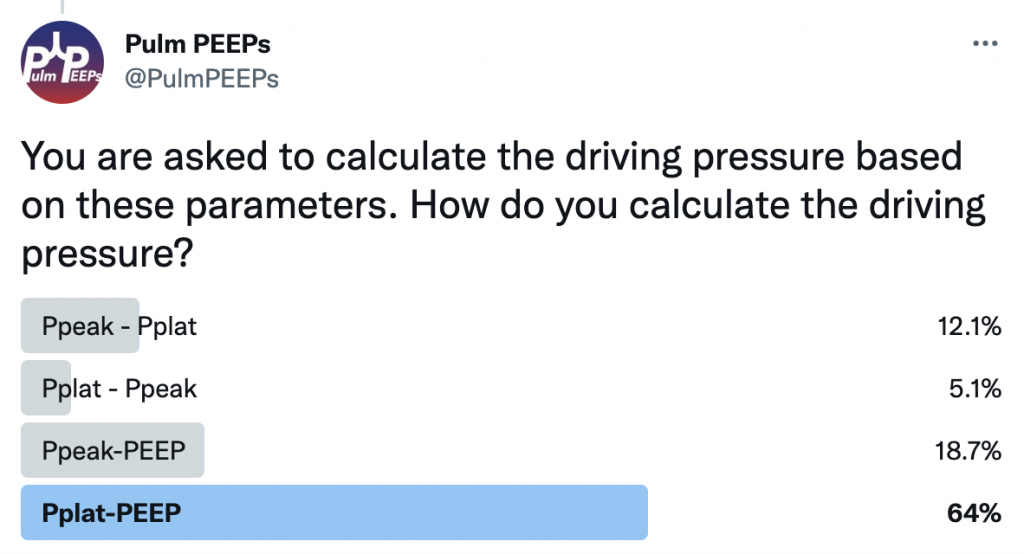
Based on these parameters, we can also calculate the driving pressure. Driving pressure is calculated by using Pplat-PEEP. In this case, Pplat (30)-PEEP (10), would give a driving pressure of 20.