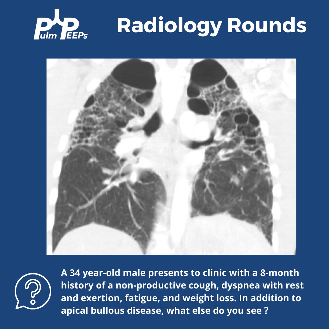
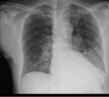
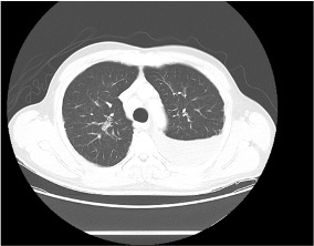
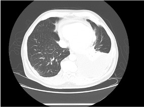
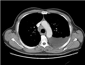
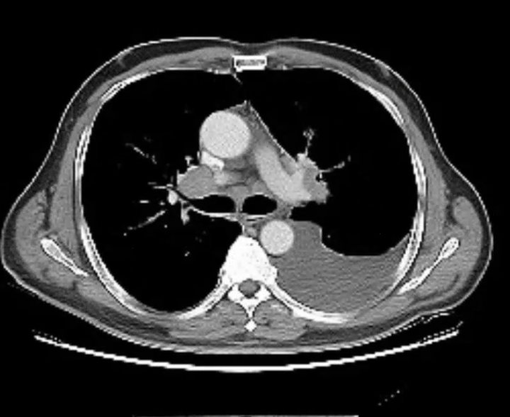
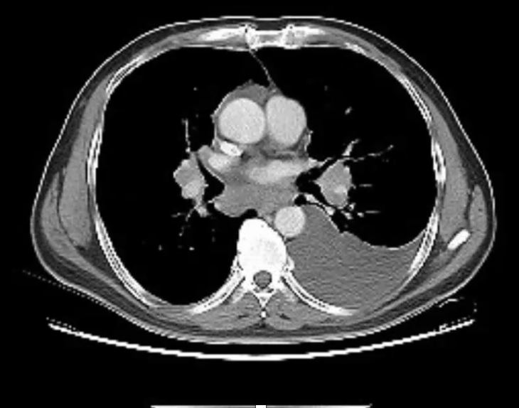
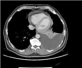
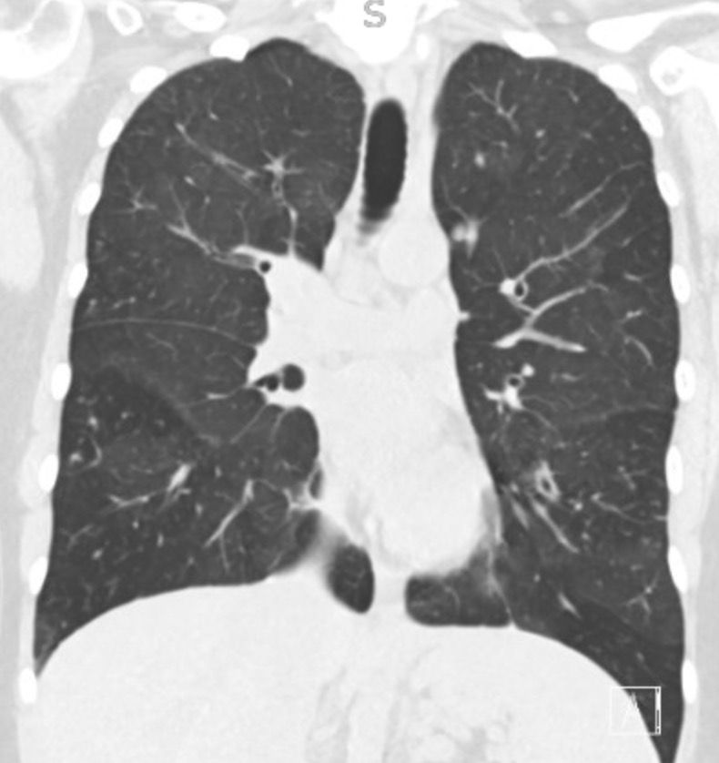
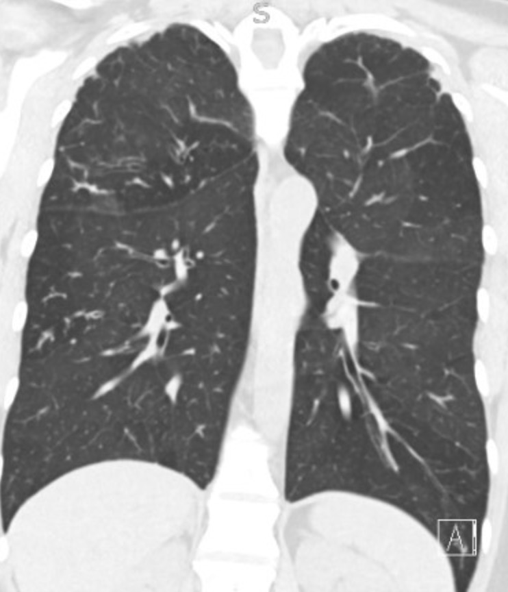
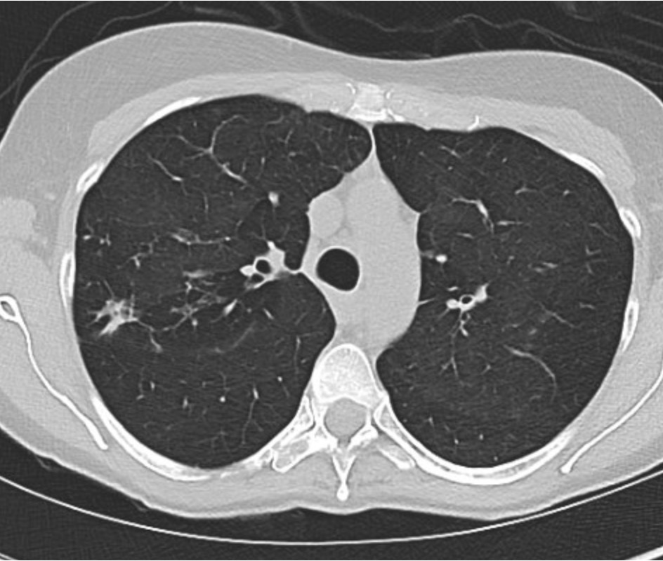
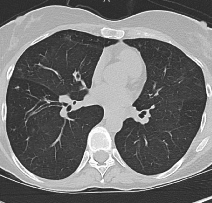
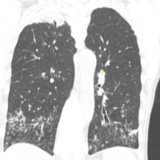
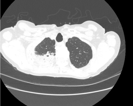
We have another #RadiologyRounds for you today! You are seeing a new patient in the clinic with dyspnea who brings in prior CT chest imaging. A representative coronal image is shown.
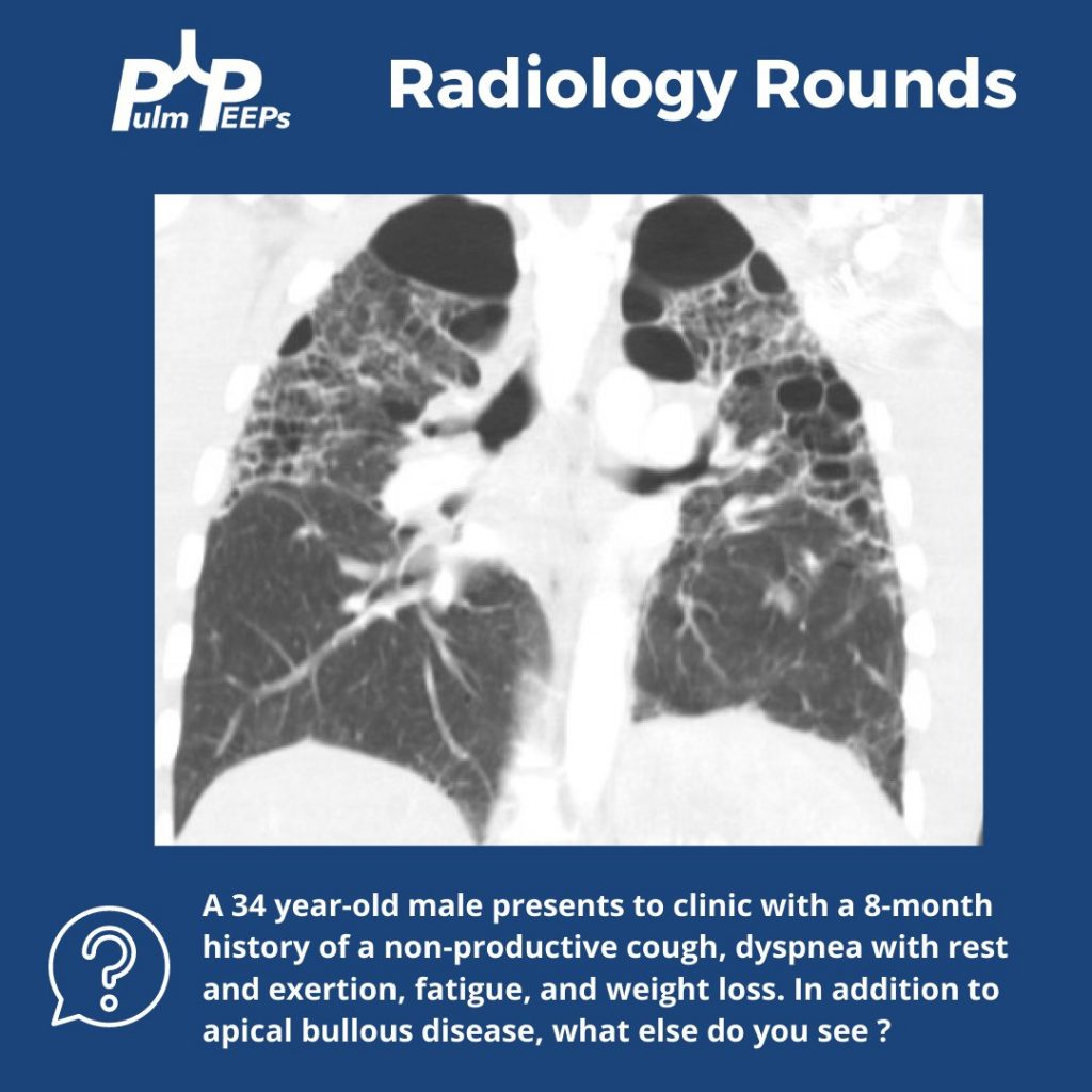
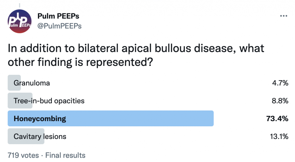
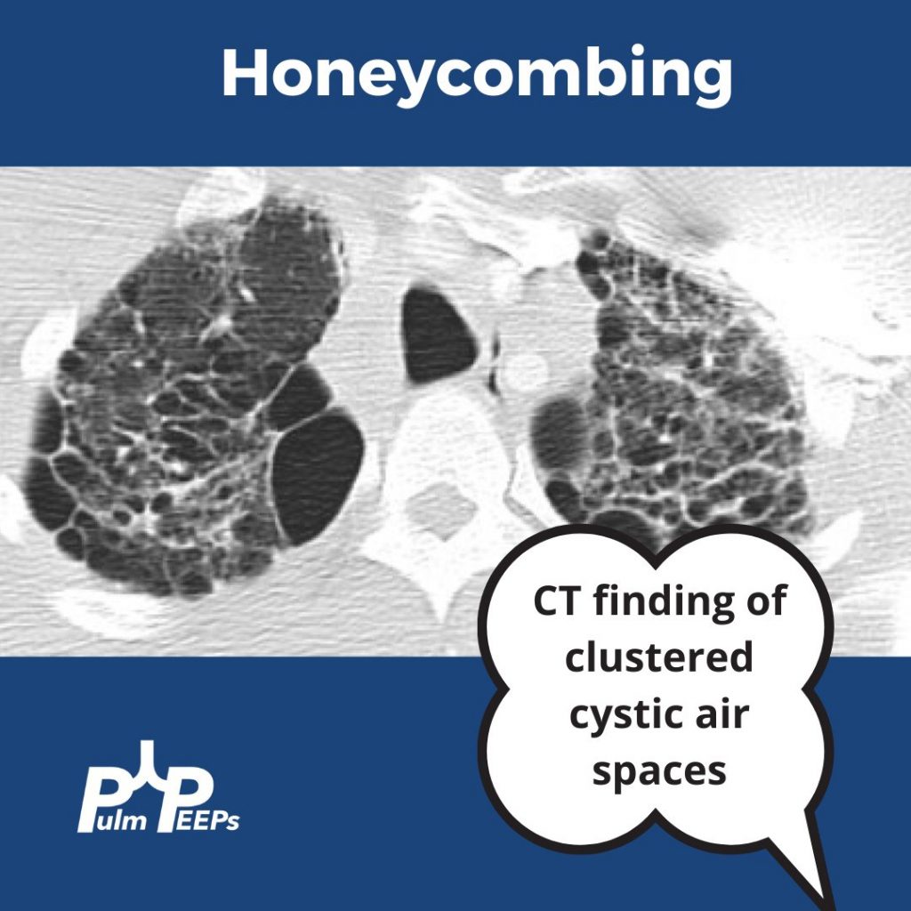
In addition to bullous disease, you see bilateral honeycombing with evidence of fibrosis primarily in the upper lung fields.
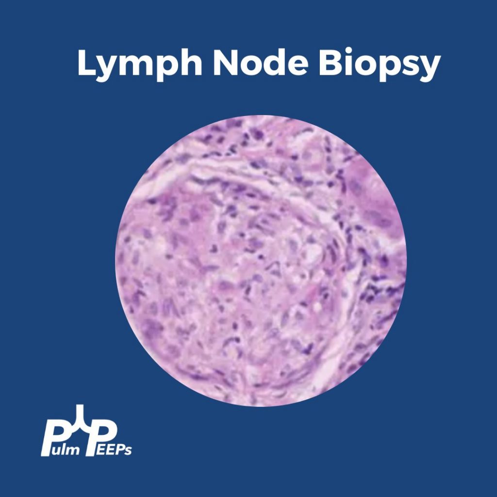
As part of your evaluation, an EBUS is performed showing the following representative lymph node tissue pathology.
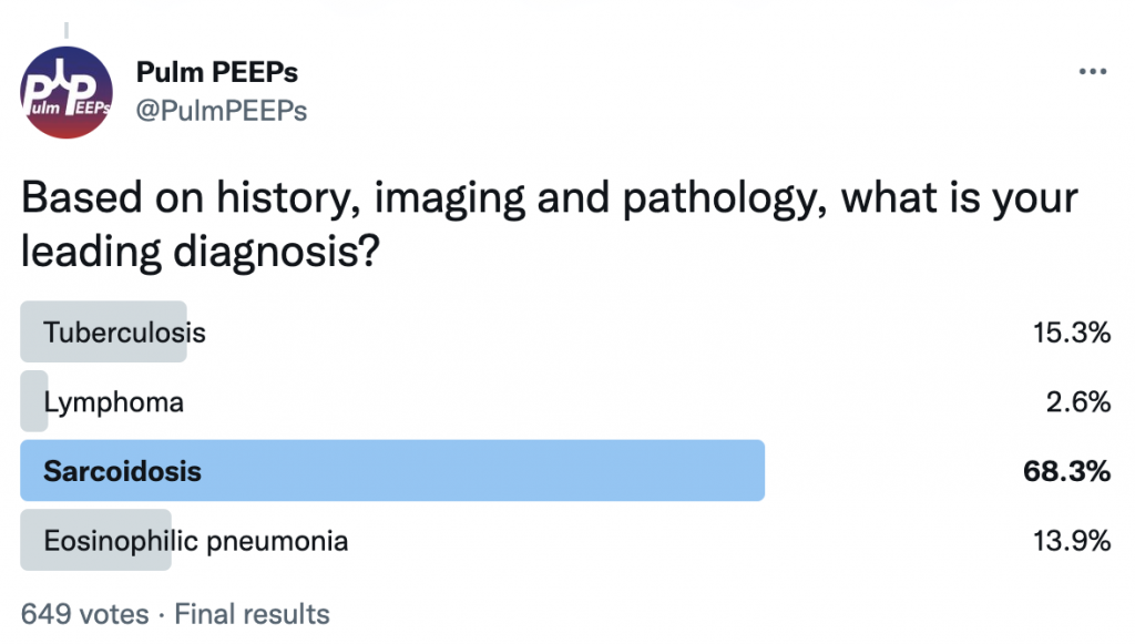
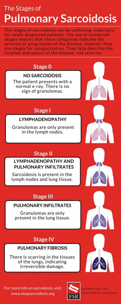
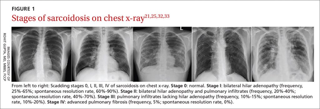
We had evidence of noncaseating granulomas, evidence of fibrocystic changes on chest imaging, and we excluded other causes of granulomatous disease. Given his symptoms and clinical context, we were concerned about Stage IV pulmonary sarcoidosis which can be categorized below.
Dave Furfaro, Kristina Montemayor, and Ansa Razzaq are back to tackle another pulmonary case! Listen in and solve the case yourself, and we’ll share some diagnostic pearls along the way. Let us know any additional thoughts on Twitter.
Patient Presentation
The is patient is 57-year-old man with hypertension and asthma who presents with dyspnea and left-sided pleuritic chest pain for 3 weeks. He was in his usual state of health until 3 weeks prior to admission, when he developed dyspnea and sharp left-sided chest pain that worsens with deep breathing. His symptoms are accompanied by unintentional 30-pound weight loss over the past several months as well as an intermittent cough that is nonproductive.
On physical exam, he is mildly tachypneic and saturating well on room air with otherwise normal vital signs. He has decreased breath sounds at the right lung base.
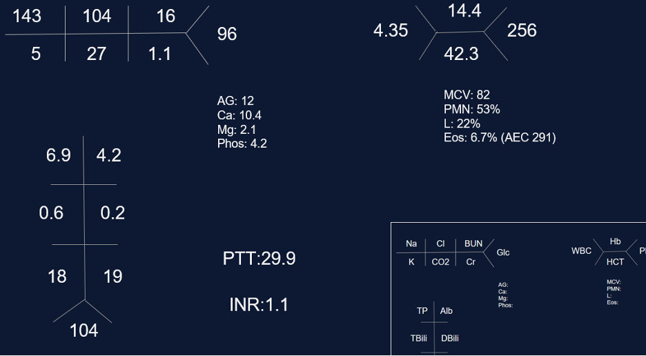
Initial labs
Key Learning Points
**Spoilers Ahead** If you want to think through the case on your own we advise listening to the episode first before looking at the infographics below
Physical Exam Pearls
Reasons for decreased breath sounds on physical exam
1. Increased thickness of chest wall
2. Reduced airflow to part of the lung
3. Overinflation to part of the lung
4. Something between the lung and chest wall — air or fluid
Determine bradypnea and tachypnea quickly by matching your breahting rate to the patient’s respiratory rate
1. Is there a compatible presentation (imaging, physical exam)
2. Detection of non-necrotizing granulomatous inflammation in one or more tissue samples
3. Exclusion of other disease that may present similarily
Pulmonary stages of sarcoidosis:
Key to remember that patients don’t always progress through these stages. The system is useful for prognosticating and determining treatment based on the risk for disease progression.
Patients with stage 1, and even many with stage 2, often don’t require treatment
The first-line agent is oral glucocorticoids and the typical starting dose is prednisone 20 – 40 mg by mouth daily. The patient should be evaluated closely, and ideally, this dose can be tapered starting at about 4 – 6 weeks. Following this, the prednisone dose is tapered slowly over 6 months – 1 year while monitoring for symptom recurrence.
Second-line steroid-sparing agents are methotrexate, azathioprine, or mycophenolate. These are often used if the patient relapses, or is on more then 10mg daily for 3 months after the initial taper with intolerance of steroids
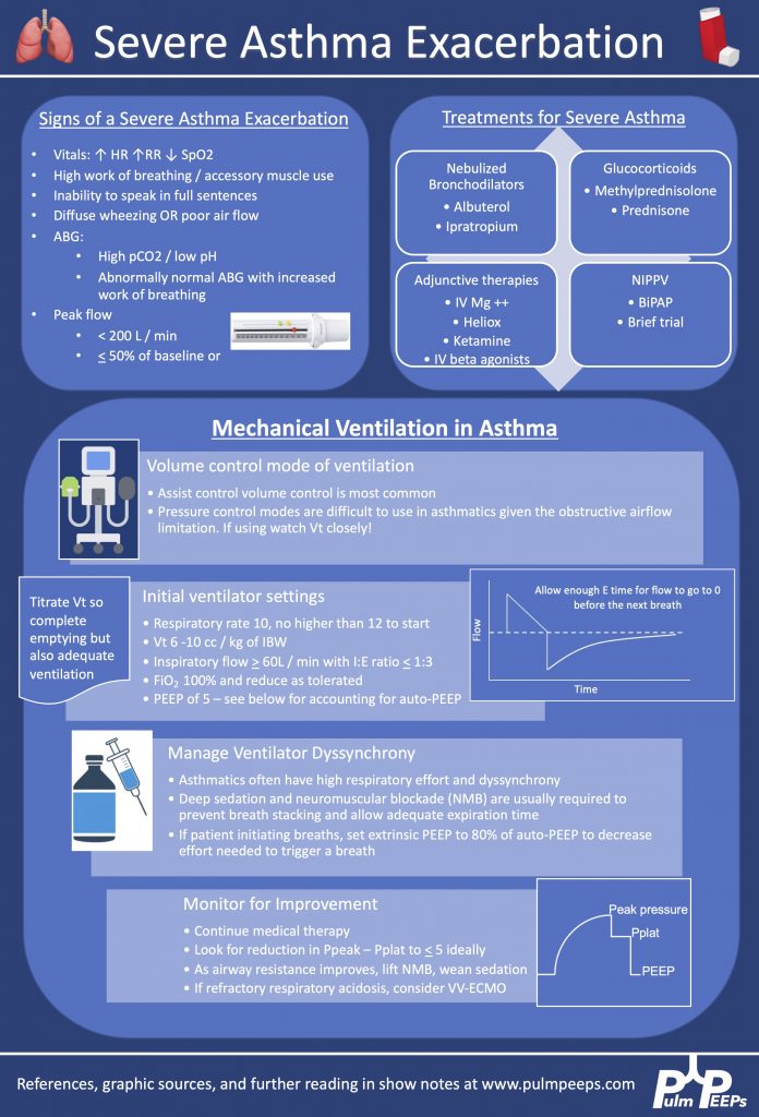
We are excited to bring you another episode in our Pulm PEEPs Top Consults series! Kristina Montemayor and David Furfaro, are joined by Sandy Zaeh to discuss the assessment and management of a patient with a severe asthma exacerbation. We’ll follow a consult patient from the emergency department to the ICU, and cover everything from the physiology of pulsus paradoxus in asthma to how to manage the ventilator in status asthmaticus. Listen today and please send any questions our way on Twitter @pulmPEEPS.
Meet Our Guests
Sandy Zaeh is an Instructor of Medicine and Pulmonary & Critical Care Medicine physician at Yale School of Medicine.
The Pulm PEEPs are joined again by Natalie West to discuss a patient who presented with a chronic, productive cough. Listen in today as we work through our differential diagnosis, interpret basic pulmonary testing, and share our clinical reasoning along the way. We have some fantastic diagnostic and treatment teaching points, so once you’ve solved the case check out the takeaways and infographics below. Please let us know any additional insights you have on Twitter!
Patient Presentation
A 50-year-old woman, who is a never smoker, with a past medical history of recurrent pancreatitis presents to the pulmonary clinic with a chronic, productive cough. Her cough has been present for 3 years and has increased in frequency to now being present daily. In the last three months, the cough has also worsened and is productive of small amounts of yellow to green sputum. She has a history of chronic post-nasal drip and sinus infections, and uses intranasal steroids, but has not noted changes in these symptoms. There is no significant family history of pulmonary disease, and an exposure history review of symptoms is negative.
On physical exam, she was a thin woman who appeared her stated age and was breathing comfortably on room air. Her exam was notable for mild expiratory wheezing, primarily on auscultation of the right posterior lung field. She had no cyanosis, clubbing, evidence of volume overload, or abdominal tenderness.
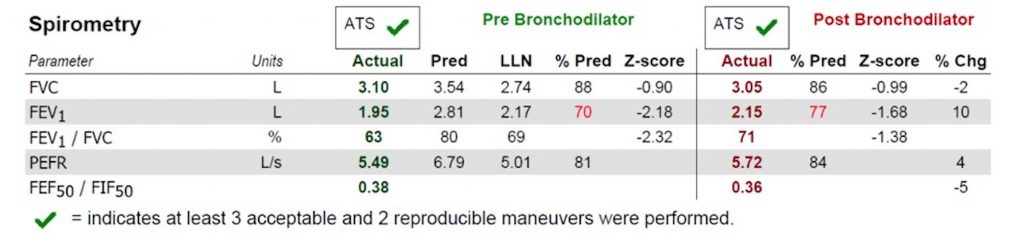
Basic Spirometry Values
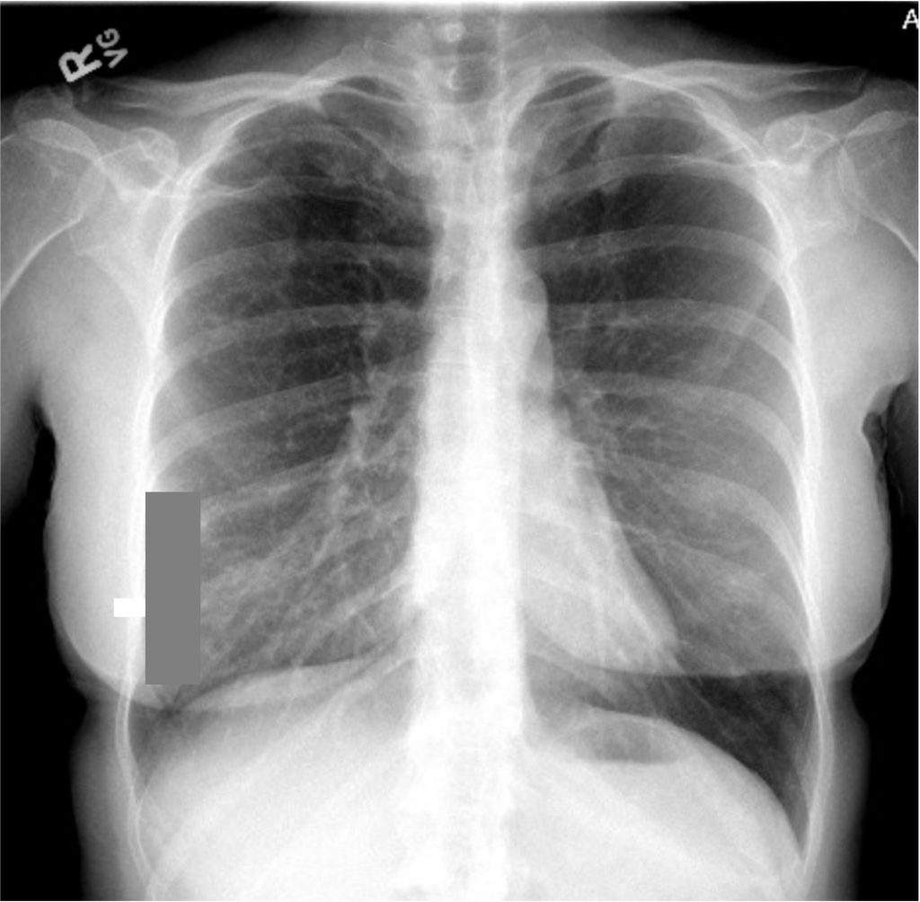
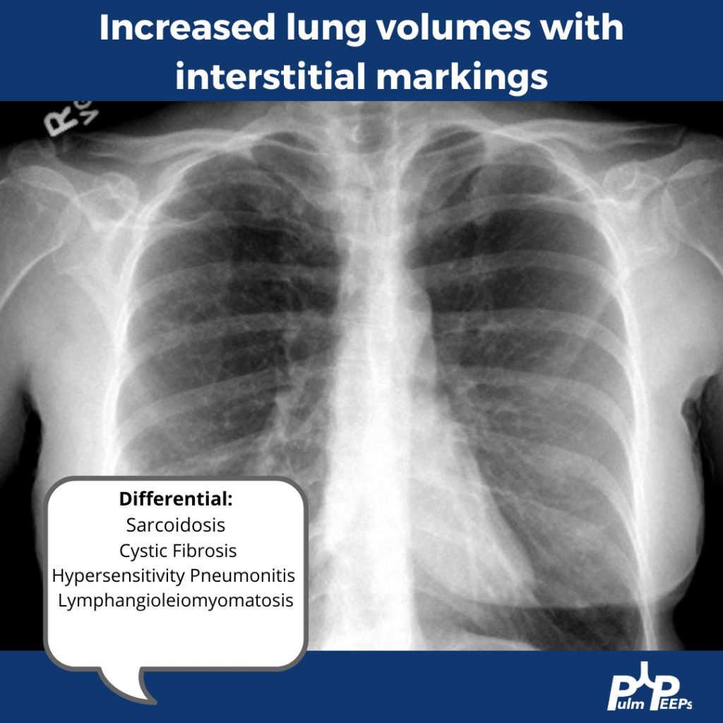
Chest X-ray
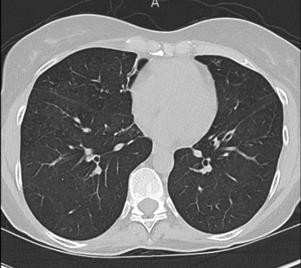
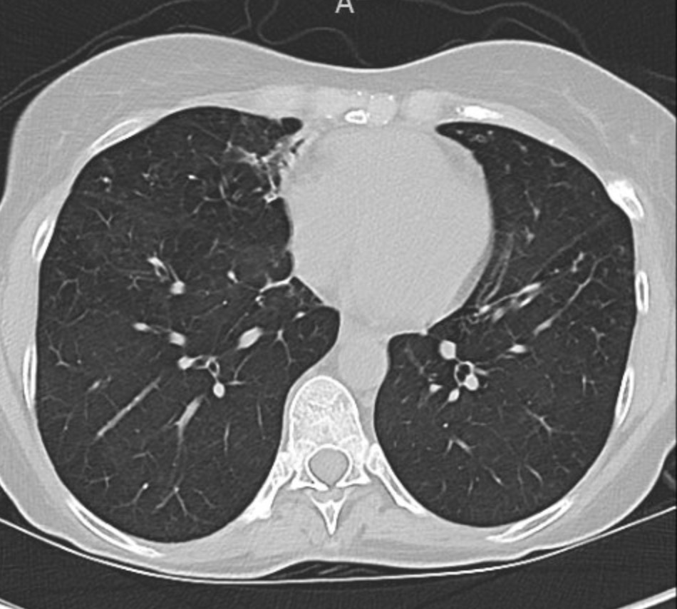
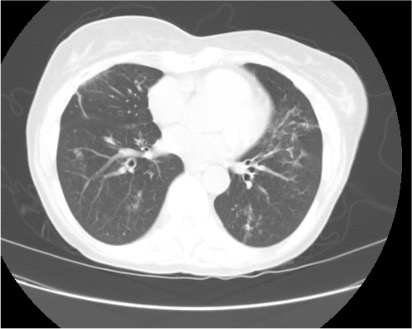
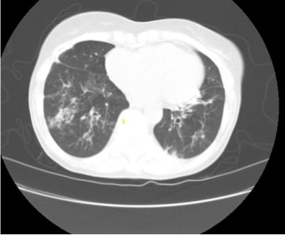
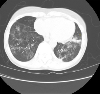
Representative Images from CT Scan
Key Learning Points
**Spoilers Ahead** If you want to think through the case on your own we advise listening to the episode first before looking at the infographics below.
Differential Diagnosis of Chronic Cough
Three most common causes: upper airway cough syndrome, GERD, cough variant asthma
Additional etiologies to consider: chronic bronchitis, post-infectious after a respiratory tract infection, bronchiectasis, ACE inhibitors, lung cancer, eosinophilic bronchitis, interstitial lung disease
Imaging Pearl
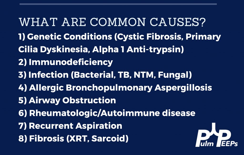
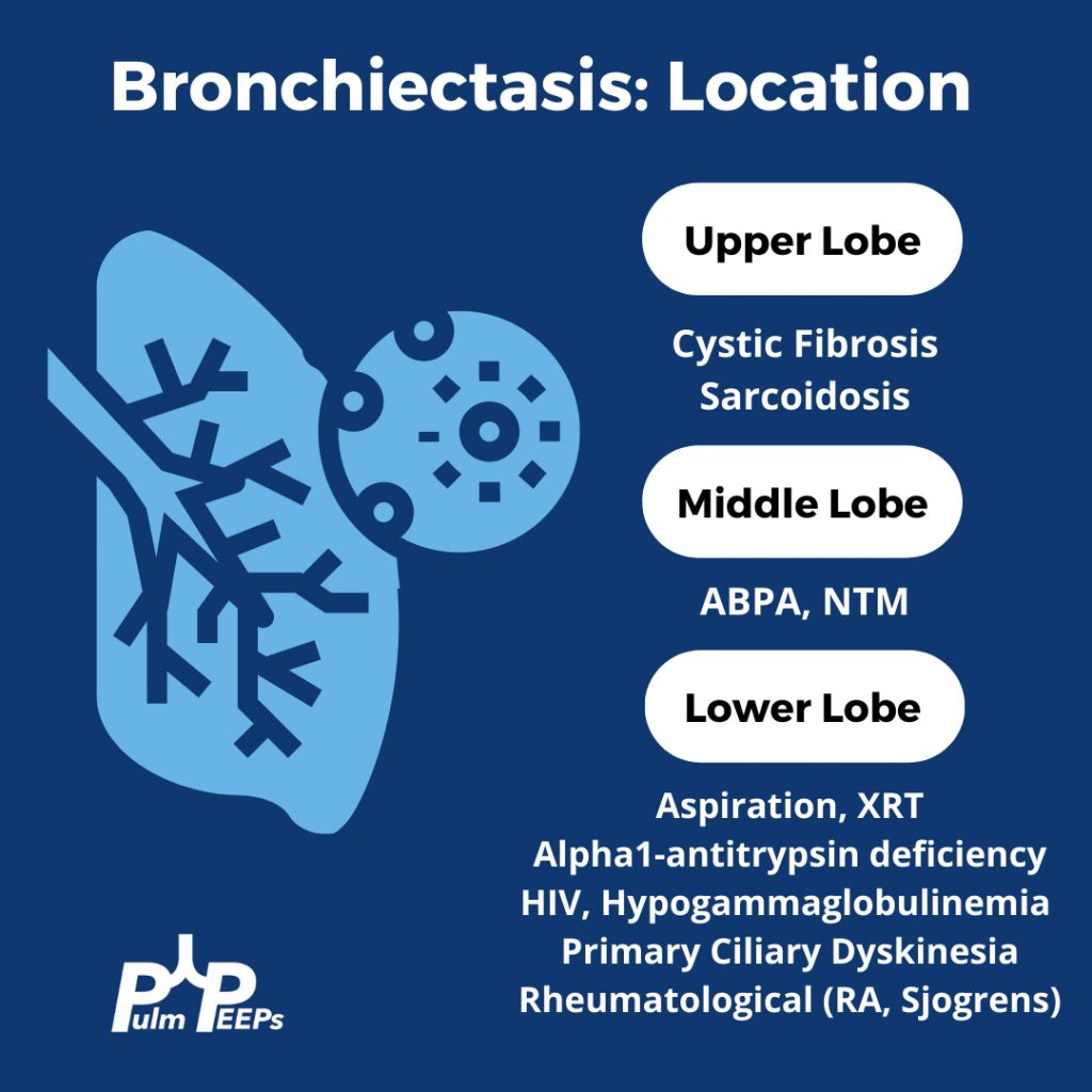
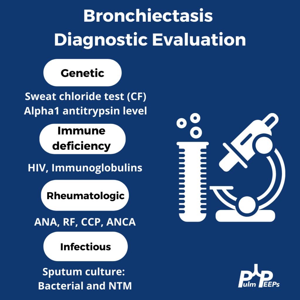
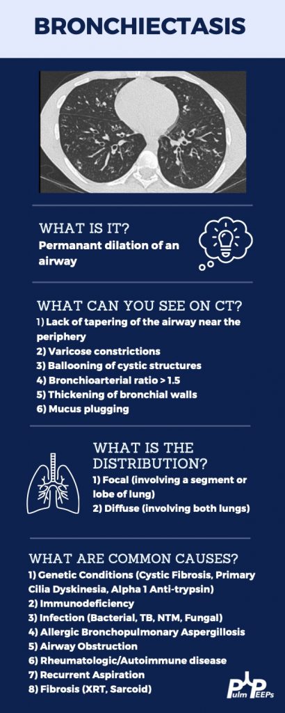
Evaluating Bronchiectasis
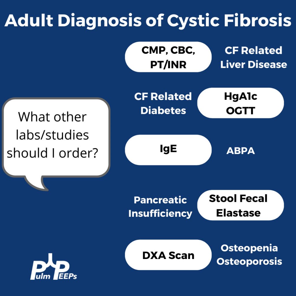
Making a New Diagnosis of Cystic Fibrosis in an Adult
Sweat testing
Sweat testing should be done in CF accredited center. Inform patients that there are no needles involved. Pilocarpine and electrical stimulation are applied to the arm or leg to stimulate the sweat gland, and then sweat is collected on filter paper, a gauze, or a plastic coil. From there, the amount of chloride in the sweat is calculated
Results
< 30 normal
31 – 60 indeterminate
> 60 is positive and Cystic Fibrosis is likely
What do you do with an Indeterminate test?
Patients with milder phenotypes of Cystic Fibrosis can have a normal or indeterminate sweat chloride level, and 10% of adults diagnosed with CF have a normal sweat chloride. If the sweat chloride test is indeterminate or normal, but suspicion is high for CF, then genetic testing for the whole array of mutations should be performed
The Pulm PEEPs are excited to bring our first mystery case! Kristina Montemayor and Dave Furfaro hear a fascinating case presentation from Pulm PEEPs senior editor Ansa Razzaq. Join us as we work through this case together to come to a diagnosis, and share our thought process along the way. Come back to these show notes afterward, or once you’ve solved the case yourself, for some key teaching pearls and representative images.
Patient Presentation
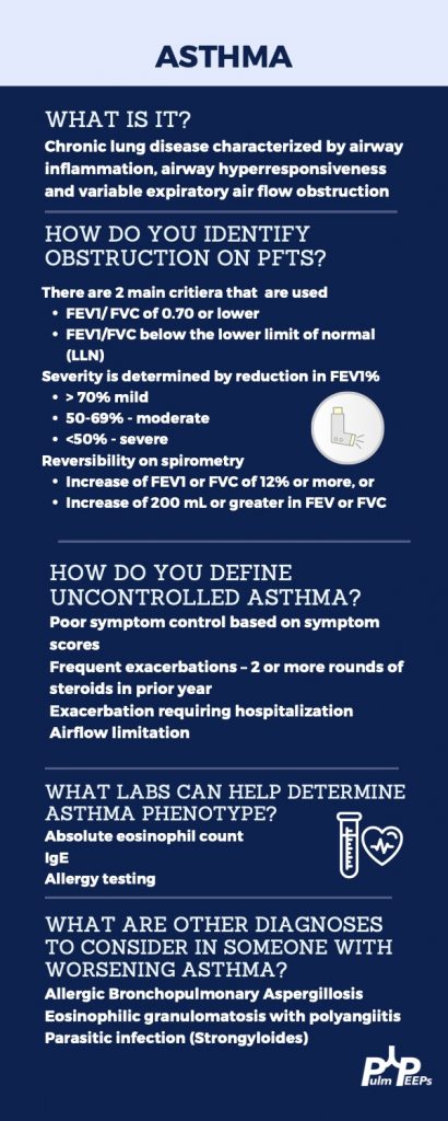
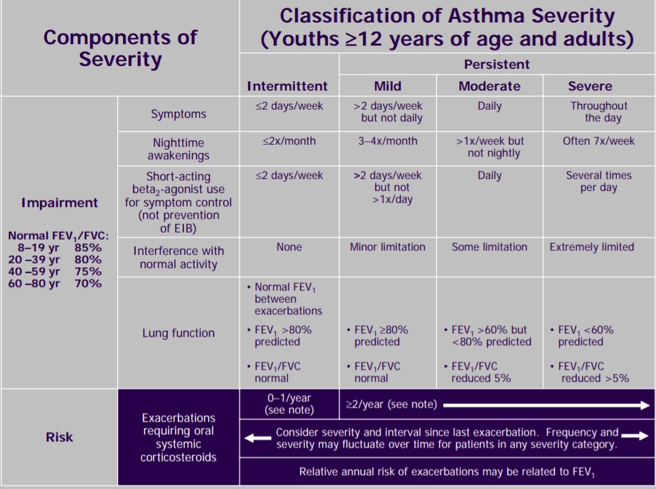
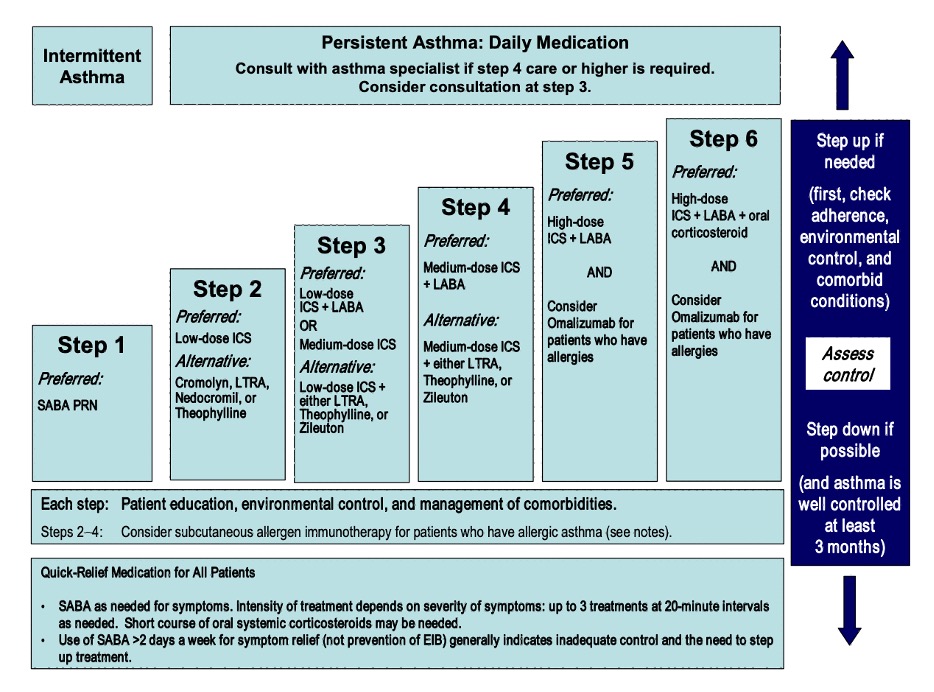
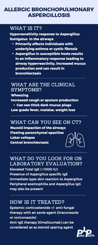
A 66-year-old woman with no smoking history and past medical history of previously well-controlled asthma is referred to pulmonary clinic after multiple recent episodes of dyspnea, wheezing, and coughing. The episodes have features consistent with asthma exacerbations; however, they are also associated with migratory infiltrates. She has been treated with multiple courses of antibiotics and steroids, and despite escalating therapy, the episodes are occurring more frequently and she was worsening overall exercise tolerance. Listen in to hear more and try to solve the case!
Representative Imaging
Key Learning Points
**Spoilers Ahead** If you want to think through the case on your own we advise listening to the episode first before looking at the infographics below.
The Pulm PEEPs (Kristina Montemayor and Dave Furfaro) host a panel of Cystic Fibrosis (CF) providers to discuss the current state of the disease, recent advances in cystic fibrosis transmembrane conductance regulator (CFTR) modulator therapies, and the evolving faces and voices of Cystic Fibrosis.
Cystic Fibrosis is an autosomal recessive disorder caused by mutations in the CFTR gene that affects over 30,000 individuals in the United States and 70,000 people worldwide. Absence or dysfunction of the CFTR protein leads to abnormal secretion of mucus, sweat, and digestive fluids, which impacts the lungs, digestive tract, and reproductive system.
From the first formal publication on Cystic Fibrosis in 1938 by Dorothy Hansine Andersen, to the discovery of the delta F508 mutation and CFTR gene in 1988 -1989 by Lap-Chee Tsui, Francis Collins, and John R. Riordan, to the approval of the first CFTR modulator therapy, Ivacaftor, in 2012, our knowledge about Cystic FIbrosis has been advancing in leaps and bounds. As therapies have improved, they have dramatically impacted the lives of patients with Cystic Fibrosis. Join us today as we explore what this evolution in care has looked like from the perspective of Cystic Fibrosis providers, and hear about the new questions and challenges on the horizon.
Meet our guests
Emily DiMango is a Professor of Medicine at Columbia University Medical Center and the Director of the John Edsall-John Wood Asthma Center and the Gunnar Esiason Adult Cystic Fibrosis Program
Terri Laguna is an Associate Professor of Pediatrics at Northwestern Medicine / Feinberg School of Medicine and the Chief of Pulmonary and Sleep Medicine in the Department of Pediatrics
Patrick Sosnay is a Senior Medical Director at Vertex Pharmaceuticals and specializes in Cystic Fibrosis
Natalie West is an Assistant Professor of Medicine at Johns Hopkins Hospital and specializes in Cystic Fibrosis.