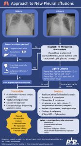
Hi Pulm PEEPs! Today we have a special episode for you. Monty and Furf were invited on the Core IM Podcast to talk about the work up and management of pleural effusions. This is a great overview and we hope you enjoy listening as much as we did recording. If you want a deeper dive into pleural effusions check out our prior series:
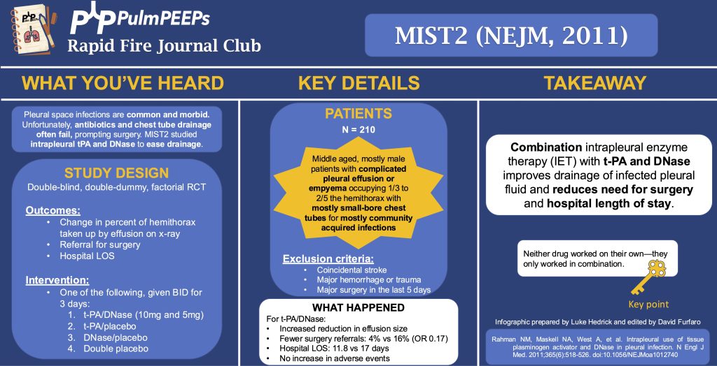
In this episode, we add another article to our Rapid Fire Journal Club. Luke Hedrick and Dave Furfaro discuss the MIST 2 trial published in NEJM in 2011 evaluating enzymatic therapy for complex parapneumonic effusions and empyemas.
Article and Reference
We are talking today about the MIST 2 trial evaluating the use of intrapleural tPa and DNase for intrapleural infections.
Infections in the pleural space are common and morbid, often requiring surgical intervention. Unfortunately, antibiotics and chest tube drainage often fail. The MIST1 trial (NEJM, 2005) of intrapleural streptokinase showed no benefit. MIST2 studied intrapleural tPA and DNase to ease drainage by breaking down septations and thinning pleural fluid.
Study Design (design, primary outcome, participants, etc)
Design:
Double-blind, double-dummy, 2×2 factorial RCT at 11 UK hospitals from 12/2005 to 11/2008
By double dummy, we mean that there was a sham placebo for each of the study drugs
Primary Outcome
Change in the percent of the hemithorax taken up by effusion on CXR at day 7 compared to day 1
Key secondary outcomes:
Referral for surgery
Hospital LOS
All cause 3 month and 12 month mortality
AEs
Participants
Inclusion:
Clinical evidence of infection (assessed by recruiting MD; EG, fever, CRP, WBC) and
Pleural fluid with any of:
Grossly purulent
Positive pleural fluid culture or gram stain
pH < 7.2
Exclusion: aiming to exclude patients with increased bleeding risk or who can’t re-expand the lung after drainage
Age < 18
Previous intrapleural fibrinolytics, DNase, or both for empyema
Allergy to tPA or DNase
Coincidental stroke (hemorrhage risk)
Major hemorrhage or trauma
Major surgery in the last 5 days
Previous pneumonectomy on the infected side
Pregnancy, lactation
Expected survival < 3 months from something other than what caused the pleural problem
Summary: Middle-aged, mostly male patients with complicated pleural effusion or empyema occupying 1/3 to 2/5 hemithorax with mostly small-bore CDs for mostly community-acquired infections
Small-bore here meant < 15 Fr
Intervention/Limitations
N = 210 (193 analyzed) randomized approximately 1:1 to one of the following 4 arms:
tPA/Dnase (10mg and 5mg)
tPA and placebo
DNase and placebo
Double placebo
Medications were given BID for 3 days with clamping of the CD for 1 hour after each dose (to keep the drug in the pleural space)
Outcomes/Safety
Power: with N = 210 (actual analysis = 193), 80% power to detect 1 in 5 more patients with a 50% reduction in pleural opacity on CXR
We’ll discuss the outcomes of tPA/DNase in combination because there was a highly significant interaction between the two (P = 0.002) for the primary outcome
Efficacy:
Primary (pleural effusion size reduction): -29.5% hemithorax vs baseline and -7.9% effusion size vs placebo (P = 0.005)
Neither drug worked on their own
Secondary:
Referral for surgery: 4% vs 16% (OR 0.17, P = 0.03)
Hospital LOS (excluding 391d outlier in placebo group): mean 11.8 vs 17 days (P = 0.006)
Mortality: no difference
Safety:
No difference in AE between groups
6 serious events across all groups, mostly related to bleeding (intra-pleural, GI, hemoptysis); other AE were made up of pain with drug administration, transient AMS, rash
Takeaway
Combination intrapleural enzyme therapy (IET) with tPA and DNase improves drainage of infected pleural fluid, and reduces need for surgery and hospital LOS
Today we’re visiting the University of New Mexico for another interesting entry in our Fellows’ Case Files.
Meet Our Guests
Neel Vahil is a second-year internal medicine resident at the University of New Mexico. He completed medical school at New York Medical College and is planning on applying to pulmonary critical care fellowship programs.
Ishan Patel is a third year PCCM fellow at the University of New Mexico and will be pursuing a second fellowship in clinical informatics this year. He completed medical school and residency in Internal Medicine at Oregon Health & Science University. His fellowship research has focused on clinical outcomes of intensivist-led ECMO programs.
Dr. Lucie Griffin completed her internal medicine residency and PCCM fellowship at the University of New Mexico and is currently the Director of the Albuquerque VA medical intensive care unit.
Case Presentation
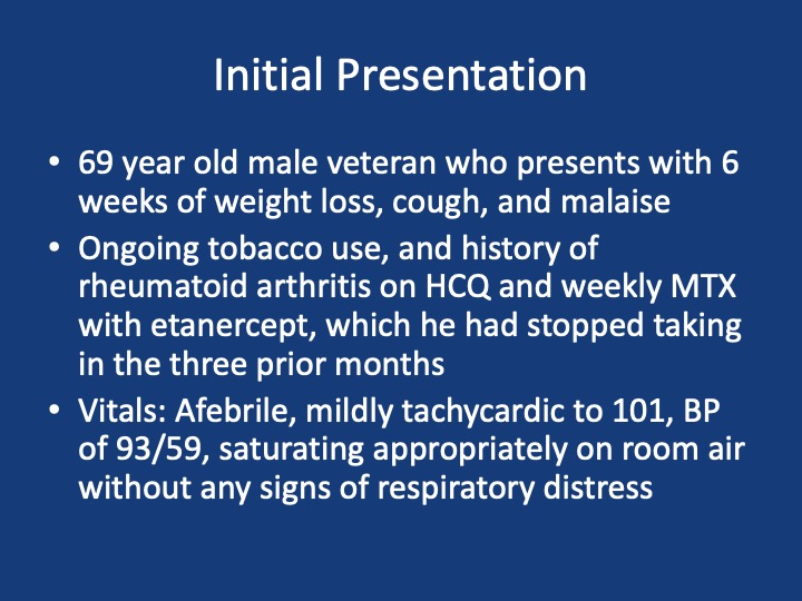
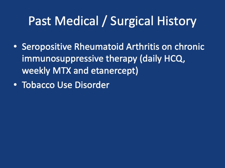
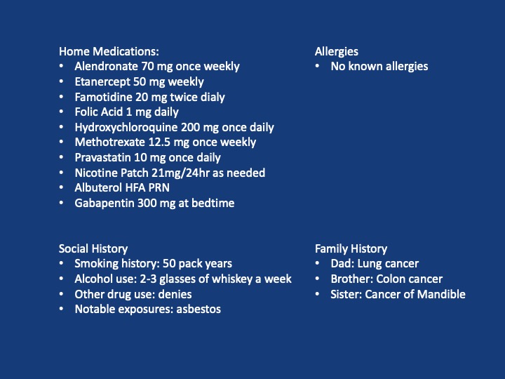
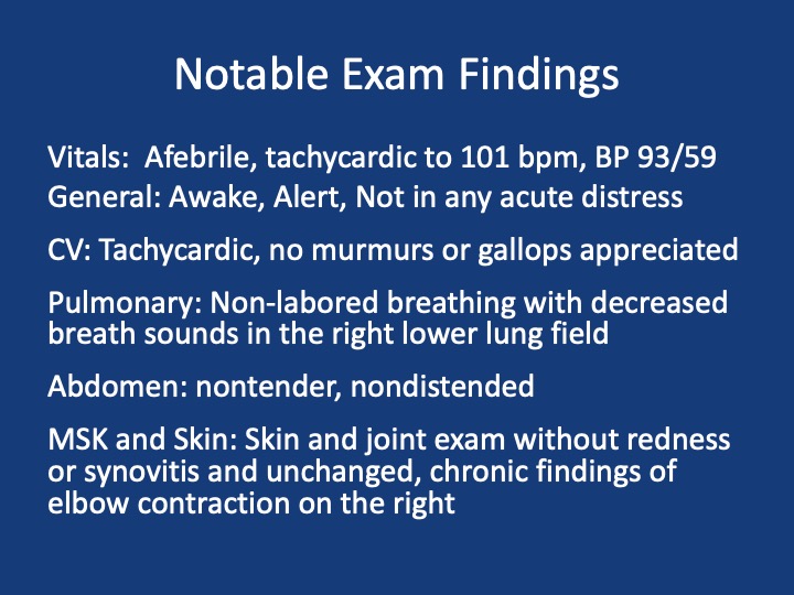
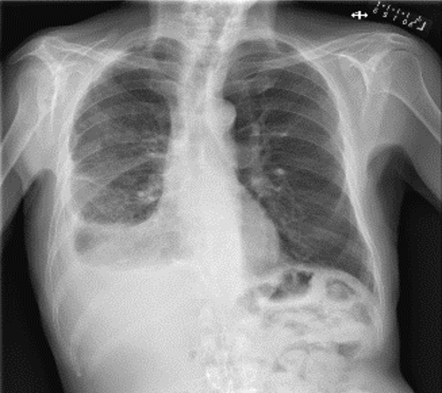
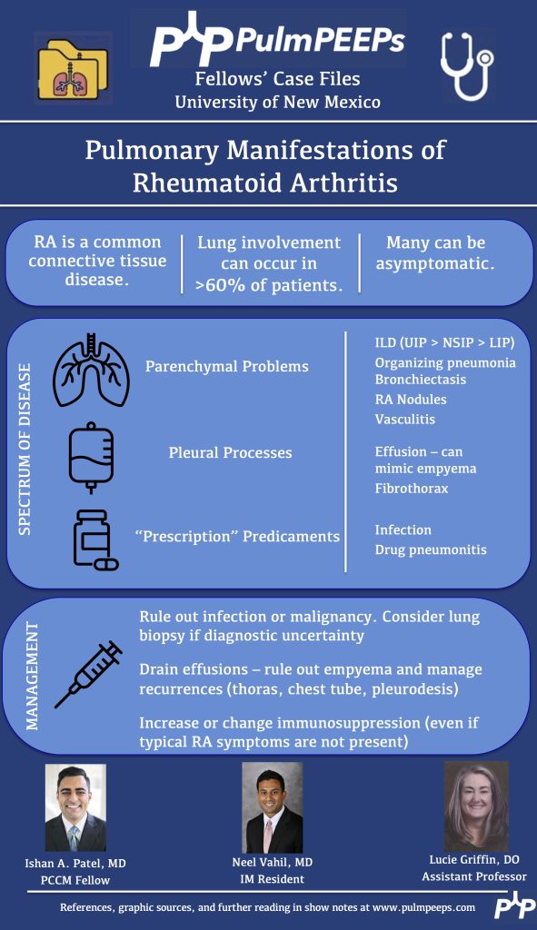
A 69 year old male veteran who presents with 6 weeks of weight loss, cough, and malaise. He has ongoing tobacco use, and history of rheumatoid arthritis on HCQ and weekly MTX with etanercept, which he had stopped taking in the three prior months. Vitals: Afebrile, mildly tachycardic to 101, BP of 93/59, saturating appropriately on room air without any signs of respiratory distress
Key Learning Points
References and Further Reading
Komarla A, Yu GH, Shahane A. Pleural effusion, pneumothorax, and lung entrapment in rheumatoid arthritis. J Clin Rheumatol. 2015;21(4):211-215.
Boddington MM, Spriggs AI, Morton JA, Mowat AG. Cytodiagnosis of rheumatoid pleural effusions. J Clin Pathol. 1971;24(2):95-106.
Balbir-Gurman A, Yigla M, Nahir AM, Braun-Moscovici Y. Rheumatoid pleural effusion. Semin Arthritis Rheum. 2006;35(6):368-378
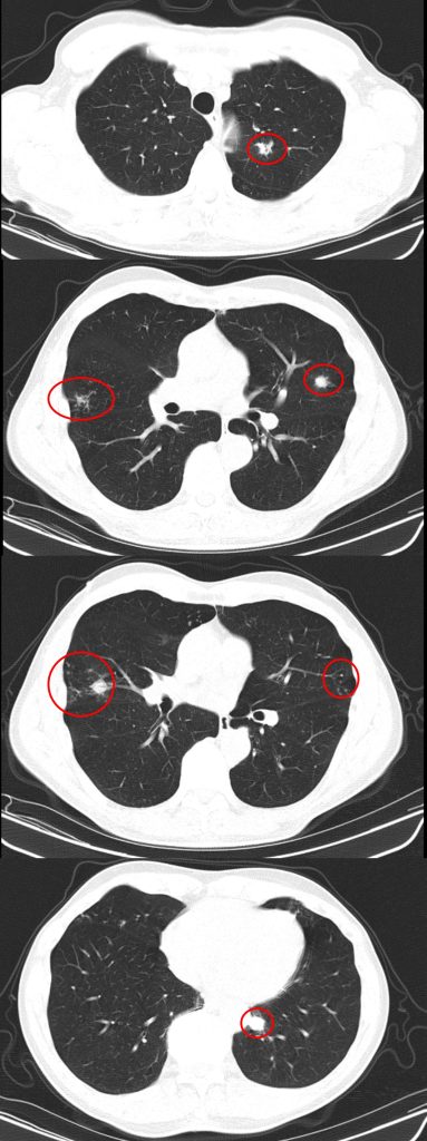
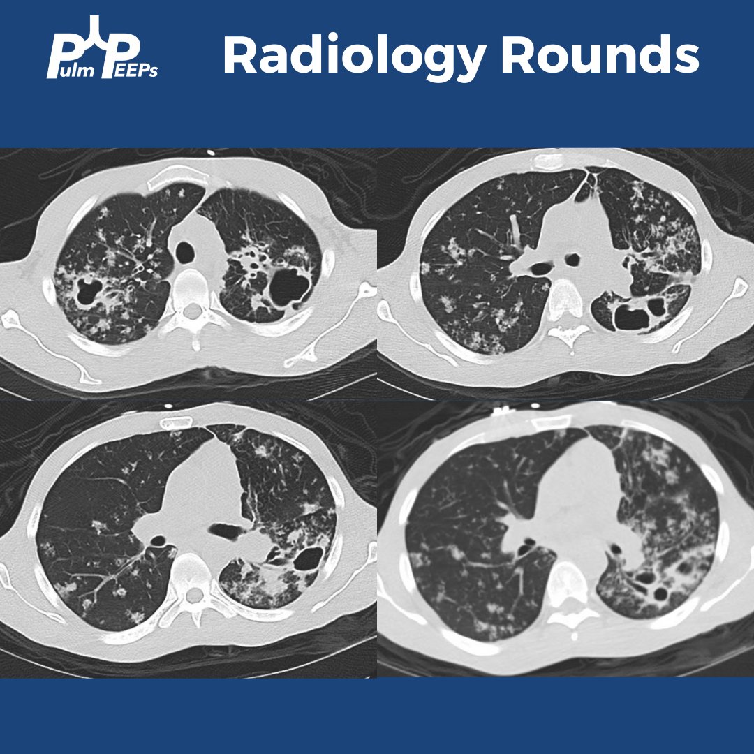
A #RadiologyRounds case with 3 different imaging modalities! A 65+ year old man never smoker, former marathoner has had 2-3 years of progressive non-productive, incessant cough with decreasing exercise tolerance. Some select CT scan slices are below
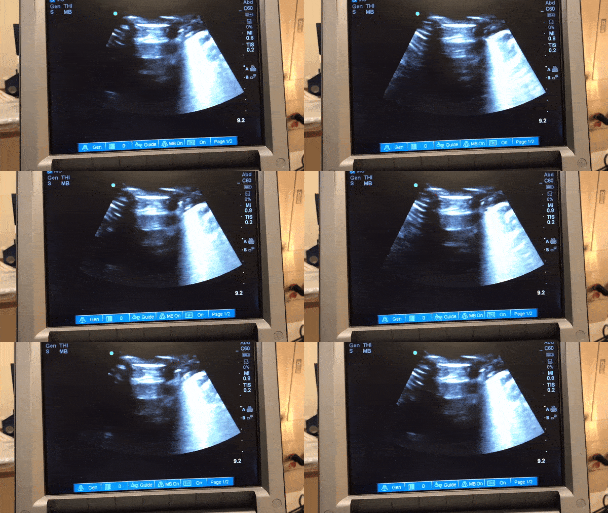
You are considering multiple etiologies including airway bleeding, pneumothorax, and hemothorax. You grab an ultrasound and perform a lung / pleural POCUS. Here is what you see
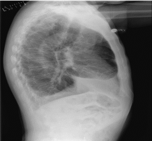
The long POCUS shows an area of lung sliding and an area without any lung sliding. This is called lung point and is diagnostic of a pneumothorax. To get a better look at this, you can use M-mode.
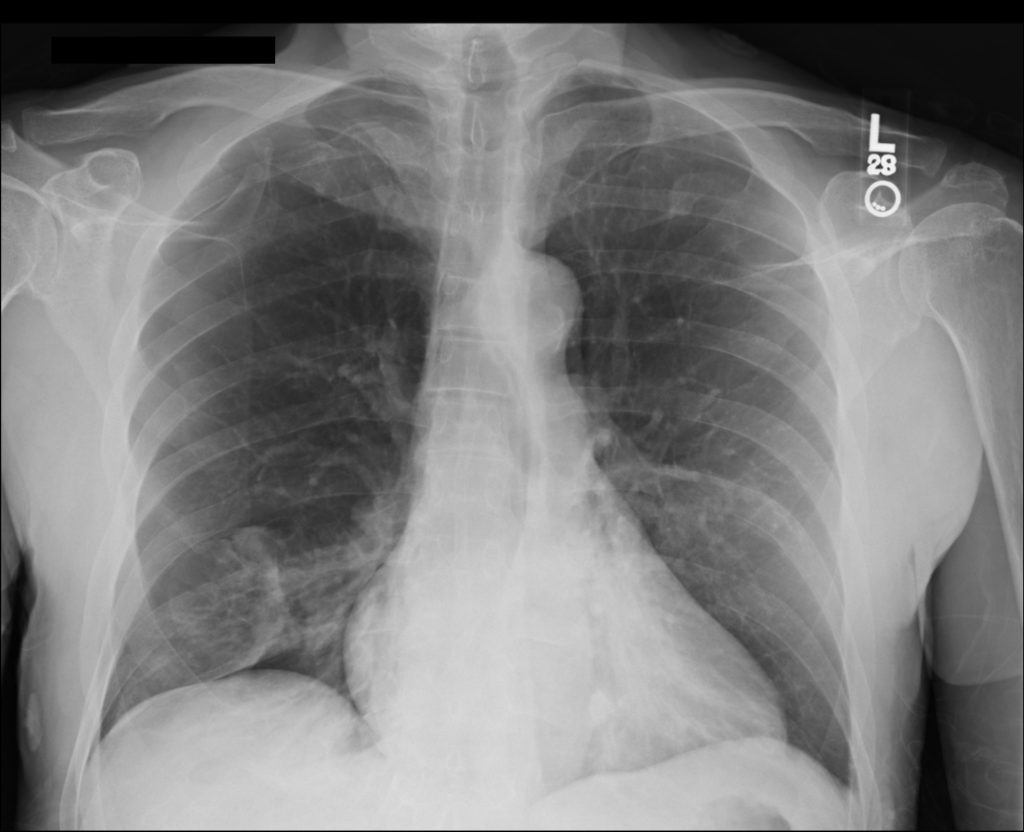
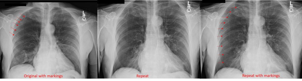
A CXR confirmed the finding of a pneumo. He was trialed on 100% oxygen but repeat CXR showed the pneumothorax was expanding. He had a chest tube placed with re-expansion of his lung and no air leak. It was able to be removed the next morning without incident
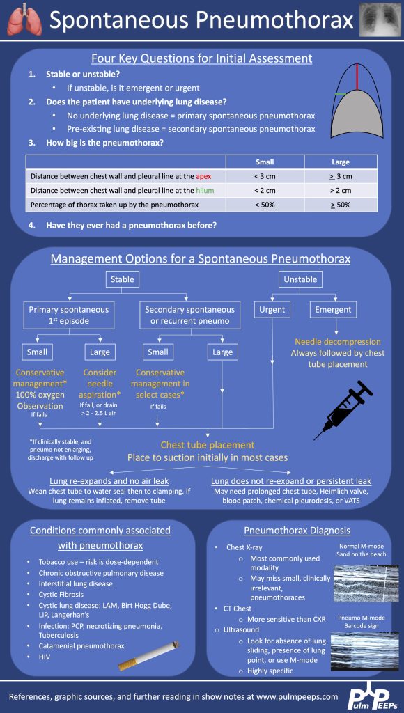
Here is our algorithm for pneumothorax!
Both BAL and tbbx returned positive for MAI complex. He was HIV neagative. Given his persistent and bothersome symptoms, he was started on treatment for pulmonary MAC with a macrolide, ethambutol and a rifamycin with plan for 6 months of therapy. He improved with this
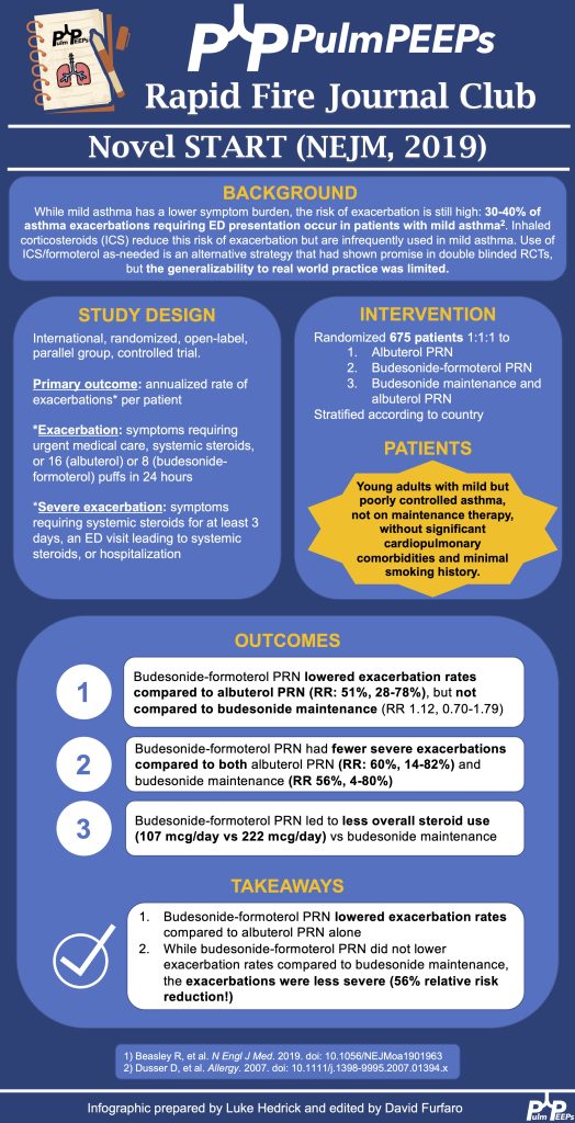
Today on our Rapid Fire Journal Club series, we’re discussing the Novel START study published in the NEJM in 2019. This study evaluated multiple strategies for the management of mild asthma with exacerbations, and it guides our current therapeutic approach. Pulm PEEPs Associate Editor Luke Hedrick walks us through the study. If you take care of asthma patients, be it in a primary care clinic, pulmonary clinic, or the hospital, make sure to listen in!
Article and Reference
Today we’re discussing the 2019 Novel START Study published in NEJM
Time for another Radiology Round by our Associate Editor Tess Litchman!
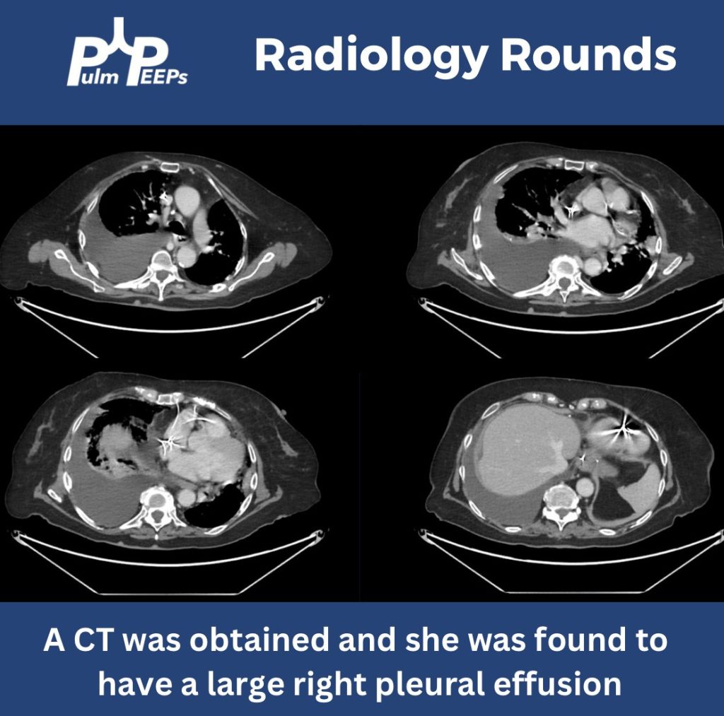
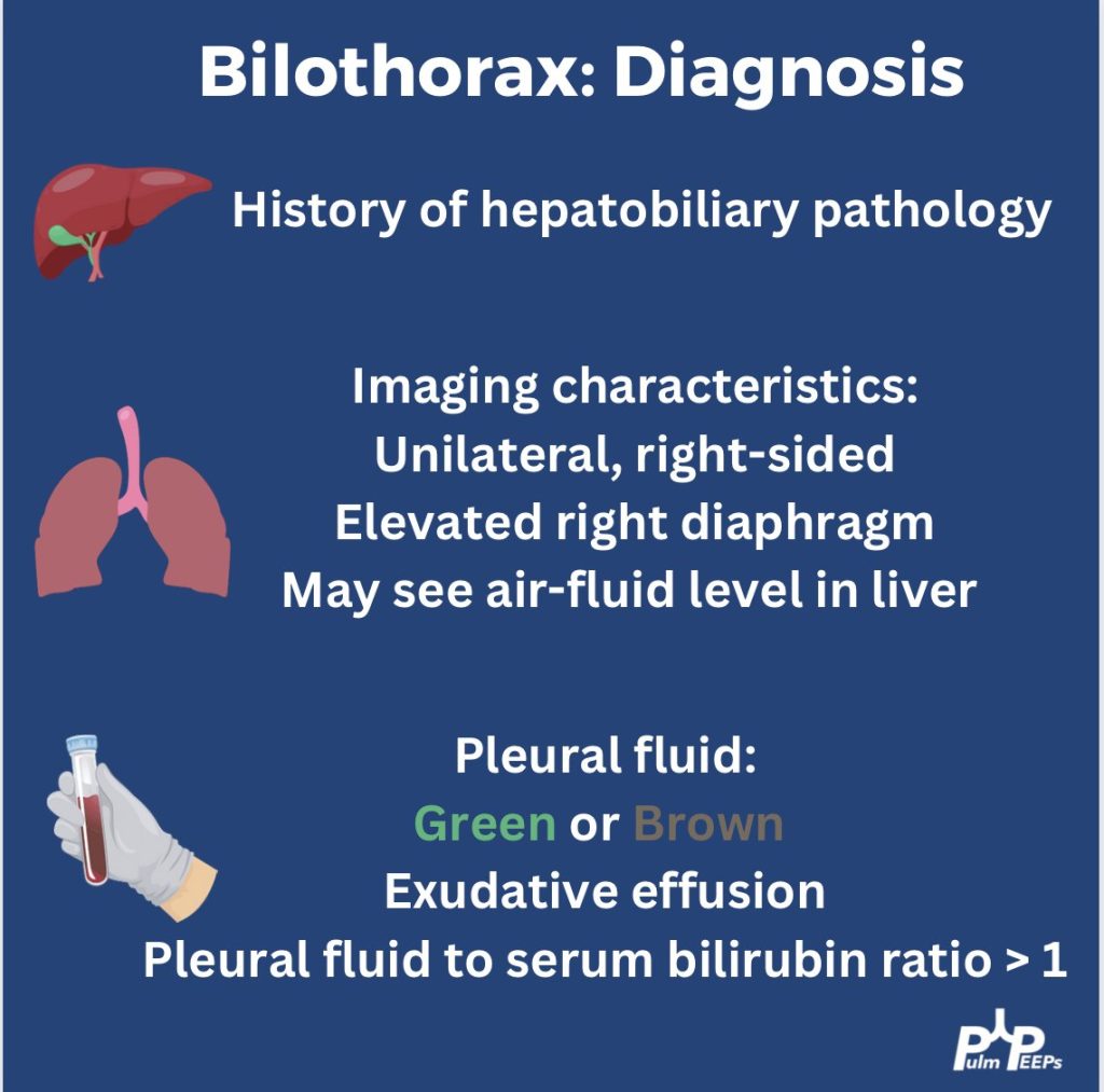
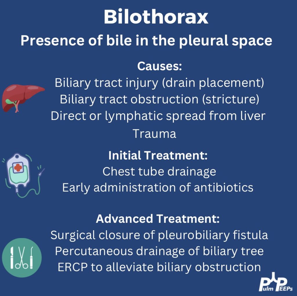
We have an older-aged woman found to have gastric and duodenal ulcers requiring multiple abdominal surgeries and transhepatic biliary drainage presenting with respiratory distress
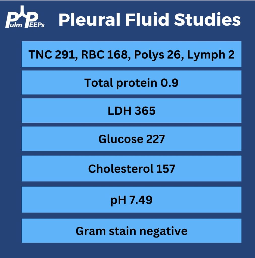
She underwent pleural fluid drainage with the following pleural fluid studies
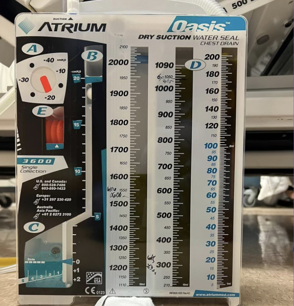
She ultimately had a right chest tube placed with 1.6 liters of green pleural fluid was drained
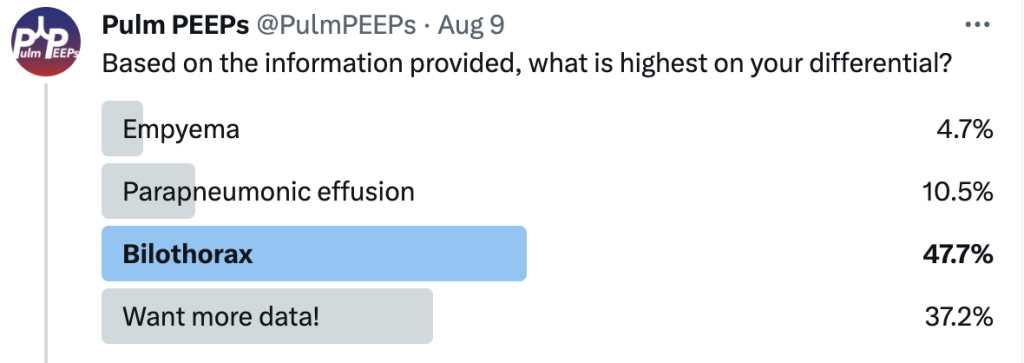
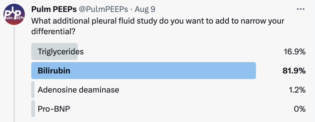
Given concern for a bilothorax, a pleural and serum bilirubin test were added. Her pleural bilirubin level was 17.2 and her serum bilirubin level was 0.5. Her chest tube was maintained, IV antibiotics were started and multi-disciplinary meetings occurred to determine next steps.
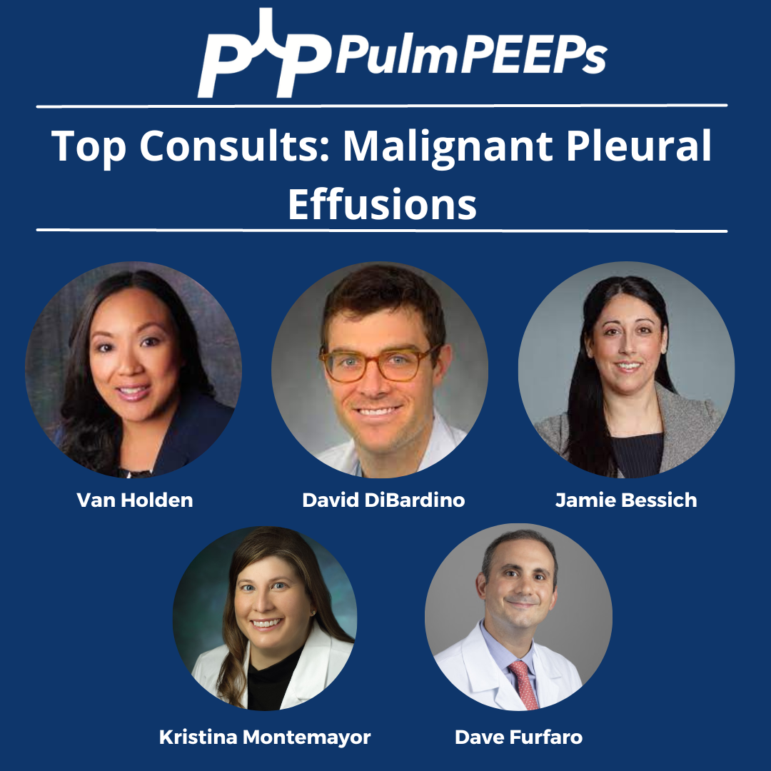
We’re diving back into some pleural fluid today (okay that is kind of gross to think about and we apologize). If you haven’t listened to our prior pleural effusion episodes and want to start from the top, then check out Episode 36 for a general approach to pleural effusions, and Episode 37 to hear about parapneumonic effusions. Malignant effusions are another common consult question and we’ll talk about everything from detection to monitoring, to definitive management.
Meet Our Guests
Dr. David DiBardino is an Assistant Professor of Medicine at the University of Pennsylvania Medicine and is the Associate Director for Clinical Research within the Section of Interventional Pulmonology. He is also the Program Director for the Interventional Pulmonary Fellowship there.
Dr. Jamie Bessich is an Assistant Professor of Medicine and Cardiothoracic Surgery at NYU Grossman School of Medicine. She is the Section Chief of Interventional Pulmonology and is the Director of Bronchoscopy at Tisch Hospital.
Dr. Van Holden is an Associate Professor of Medicine at the University of Maryland School of Medicine and is the Pulmonary and Critical Care Fellowship Program Director there. She was last on the show for our very first Fellows’ Case Files when we discussed a fascinating case of pulmonary alveolar proteinosis.
Case Presentation
The page: 72M smoker, new effusion, concern for malignancy, tap?
Further history: 72 year old man with PMH of GOLD B COPD, tobacco use (55 pack years), HTN, HLD, and diabetes. He presented to the ED with progressive dyspnea and fatigue. He is on LAMA/LABA for his COPD, and he does not frequently have exacerbations. He has no increased sputum production or wheezing, but he has been feeling progressively fatigued and lethargic. H Over the past few weeks he has had more dyspnea on exertion, and now has it at rest too. It is a bit worse when he lies flat. He has had no weight gain or edema in his legs and has actually lost 10-15 pounds in the last 3 months. In the ED, he is newly requiring 3-4L NC, has decreased breath sounds on the right, and a CXR shows a large right-sided pleural effusion, as well as a large apical nodule with some spiculation, both of which are new from prior.
Key Learning Points
Causes of malignant pleural effusion
–Lung cancer is the most common in men
–Breast cancer is the most common cause in women
–Lung and breast cancer account for > 50% of all malignant pleural effusions
–Other less common causes are lymphoma, GU or GI tract cancer
–Remember to consider mesothelioma
Prognosis of MPE
–Malignant pleural effusion means the cancer is advanced and stage 4 by definition
–The average life expectancy after a diagnosis of MPE is 3-12 months, depending on the patient and the malignancy
Imaging and MPE
–Make sure to get a CT scan after drainage so no lesions are missed
–Ultrasound can be helpful to look for disruptions of the pleural line, loculated fluid, or pleural nodules
Pleural fluid analysis
–Make sure to send common labs (gram stain, culture, pH)
–Cell count is very important as most MPE are lymphocyte-predominant
–Triglycerides can be helpful as well (more on chylothorax in future episodes)
–Cytology is essential and makes the diagnosis. The sensitivity is 65 – 75 percent so repeat taps may be needed and you need to send at least 60 cc of fluid (often more)
–If you have high suspicion and no diagnosis after two taps, pleuroscopy and pleural biopsy is warranted
Management of MPE
–First you need to determine if the MPE is recurrent. This requires drainage and then monitoring
— The main options are repeat thoracenteses, pleurodesis, or indwelling pleural catheter. A combination can often be used, and shared decision making is essential to the determining the best option
–The three things to consider with recurrent malignant pleural effusion are:
Did the patient feel better after drainage?
Did the lung fully re-expand?
What is the best option for this specific patient to optimize quality of life and time outside of the hospital?
–In AMPLE, pleural cetehters and talc pleurodesis were compared, and both are reasonable options with equivalent outcomes on quality of life; although pleural catehters had fewer hospital days overall.
It is Tuesday #RadiologyRounds time! We are in a pleural state of mind here at Pulm PEEPs. This is another great case authored by rockstar-associate editor @TessLitchman
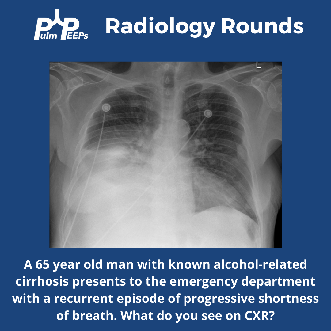
A 65-year-old man with cirrhosis presents to the ED with progressive shortness of breath:
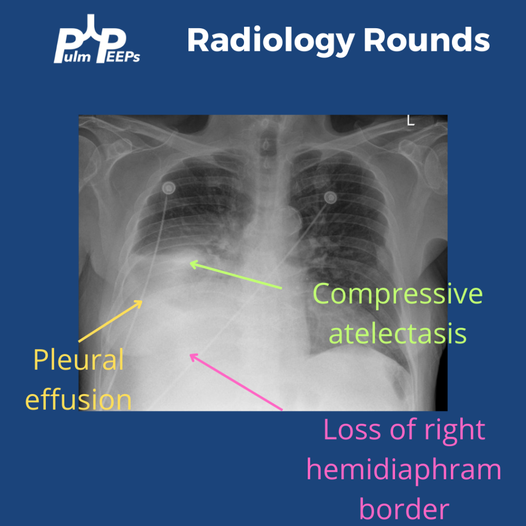
The CXR has a right lower opacity decreasing in density that is silhouetting the right hemi-diaphragm without signs of volume loss on the right concerning for pleural effusion. There is also associated atelectasis adjacent to the effusion
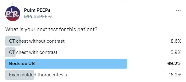
What is your next step for this patient?
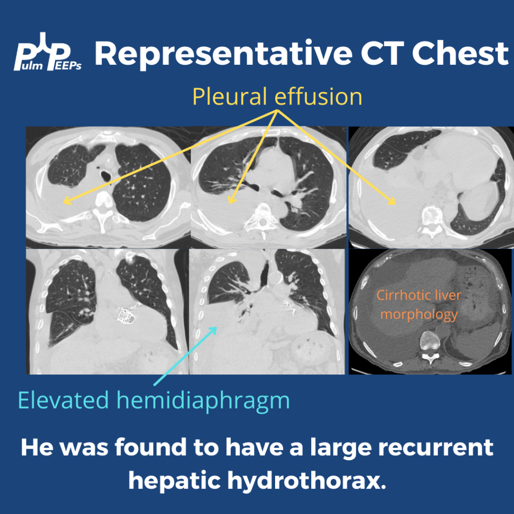
This patient had a bedside POCUS revealing a simple pleural effusion and abdominal ascites. He also had a CT scan performed:
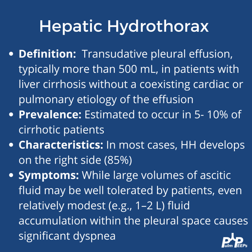
Based on his imaging and history, the most likely diagnosis on the differential was a hepatic hydrothorax. Here is some more information on hepatic hydrothoraces:
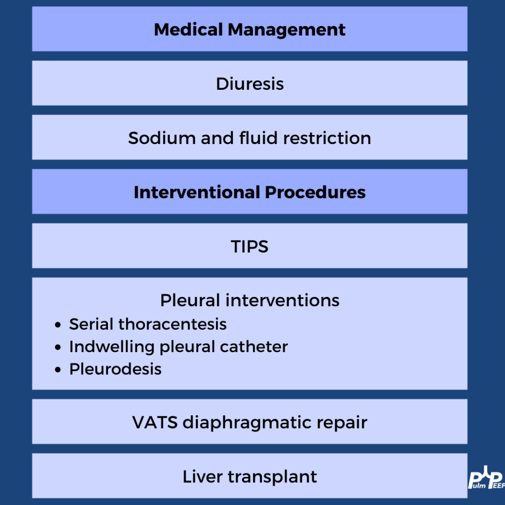
A transudative effusion was confirmed on thoracentesis, and no other clear etiologies were identified The treatment of hepatic hydrothorax should always start with medical management of volume overload in cirrhosis. Pleural procedures can be used for disease that is refractory
We continue our Top Consult Series on Pleural Disease and bring you a dedicated episode on Parapneumonic effusions. We are joined by two guest experts, Dr. David Feller-Kopman and Dr. Mihir Parikh. Listen in as we discuss the spectrum of parapneumonic effusions, including simple parapneumonic effusions, complicated parapneumonic effusions, and empyema. You will hear what to look for on imaging, what tests to send with pleural drainage as well as discuss the need for surgical consultation.
Meet our Guests
Dr. Mihir Parikh is currently an Assistant Professor of Medicine and academic interventional pulmonologist at Beth Israel Deaconess Medical Center. He is a highly esteemed educator and has worked to incorporate simulation training to improve procedural training for trainees and is a master of pleural disease.
Dr. David Feller-Kopman is a Professor of Medicine and the Section Chief of Pulmonary and Critical Care Medicine at Darmouth whose clinical and research expertise span the field of interventional pulmonology. Dr. Feller-Kopman is a true master of pleural disease, and has authored more than 225 peer-reviewed manuscripts and has been a leader for both ATS and CHEST committees.
Today the PulmPEEPs are joined by two amazing educators as we start off our Top Consult series on Pleural Disease. Join us today as we go through cases to learn a systematic approach for evaluation and management of pleural effusions.
Meet our Guests
Dr. Mira John received her medical degree from Tulane University School of Medicine in New Orleans and completed internal medicine residency at Icahn School of Medicine at Mount Sinai. She is currently a second-year pulmonary and critical fellow at the University of Washington.
Dr. Ylinne Lynch completed her fellowship training at the University of Washington and is currently a Clinical Instructor at the UW. She is a great medical educator and spends her clinical time on the pulmonary consult service as well as in the ICU.