Today we’re joined by Dr. Molly Hayes and talking about an upcoming CME course: Principles of Critical Care Medicine for Non-Intensive Care Specialists. This is a wonderful education course covering a wide array of topics in critical care in a case-based, systematic, and peer-reviewed fashion. The course is interactive and all resources will be available for attendees afterwards for their review. You can sign up at https://learn.hms.harvard.edu/criticalmed and save using the discount code “PulmPEEPS50”!
Meet Our Guest
Dr. Molly Hayes is an Associate Professor of Medicine at Beth Israel Deaconess Medical Center and Harvard Medical School, the Director of the MICU at BIDMC, and the Director of External Education at the Carl J Shapiro Institute for Education and Research. She additionally is a co-founder of the BIDMC Center for Humanizing the ICU.
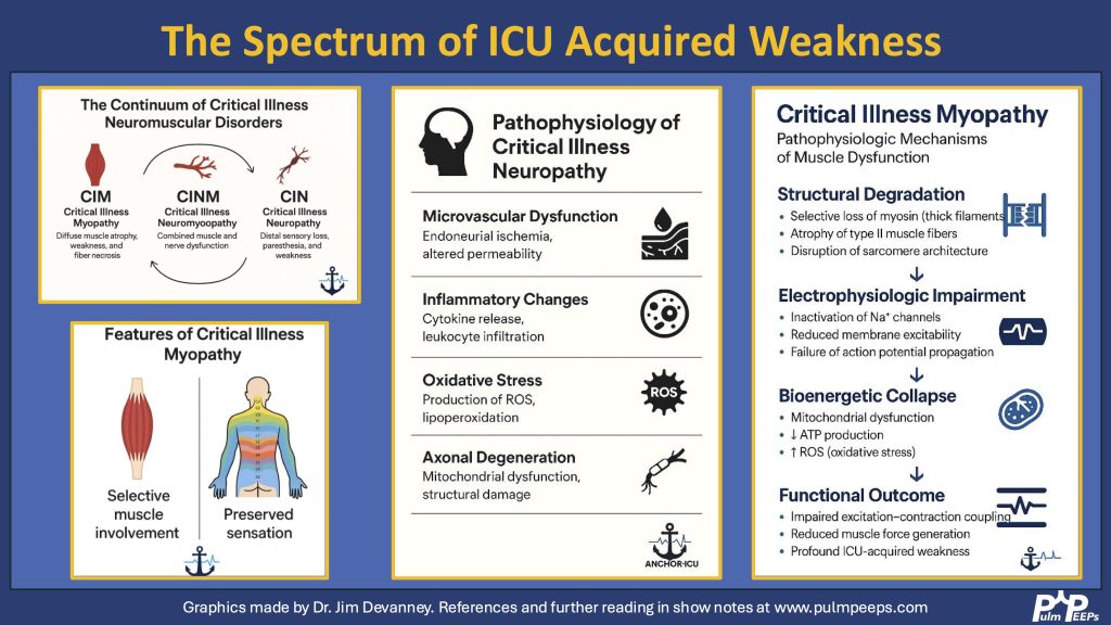
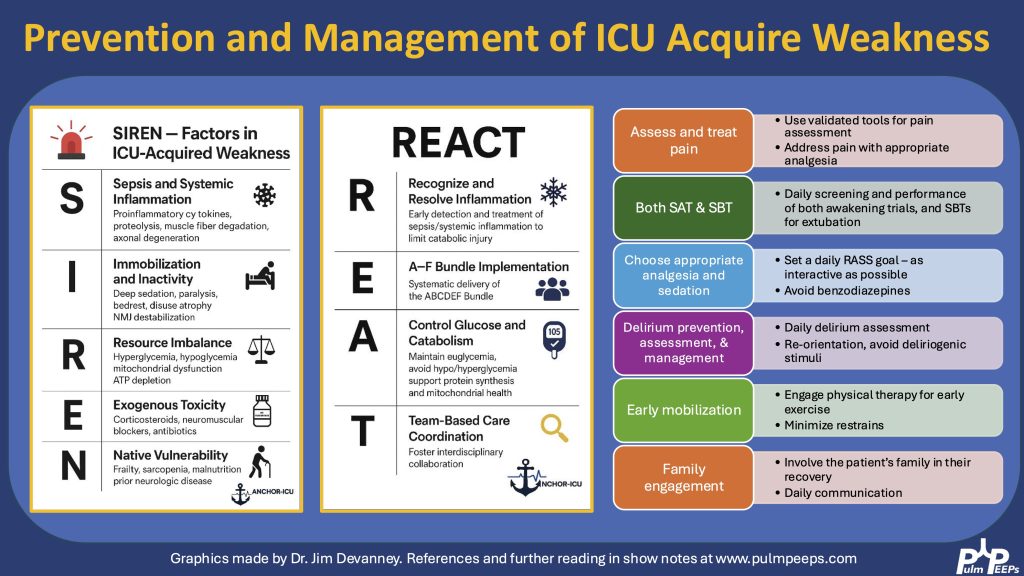
Today we’re talking about a topic that is relevant for all critical care physicians but under-examined: ICU Acquired Weakness. We are joined by two excellent guests to walk through a case and discuss the diagnosis, pathophysiology, prevention, and treatment of ICU Acquired Weakness. Check out our associated infographics and key learning points below.
Meet Our Guests
Jim Devanney is a Physiatrist who just completed a neurocritical care fellowship at BIDMC. He is transitioning to a clinical associate position at University Health Network – University of Toronto where he will be working as a PM&R consultant within the ICU.
Kalaila Pais is a third year internal medicine resident at BIDMC, interested in pulmonary and critical care and medical education and is returning for her third Pulm PEEPs episode.
Key Learning Points
Definition & Clinical Presentation
ICU-AW refers to new-onset, generalized muscle weakness that arises during critical illness, not explained by other causes.It typically presents as:
Symmetric, proximal > distal weaknessRespiratory muscle involvementPreserved cranial nerve functionNo sensory deficits in myopathy (sensory loss points toward neuropathy)
Differential Diagnosis Using Neuroanatomical ApproachAn anatomical approach (central → peripheral) helps localize the etiology weakness
Today we have a mini-episode / clinical pearl. We previously discussed the PROSEVA trial and the evidence for prone positioning in ARDS. In that trial, patients with elevated intracranial pressure (ICP) were excluded. We are joined now by Dr. Jon Rosenberg, a neuro intensivist, to discuss his how prone positioning can still be employed for patients with neurologic injuries and elevated ICP.
Meet Our Guest
Dr. Jon Rosenberg is an assistant professor of neurology and neurosurgery at Westchester Medical Center, New York Medical College. He’s also the associate program director of the Neurocritical Care Fellowship at Westchester Medical Center and a frequent contributor to the Neurocritical Care Society podcast.
Key Learning Points
Elevated Intracranial Pressure (ICP) and Proning: A Common Misconception
Elevated ICP is often considered a contraindication to proning, but this is more of a relative caution rather than an absolute contraindication.
Many neuro ICUs have successfully proned patients with elevated ICP, particularly since the COVID-19 pandemic, when critical care units had to manage both respiratory failure and neurological conditions simultaneously.
Patient Selection for Proning with Elevated ICP
Most patients with elevated ICP can still be proned, including those with:
Global cerebral edema (e.g., post-anoxic brain injury, liver failure)
Situations where proning might be more concerning:
Severe hemodynamic instability (multi-pressor shock)
Morbid obesity (e.g., >300 lbs), where physically flipping the patient is a major challenge
Theoretical Concerns with Proning in Elevated ICP
Loss of neurological exam access (sedation + flipped position makes pupil and motor exam difficult)
Jugular venous compression (especially if the head is turned to one side)
Cerebrospinal fluid (CSF) flow obstruction, depending on the lesion
Risk of increased ICP if venous outflow is impaired or head positioning is not optimized
Best Practices for Proning Patients with Elevated ICP
Patients with invasive ICP monitors vs. without monitors:
If possible, placing an ICP monitor (EVD or parenchymal bolt) before proning provides better guidance.
Without a monitor, providers must rely on other practices like maintaining strict MAP goals and sodium targets, and indirect signs of increased ICP.
Positioning considerations:
Keep the head midline to prevent jugular venous compression.
If head positioning is not neutral, place the dominant/internal jugular facing upward to maintain venous drainage.
Maintain the head of the bed elevated even while prone (reverse Trendelenburg positioning).
Hemodynamic management:
Target a higher MAP (e.g., 70–75 mmHg, sometimes 75–80 mmHg) to ensure adequate cerebral perfusion pressure (CPP) if there is no ICP monitor
Avoid hypotension, as MAP – ICP = CPP, and low MAP could critically reduce cerebral perfusion.
A normal intracranial pressure is 7 – 15 mmHg
The recommended CPP is between 60 – 70 mmHg
Sedation & Sodium Management:
Consider deep sedation (RASS -5) to reduce metabolic demand and intracranial blood volume.
Consider keeping sodium >145 mmol/L prophylactically to mitigate brain swelling if no ICP monitor in place
When to Reconsider Proning (i.e. when to supinate)
If a patient’s ICP spikes significantly (e.g., from 20 to 60 mmHg) despite medical management (hypertonic saline, sedation, paralysis, etc.).
If new signs of neurological deterioration emerge (e.g., changes in pupil exam once patient is repositioned).
Hemodynamic instability that is unmanageable in the prone position.
Literature and Future Considerations
Small case series have demonstrated success in proning patients with traumatic brain injury (TBI) and aneurysmal subarachnoid hemorrhage.
While more formal research is needed, the neurocritical care community has begun embracing proning for neuro patients, provided that proper precautions are taken.
Bottom Line
Proning is not an absolute contraindication for patients with elevated ICP—it can be done safely with proper monitoring, patient selection, and precautions.
Having an ICP monitor makes the process more controlled and allows clinicians to adjust treatment in real time.
Key considerations: Maintain cerebral perfusion, optimize head positioning, monitor hemodynamics, and have a plan for reversing if ICP becomes unmanageable.
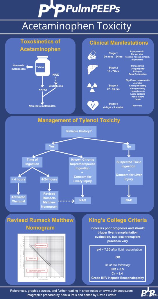
This week we’re talking about a case as a lens for discussing Tylenol toxicity and Acute Liver Failure. These relatively common critical care presentations are essential knowledge for anyone practicing in the ICU. Listen in for some key discussion both about toxicology and the diagnosis and management of acute livery injury and failure.
Meet Our Guests
Kalaila Pais received her MD from Howard University College of Medicine and is currently a second year internal medicine resident at BIDMC. She is interested in pulmonary and critical care, as well as medical education. She also had the idea for this episode and was essential in its writing and production.
Hima Veeramachaneni received her MD from University of Missouri-Kansas City School of Medicine, and her residency at Emory where she was also a Chief Resident at Grady Memorial Hospital. She is a gastroenterologist and completed her GI and transplant hepatology training at Emory. She is also now doing a critical care medicine fellowship year.
Case Presentation
Presentation: Patient found down, surrounded by liquor bottles, with coffee-ground emesis, hemodynamic instability, scleral icterus, and metabolic derangements.
Key Lab Findings:
Severe transaminitis (AST >10,000, ALT ~3,000).
Elevated bilirubin (5.8), lactate (16), and INR (>2).
Metabolic acidosis with a pH of 7.04.
Tylenol level: 41 (slightly elevated but inconclusive without ingestion timing).
Key Learning Points
Infographic:
Acute Liver Injury vs. Acute Liver Failure
Acute Liver Injury (ALI): Elevated liver enzymes without encephalopathy or significant synthetic dysfunction.
Acute Liver Failure (ALF): Defined by:
Presence of encephalopathy.
Coagulopathy (elevated INR).
Rapid onset (<26 weeks) in patients without pre-existing liver disease.
ALF often leads to complications such as cerebral edema, which necessitates aggressive management.
Tylenol Toxicity and Interpretation
Pathophysiology:
Tylenol overdose overwhelms liver glutathione, leading to accumulation of NAPQI, which causes hepatocyte necrosis.
Interpretation of Tylenol Levels:
Timing of ingestion is critical to interpreting levels.
The Rumack-Matthew Nomogram is used for acute ingestions but requires a known ingestion time.
Management:
N-acetylcysteine (NAC): Standard of care; acts as a glutathione precursor and mitigates liver damage.
Early use is recommended in suspected cases of Tylenol toxicity, even if ingestion timing is unclear.
Critical Management Principles
Stabilization: Focus on airway, hemodynamics, and perfusion.
Monitor for signs of cerebral edema (e.g., pupillary changes, seizures).
In select patients, use hypertonic saline to maintain sodium levels (145–150 mmol/L) to mitigate cerebral edema risks.
CRRT and Plasma Exchange:
Continuous renal replacement therapy (CRRT) for hyperammonemia and acidosis.
Plasma exchange (PLEX) may stabilize cytokine storms and improve survival.
Organ-Specific Considerations:
Renal failure: Common due to hepatorenal syndrome; requires CRRT.
Coagulopathy: Managed with blood products as needed but indicates worsening liver synthetic dysfunction.
Prognosis and Transplant Considerations
King’s College Criteria: Guides transplant listing for ALF patients.
Welcome to our second episode of ATS 2024 highlighting content featured through the ATS Critical Care Assembly. Today we are going to be talking about one of the Critical Care Assembly Symposiums entitled: “Care of Dying in the ICU: End of Life Care in 2024 and Beyond”
Meet our Guests
Dr. Theodore “Jack: Iwashyna is a Bloomberg Distinguished Professor at Johns Hopkins School of Medicine and the Johns Hopkins Bloomberg School of Public Health. Jack is a critical care physician and has a broad focus on research that understands the broader context of critical illness, and the long term impact on patients’ lives. He is an enormously productive and successful researcher with numerous publications in the field of critical care, and is a pioneer in the field of ICU survivorship. He is a devoted mentor and has received accolades from numerous societies
Dr. Molly Hayes is an Associate Professor of Medicine at Beth Israel Deaconess Medical Center and Harvard Medical School, the Director of the MICU at BIDMC, and the Director of External Education at the Carl J Shapiro Institute for Education and Research. She additionally is a co-founder of the BIDMC Center for Humanizing the ICU. Molly has been extensively involved with ATS with leadership roles in the Critical Care Assembly, and the newly minted Steering Committee on the Advancement of Learning.
Meet our Collaborators
The American Thoracic Society Critical Care Assembly is the largest Assembly in the American Thoracic Society. Their members include a diverse group of intensivists and care providers for both adult and pediatric critically ill patients. The primary goal of the Critical Care Assembly is to “improve the care of the critically ill through education, research, and professional development.”
Welcome to our first episode of ATS 2024 highlighting content featured through the ATS Critical Care Assembly. Today we are going to be talking about one of the Critical Care Assembly Symposiums entitled: “A New Reality for Critical Care after Dobbs.”
Meet our Guests
Dr. Katie Hauschildt is a Faculty Research Associate at The Johns Hopkins University School of Medicine where she conducts research on equity in healthcare and critical illness recovery. She has her PhD in Sociology from the University of Michigan and an Advanced Fellowship in Health Services Research from the VA Ann Arbor Healthcare System, and is a board certified patient advocate.
Dr. Kathleen Akgün is an Associate Profess or Medicine at the Yale School of Medicine. She is the Association Section Chief for the VA section of Pulmonary, the Co-Director of the Network of Dedicated Enrollment Sites Program, the director of the MICU at the VA Connecticut health care system, and a member of the DEI Working Group at Yale.
Meet our Collaborators
The American Thoracic Society Critical Care Assembly is the largest Assembly in the American Thoracic Society. Their members include a diverse group of intensivists and care providers for both adult and pediatric critically ill patients. The primary goal of the Critical Care Assembly is to “improve the care of the critically ill through education, research, and professional development.”
MacDonald A, Gershengorn HB, Ashana DC. The Challenge of Emergency Abortion Care Following the Dobbs Ruling. JAMA. 2022;328(17):1691-1692. doi:10.1001/jama.2022.17197
Ashana DC, Chen C, Hauschildt K, et al. The Epidemiology of Maternal Critical Illness Between 2008-2021. Ann Am Thorac Soc. Published online June 14, 2023. doi:10.1513/AnnalsATS.202301-071RL
Sonntag E, Akgun KM, Bag R, et al. Access to Medically Necessary Reproductive Care for Individuals with Pulmonary Hypertension. Am J Respir Crit Care Med. Published online June 13, 2023. doi:10.1164/rccm.202302-0230VP
Griffin KM, Oxford-Horrey C, Bourjeily G. Obstetric Disorders and Critical Illness. Clin Chest Med. 2022;43(3):471-488. doi:10.1016/j.ccm.2022.04.008
We’re super excited to have a joint episode this week with Dr. Cyrus Askin and Dr. Nick Mark from Critical Care Time! We discuss all the ways that bronchoscopy can be your best friend in the ICU and how to be prepared for the unexpected scary situations that arise in the ICU. This ranges from airway bleeds, difficult intubations, lobar collapse, and trach emergencies. Don’t miss this great discussion!
Key Learning Points
Utility of bronchoscopy in people with critical illness
Bronchoscopy can be both diagnostic and therapeutic; both are potentially lifesaving.
General situations where bronchoscopy is useful in the ICU:
Placing (or confirming placement of) an endotracheal tube or tracheostomy tube
Removing a foreign body or mucous plugs from the lungs
Localizing the source of pulmonary hemorrhage or performing interventions to stop/contain the bleed
Diagnosing certain rare conditions, particularly those where the diagnosis can substantially change management (e.g. DAH, AEP, rare infections, etc).
Proficiency with bronchoscopy is important to realize the benefits. Simply “having the equipment” is insufficient, regular practice/simulation is essential
Anesthesiologists, emergency physicians, and other specialists may have limited experience with bronchoscopy in training. Even experienced pulmonologists, who may be good at diagnostic bronchoscopy often have limited experience deploying bronchial blockers, using retrieval baskets, etc.
Remember: “People don’t rise to the occasion, they sink to the level of their training.”
If you haven’t regularly practiced with a bronchoscope, you are not going to be able to use it effectively under stress when performing high acuity low occurrence (HALO) procedures such as in emergent airways, deploying bronchial blockers, retrieving foreign bodies, etc.
Two broad situations where a bronchoscope is generally used:
Awake intubation in the anticipated difficult airway (e.g. someone with abnormal anatomy, airway tumor, etc)
Rescue method in the unanticipated difficult airway (e.g. very anterior cords, difficulty with Bougie, etc)
Nasal vs Oral approach:
Oral approach is usually used in an unanticipated difficult airway
Nasal approach: More common if performing an awake intubation. Nasal is often better tolerated however epistaxis can make a difficult airway almost impossible.
Sedation strategy:
Full topicalization: lidocaine vs cocaine (equally effective and lidocaine is normally preferred, however the vasoconstriction action of cocaine may be helpful in preventing epistaxis).
Gurgling (Nick: from personal experience lidocaine is super gross)
Remember total dose of lidocaine: < 8 mg/kg
Ketamine
Ideal because it’s dissociative and analgesic, maintains respiratory drive and (maybe) airway reflexes
Consider scopolamine patch to reduce oral secretions
Dexmedetomidine
Great adjunct
One vs two operator
Especially in unanticipated difficult airways; the second operator can use VL/DL to facilitate visualization of the vocal cords.
Second operator can also be preparing for a surgical airway.
Equipment considerations:
Preload the endotracheal tube onto the bronchoscope. Use the bronchoscope as a bougie to guide the ETT through the vocal cords.
Suction! You want two – one connected to the bronch and one connected to a yankuer.
Disposable vs “good” scope
Remember to load the tube first!
Also remember to lube the tube!
Tracheostomy troubleshooting
Similarly to intubation, bronchoscopy can be very useful to confirm placement
Mechanics are similar to above
Goal is to avoid inadvertent placement of the tracheostomy tube into the soft tissues of the neck and to avoid putting air into those tissues (false lumen).
Advanced trick for exchanging tubes: You can use a disposable bronchoscope to exchange tubes: you can get it in, confirm placement, then cut it with trauma shears! Now you can slide the old tube out and put a new one in. (Don’t try this on a $40,000 fiberoptic bronchoscope!)
Ideally you should load the ETT onto the bronchoscope in advance (red arrow). If necessary however, you can cut the ETT and turn the disposable bronchoscope into a improvised exchange catheter. This technique is very useful for exchanging tracheostomy tubes.
Foreign Body Removal from airways
Bronchoscopy is invaluable for both diagnosis and treatment of foreign body aspirations.
Most commonly these aspirations are food (nuts, seeds, etc), teeth, pills, etc
Intubated is harder in many cases: no cough to help, hard to get foreign body out of the ETT.
Flexible vs rigid
Most objects can be retrieved using flexible bronchoscope; however 15-20% require rigid bronchoscopy
Flexible can reach smaller foreign bodies that are lodged more distally.
Rigid bronchoscopy is usually done if flexible bronchoscopy fails; an interventional pulmonologist wielding a rigid is superior but more invasive (requires GA)
Cryoprobe can be especially useful for frangible materials (e.g. food)
Mucous Plugs & Lobar collapse
Presentation can be subtle or dramatic.
Bronchoscopy can remove mucous plugs and help re-expand collapsed lung areas, which is potentially life saving.
Additionally, bronchoscopy can permit diagnosis of tracheal bronchus (bronchus sui)
Pig bronchus – 1-3% of people – have a RUL bronchus that comes off the trachea.
Often presents with RUL collapse in an intubated person.
Suction considerations and bronchoscope size
Remember that suctioning force is highly dependent (i.e. radius raised to the fourth power!) upon the working channel size. Use the largest size bronchoscopy possible when suctioning.
Remember that other interventions: regular inline suctioning, chest PT, adequate hydration, mucolytics are also important to prevent recurrent mucous plugging.
Localization & Isolation of Pulmonary Hemorrhage
Pre-bronch interventions
Stabilization
Nebulized TXA
Bad side down → counter-intuitive because shifting blood flow, but also the goal is to protect the non-bleeding lung.
etc
Bronch can localize the bleeding site. Bronch can also perform interventions such as:
Cold saline
Epinephrine 1:100,000
Bronchial blockers – comparison of types
CRE balloon
Fogarty
Cryo probe – great for removing clots
Delivering ETT to contralateral side → single lung ventilation
Making “bronchoscopy only” diagnoses
Diffuse Alveolar Hemorrhage (DAH)
Finding: Increasingly bloody returns on serial lavages
1) an unexpected diagnosis was made and followed by a management change (as the most rigorous estimate of utility)
13% of the time a diagnosis was only made because of bronchoscopy which changed management
2) the post-bronchoscopy diagnosis was discordant from the leading diagnosis considered before this procedure and was followed by a management change
48% of the time pre and post procedure leading diagnoses were different
26% of the time the change in leading diagnosis led to a change in therapy
3) a change in management was made following bronchoscopy regardless of whether the diagnosis was expected or considered.
32% escalation of antibiotics
30% de-escalation of antibiotics
9% addition of steroids
2% mold → surgery
Remember that in critically ill patients whose symptoms are unexplained or failing to resolve with therapy, diagnostic flexible bronchscopy can provide useful insights.
Hi everyone, we’re here with another Fellows’ Case Files. Today, we’re going virtually to Emory University School of Medicine. We’re joined by Associated Editor Luke Hedrick to dive into a critical care case. Listen in and let us know if you have any additional thoughts or questions!
Meet Our Guests
Luke Hedrick is a first-year pulmonary and critical care fellow at Emory University. He did his internal medicine residency at BIDMC in Boston. He is also one of our amazing Associate Editors here at Pulm PEEPs
Shirine Allam is an Associate Professor of Medicine at the Emory University School of Medicine where she is the Program Director of both the Pulmonary and Critical Care Medicine fellowship as well as the Critical Care Medicine fellowship. She completed her PCCM training at the Mayo Clinic in Rochester, followed by a Sleep Medicine fellowship at Stanford. She has received multiple teaching awards throughout her career
Case Presentation
A 32-year-old male is brought in by his coworkers unresponsive. He is a construction worker and was his usual self in the morning at the start of the day, but when they broke for lunch they noticed he was acting different—his arms were drooping, and while he initially was able to answer yes/no, he soon started babbling, then grunting, then vomited and became unresponsive. They laid him flat, threw cold water on him because it was 110 degrees and humid outside that day, and brought him to the ED.
When they arrive in the ED, he is unresponsive and warm to the touch. His vitals are notable for an oral temperature of 105, HR in the 160s, BP 76/34, a RR in the high 30s, and an SpO2 100% RA. His exam is relatively unremarkable other than for significant diaphoresis and both bowel and bladder incontinence.
Key Learning Points
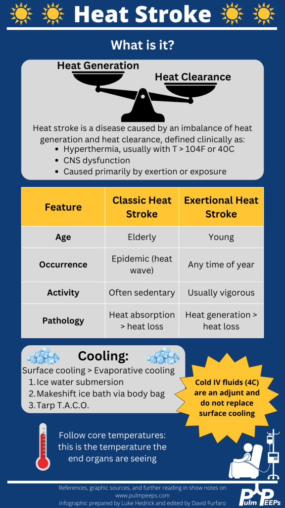
Definition and recognition of heat stroke: Heat stroke is characterized by hyperthermia (>104°F or 40°C) accompanied by CNS dysfunction, primarily caused by exertion or exposure. Encephalitis without significant heat load does not constitute heat stroke.
Management priorities: Rapid cooling is paramount to minimize long-term complications and organ failure. Cooling should be initiated as soon as possible, even before transportation to a hospital, particularly in cases of exertional heat stroke.
Cooling methods: Surface cooling, such as immersion in ice water, is the most effective way to cool heat-stroke patients. Alternative methods include the TACO method and evaporative cooling, although they are less efficient. Refrigerated IV fluids can be used as an adjunct, but they do not replace the need for surface cooling.
Monitoring and goals: Shivering during cooling should be monitored to prevent excessive heat generation. The goal is to reach a normal core body temperature (~38°C or 100.4°F). Traditional antipyretics like aspirin and acetaminophen should be avoided due to ineffectiveness and potential toxicity.
Approach to endotracheal tube (ETT) exchange: ETT exchange requires preparation for potential complications. This includes ensuring the availability of airway equipment, sedation of the patient, and having additional personnel for assistance. Direct visualization using a video laryngoscope is recommended, along with measuring and marking the exchange catheter for proper insertion depth.
The following infographic can be downloaded from our website:
This week on Pulm PEEPs, we are excited to be cross-posting an episode that Dave Furfaro did on the ATS Breathe Easy Podcast. Listen to hear a discussion about the best way to create a positive learning environment in the ICU, and how to effectively prepare bedside teaching for learners of all levels.
Meet The Host
Matthew Stutz hosted this episode of the ATS Breathe Easy Podcast. He is an Attending Pulmonary and Critical Care physician at Cook County Health and an Assistant Professor at Rush University. He is a dedicated educator and an active member of the American Thoracic Society.
Key Learning Points
Empowerment: It’s crucial to empower both learners and teachers in an educational setting.
Open Communication: Learners should be encouraged to express their discomfort or challenges in learning. This will allow teachers to adapt and create a more effective learning environment.
Self-awareness and Continuous Improvement: Teachers should be self-aware and continuously strive for improvement. If a teacher knows their weak points or areas they want to enhance, such as bedside teaching or teaching on rounds, they should communicate this to their team. This will make the team more observant and supportive in giving feedback.
Honesty: A genuine and honest dialogue helps in building a strong and trusting educational relationship. It’s beneficial for both the teacher and learner to be candid about their needs and challenges.
Feedback Mechanism: Constructive feedback is an essential part of growth. By informing team members of areas you’re working on, you can receive specific and helpful feedback at the end of a rotation or session.
Appreciation: It’s important to appreciate and acknowledge contributions in an educational or collaborative setting.
We are thrilled to spend time with Dr. Jessica Zitter who is an expert in critical care and palliative care medicine, author, speaker, and documentary filmmaker. We are honored to have her join the show today to discuss her work on improving end-of-life and humanistic care as well as promoting conversations about death and dying.
Meet our Guest
Dr. Jessica Zitter received her medical degree from Case Western Reserve University Medical School, and her MPH from UC Berkeley. She completed her IM residency at BWH, her pulmonary and critical care training at UCSF, and is additionally board certified in Palliative Care. She is an author of multiple essays and articles that have appeared in publications ranging from the NY Times to the Journal of the American Medical Association, has authored a book, and is a documentarian whose work is Emmy and Oscar nominated.
Dr. Zitter’s first book, Extreme Measures: Finding A Better Path to the End of Life, offers an insider’s view of intensive care in America today and its impact on how we die. Her work is featured in the Oscar and Emmy-nominated short documentary Extremis, available to view on Netflix. She has also produced and directed the award-winning, 2020 short documentary Caregiver: A Love Story. Dr. Zitter is currently working on her third film, The Chaplain of Oakland, which explores the devastating problem of racial healthcare inequities at the end of life. She has also founded the production and education non-profit, Reel Medicine Media.