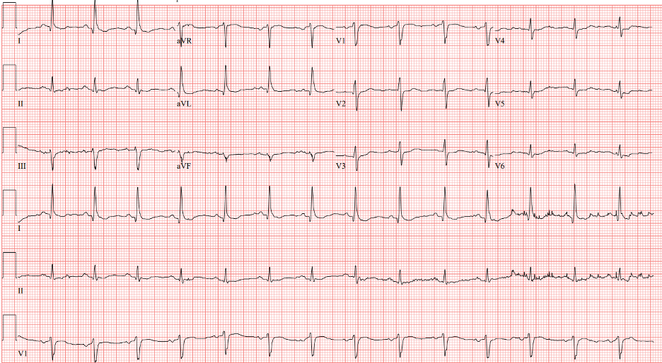
21. Post Intensive Care Syndrome (PICS)

Today on Pulm PEEPs, we are joined by two pioneers in the field of post-intensive care outcomes and delirium research. Drs. Dale Needham and Wes Ely talk to us all about the Post Intensive Care Syndrome (PICS) and cover everything from how it was first recognized, to the impact it has, and, most importantly, what we can do to prevent it. This is a huge topic in the field of critical care and we’re thrilled to be delving into it with such knowledgeable guides.
Meet Our Guests

Wes Ely is the Grant W. Liddle Chair in Medicine and a Professor of Medicine at Vanderbilt University Medical Center. He is also the Associate Director of Aging Research at the VA Tennessee Valley Geriatric Research and Education Clinical Center and the co-director of the Critical, Illness, Brain Dysfunction and Survivorship Center. He has published 100s of manuscripts on critical illness survivorship and delirium. He also published a book called “Every Deep-Drawn Breath” about his and his patients’ experiences in the ICU and about the ramifications of critical illness. All net proceeds for the book are going to the CIBS Center Endowment for Survivorship

Dale Needham is a Professor of Medicine at Johns Hopkins, where he is also the Medical Director of the Critical Care Physical Medicine and Rehabilitation Program and the Director of the Outcomes After Critical Illness and Surgery Group. He is the author of 100s of publications focusing on post-ICU outcomes and has received numerous research grants from the NIH and other organizations.

Key Learning Points

Visit our website www.pulmpeeps.com to see the key learning points from this episode summarized in two infographics.
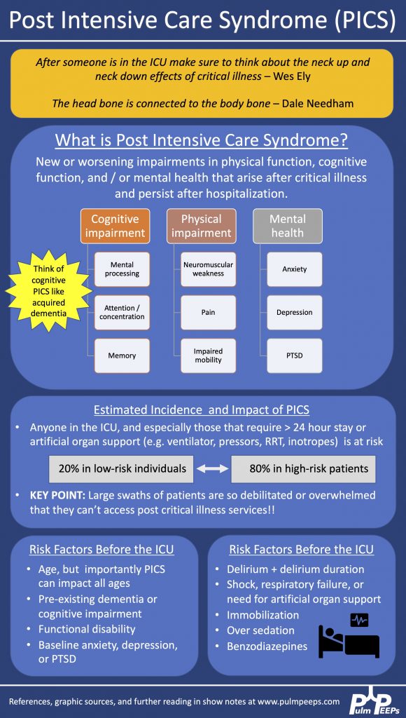
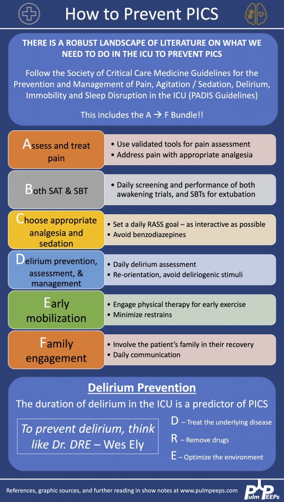
References and links for further reading

  1. Devlin JW, Skrobik Y, Gélinas C, et al. Executive Summary: Clinical Practice Guidelines for the Prevention and Management of Pain, Agitation/Sedation, Delirium, Immobility, and Sleep Disruption in Adult Patients in the ICU. Critical Care Medicine. 2018;46(9):1532-1548. doi:10.1097/CCM.0000000000003259
  2. Ely EW. The ABCDEF Bundle: Science and Philosophy of How ICU Liberation Serves Patients and Families. Crit Care Med. 2017;45(2):321-330. doi:10.1097/CCM.0000000000002175
  3. Mikkelsen ME, Still M, Anderson BJ, et al. Society of Critical Care Medicine’s International Consensus Conference on Prediction and Identification of Long-Term Impairments After Critical Illness. Crit Care Med. 2020;48(11):1670-1679. doi:10.1097/CCM.0000000000004586
  4. Needham DM, Sepulveda KA, Dinglas VD, et al. Core Outcome Measures for Clinical Research in Acute Respiratory Failure Survivors. An International Modified Delphi Consensus Study. Am J Respir Crit Care Med. 2017;196(9):1122-1130. doi:10.1164/rccm.201702-0372OC
  5. Needham DM, Wozniak AW, Hough CL, et al. Risk Factors for Physical Impairment after Acute Lung Injury in a National, Multicenter Study. Am J Respir Crit Care Med. 2014;189(10):1214-1224. doi:10.1164/rccm.201401-0158OC
  6. Semler MW, Bernard GR, Aaron SD, et al. Identifying Clinical Research Priorities in Adult Pulmonary and Critical Care. NHLBI Working Group Report. Am J Respir Crit Care Med. 2020;202(4):511-523. doi:10.1164/rccm.201908-1595WS
  7. Spruit MA, Holland AE, Singh SJ, Tonia T, Wilson KC, Troosters T. COVID-19: Interim Guidance on Rehabilitation in the Hospital and Post-Hospital Phase from a European Respiratory Society and American Thoracic Society-coordinated International Task Force. Eur Respir J. Published online August 13, 2020:2002197. doi:10.1183/13993003.02197-2020
  8. Turnbull AE, Sepulveda KA, Dinglas VD, Chessare CM, Bingham CO, Needham DM. Core Domains for Clinical Research in Acute Respiratory Failure Survivors: An International Modified Delphi Consensus Study. Crit Care Med. 2017;45(6):1001-1010. doi:10.1097/CCM.0000000000002435
  9. Ward DS, Absalom AR, Aitken LM, et al. Design of Clinical Trials Evaluating Sedation in Critically Ill Adults Undergoing Mechanical Ventilation: Recommendations From Sedation Consortium on Endpoints and Procedures for Treatment, Education, and Research (SCEPTER) Recommendation III. Crit Care Med. 2021;49(10):1684-1693. doi:10.1097/CCM.0000000000005049
  10. Ozga D, Krupa S, Witt P, Mędrzycka-Dąbrowska W. Nursing Interventions to Prevent Delirium in Critically Ill Patients in the Intensive Care Unit during the COVID19 Pandemic—Narrative Overview. Healthcare. 2020;8:578. doi:10.3390/healthcare8040578

18. A Case of Severe Weakness in the ICU

We are thrilled here @PulmPEEPS to have our first episode with one of our new Associate Editors Luke Hedrick, and our first nephrology consultant Jeff William. Luke will walk us through an interesting case presentation, and we will discuss an approach to severe weakness in our patient in the ICU.

Meet Our Guests

Jeff William is an Assistant Professor of Medicine at Harvard Medical School and Beth Israel Deaconess Medical Center, where he is also the Associate Director of the Nephrology Fellowship Program. He completed a Medical Education Research Fellowship at Harvard Medical School, and is very involved in residency, fellowship and medical student education.

Patient Presentation

We have a man in his 40s with a past medical history of asthma, hypertension, and acid reflux who was brought in by EMS with back pain and profound proximal lower extremity weakness. He reports mild weakness in his legs which started 2 days ago, but this morning his weakness acutely worsened to the point that he can’t lift his legs out of the bed. He also has some cramping pain in his thighs. He additionally has had mild shortness of breath and yesterday went to an urgent care where he was given steroids and swabbed for COVID (which was negative).

Key Learning Points

**Spoilers Ahead** If you want to think through the case on your own we advise listening to the episode first before looking at the infographics below

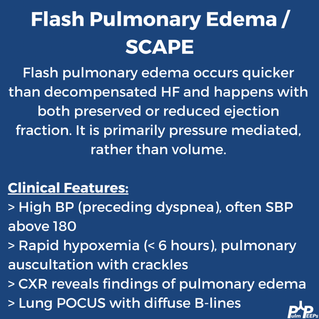
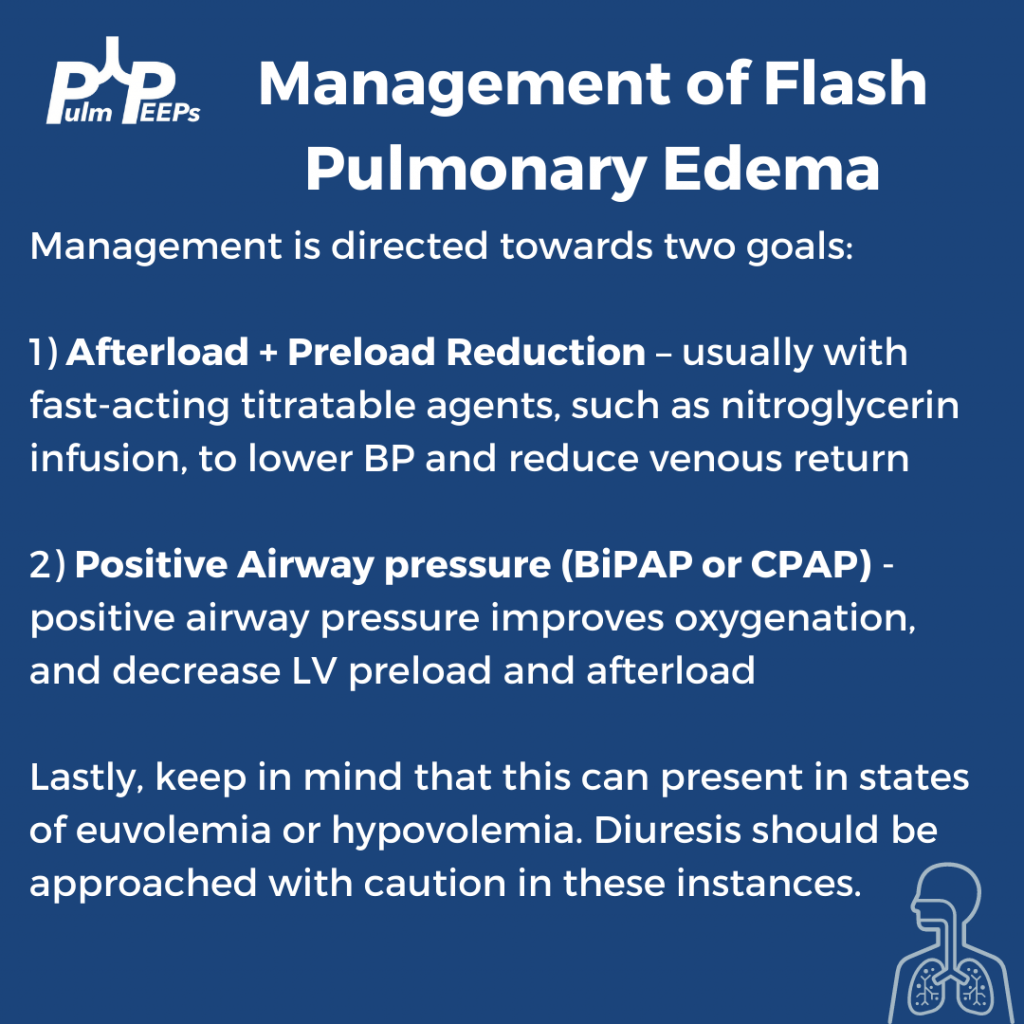
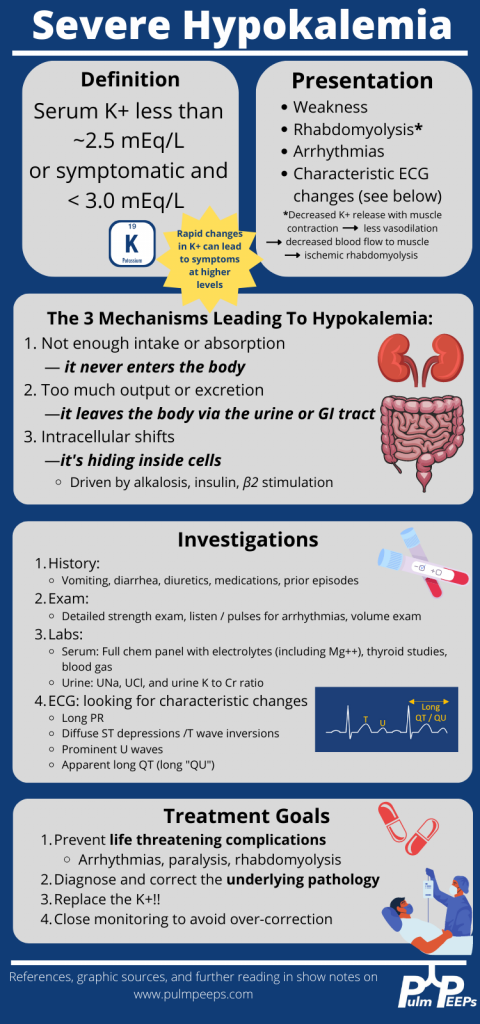
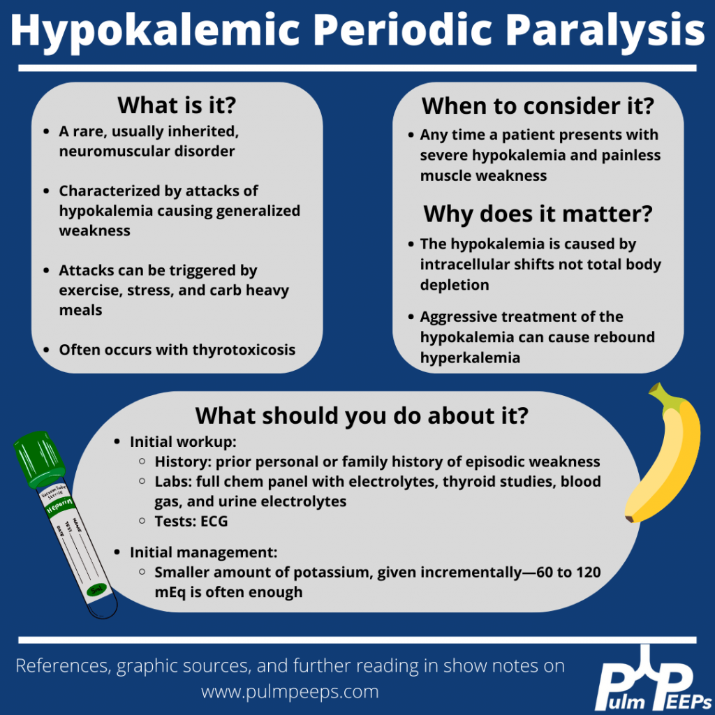
Although our patient’s etiology of severe hypokalemia was thought to be secondary to thiazide diuretic use, it is important to be familiar with hypokalemic periodic paralysis.

References

  1. Knochel JP, Schlein EM. On the mechanism of rhabdomyolysis in potassium depletion. J Clin Invest. 1972 Jul;51(7):1750-8. doi: 10.1172/JCI106976.
  2. Wang X, Han D, Li G. Electrocardiographic manifestations in severe hypokalemia. J Int Med Res. 2020 Jan;48(1):300060518811058. doi: 10.1177/0300060518811058.
  3. Venance SL, Cannon SC, Fialho D, Fontaine B, Hanna MG, Ptacek LJ, Tristani-Firouzi M, Tawil R, Griggs RC; CINCH investigators. The primary periodic paralyses: diagnosis, pathogenesis and treatment. Brain. 2006 Jan;129(Pt 1):8-17. doi: 10.1093/brain/awh639.
  4. Lin SH, Lin YF, Halperin ML. Hypokalaemia and paralysis. QJM. 2001 Mar;94(3):133-9. doi: 10.1093/qjmed/94.3.133. 
  5. Lin SH, Lin YF, Chen DT, Chu P, Hsu CW, Halperin ML. Laboratory tests to determine the cause of hypokalemia and paralysis. Arch Intern Med. 2004 Jul 26;164(14):1561-6. doi: 10.1001/archinte.164.14.1561.

Radiology Rounds 13: Mediastinal Widening – Aortic Dissection

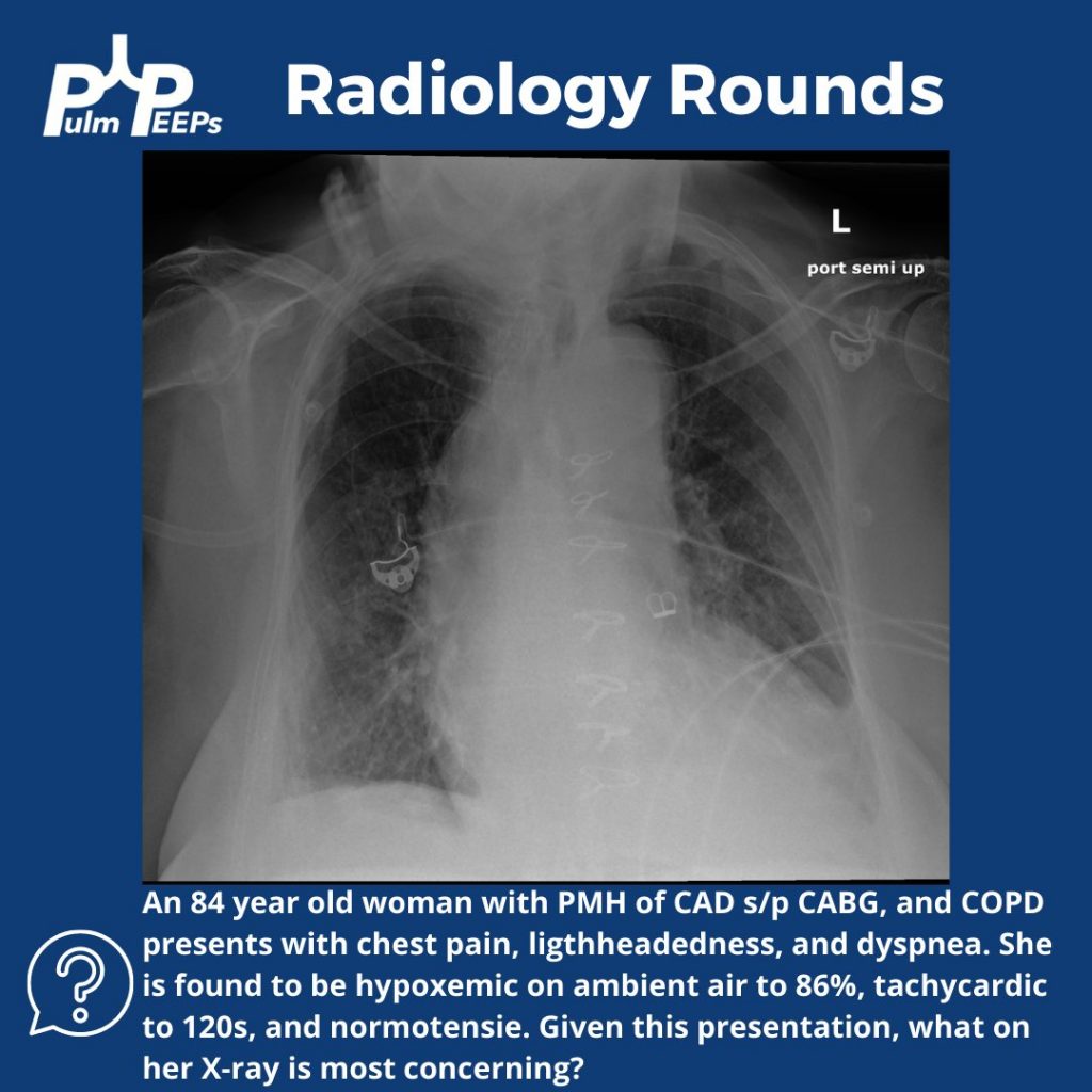
This week on #RadiologyRounds we have a patient with some key clues on the initial X-ray that help lead to the ultimate diagnosis.

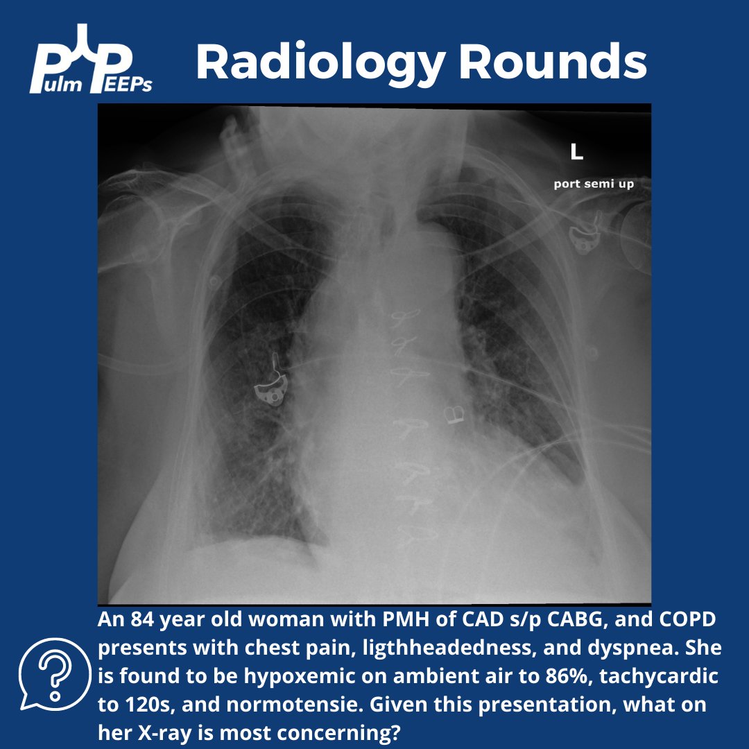
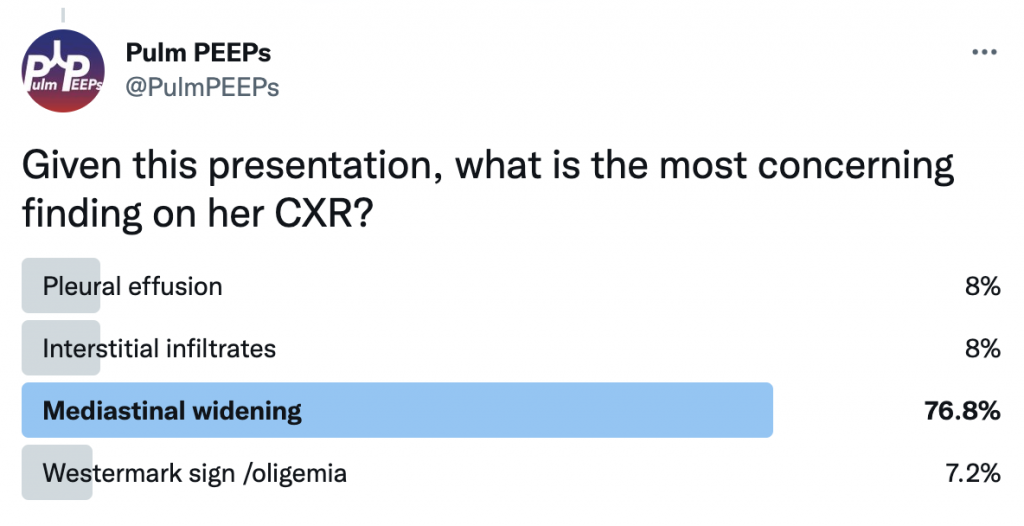
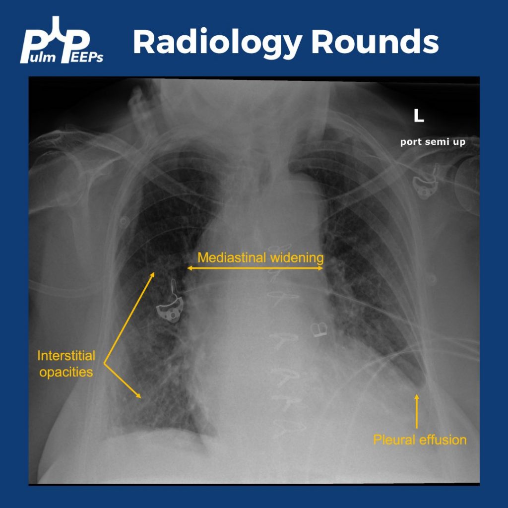
While the patient has multiple findings on the X-ray, the mediastinal widening is the most concerning finding in the setting of acute chest pain, shortness of breath, and lightheadedness.

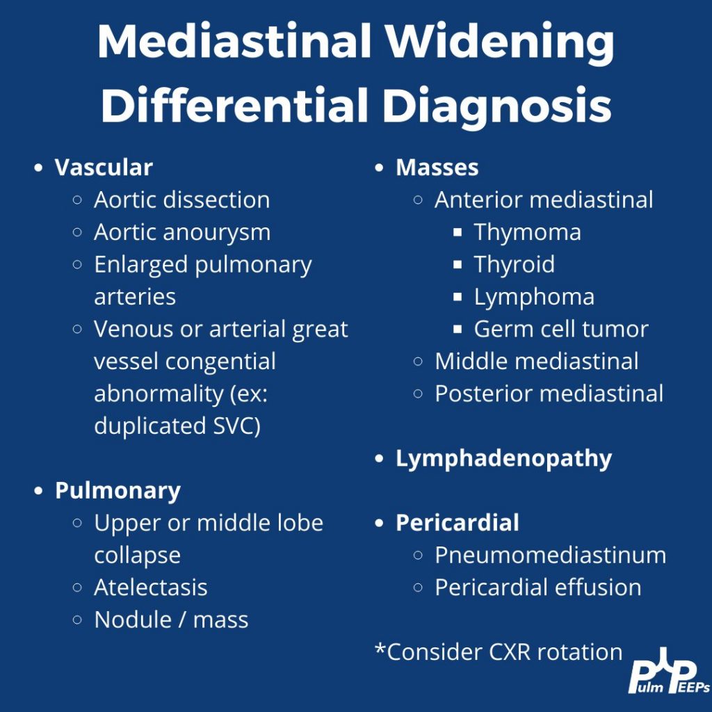
Mediastinal widening has a broad differential, which includes some can’t miss and life-threatening diagnoses

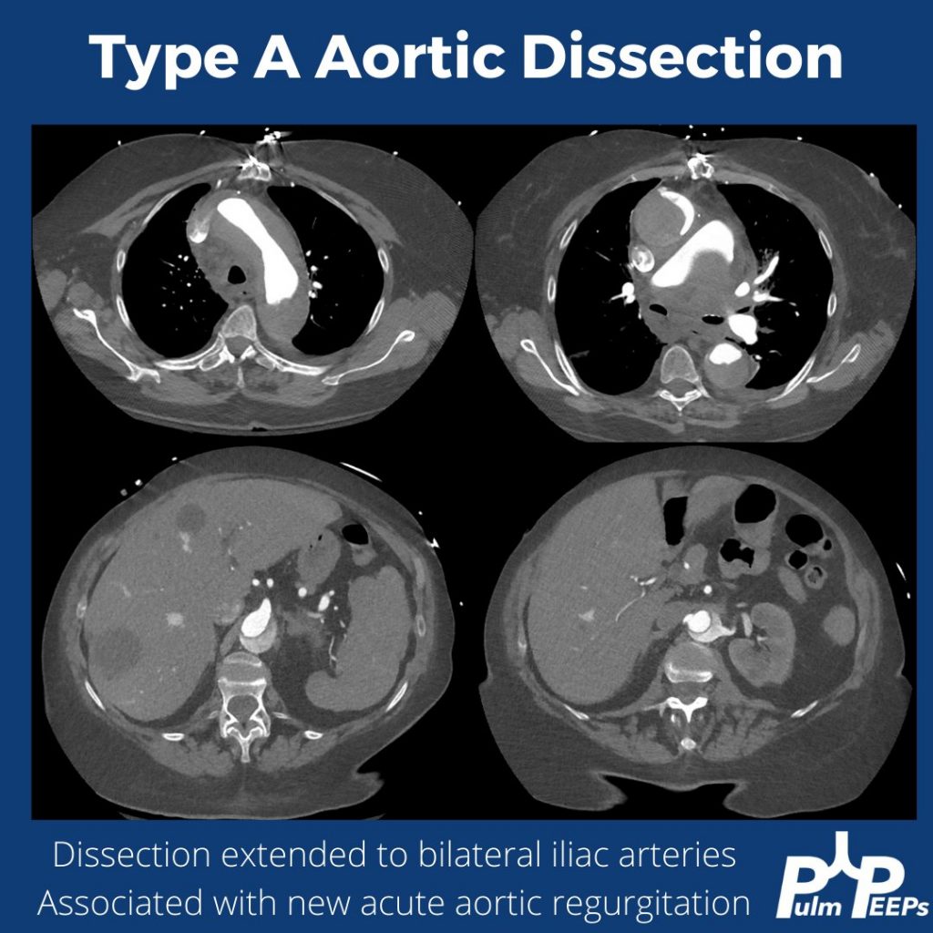
The key point is that aortic dissection, or ruptured thoracic aortic aneurysm, is high on the differential for this patient. Cross-sectional imaging is warranted and should be timed appropriately to evaluate the aorta. A triple rule-out CT would accomplish this as well. This patient had a CTA of the chest and was found to have a large type A aortic dissection with significant extension.

12. Undifferentiated Shock Roundtable

This week the Pulm PEEPs, David Furfaro and Kristina Montemayor, are joined by three outstanding critical care doctors and medical educators to discuss the evaluation of patients with undifferentiated shock. We cover everything from the basics about defining shock, to advanced POCUS techniques to clarify the etiology of shock. Listen today and let us know your favorite technique for evaluating shock in the ICU.

Meet Our Guests

Molly Hayes is an Assistant Professor of Medicine at Beth Israel Deaconess Medical Center and Harvard Medical School, the Director of the MICU at BIDMC, and the Director of External Education at the Carl J Shapiro Institute for Education and Research. She is also a course director for a yearly CME course on principles of critical care medicine run by BIDMC and HMS.

Nick Mark is a Pulmonologist and Intensivist at Swedish Medical Center in Seattle, Washington. He is also the founder of ICU One Pager, which produces high yield critical care education one-page guides that have been downloaded by thousands of learners.

Matt Siuba is an Assistant Professor of Medicine and intensivist at the Cleveland Clinic, where he is the associate program director for the Critical Care Medicine fellowship. He founded and runs the website Zentensivist.com, has his own associated podcast, and is a senior editor at CriticalCareNow.com.

Key Learning Points

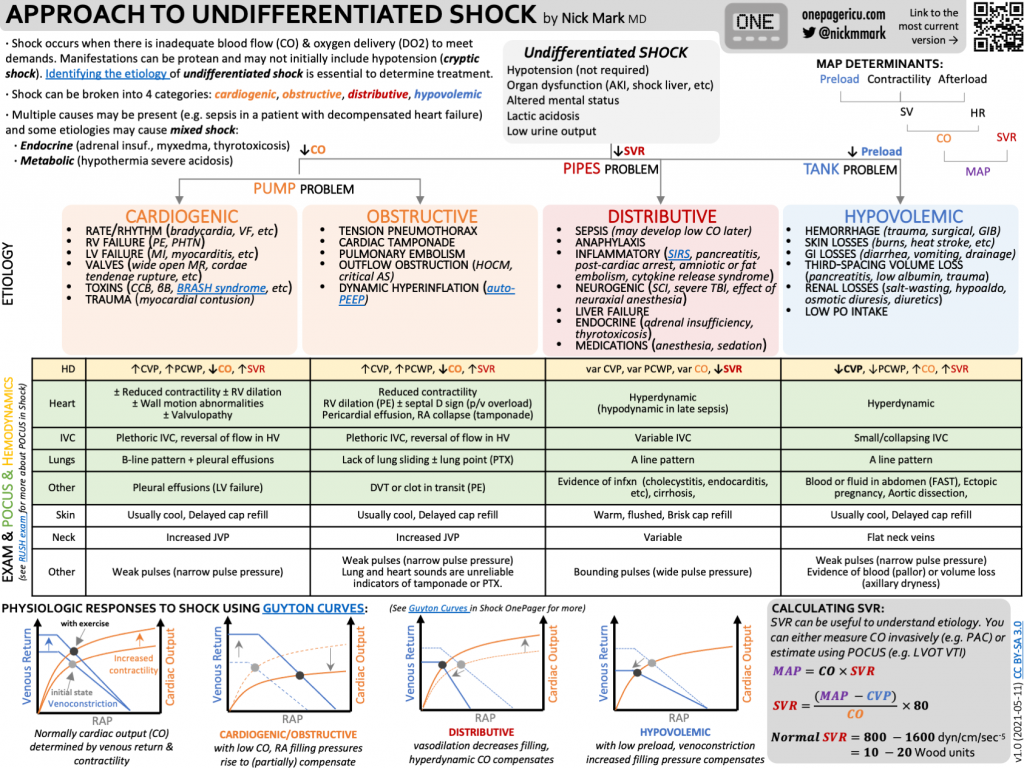
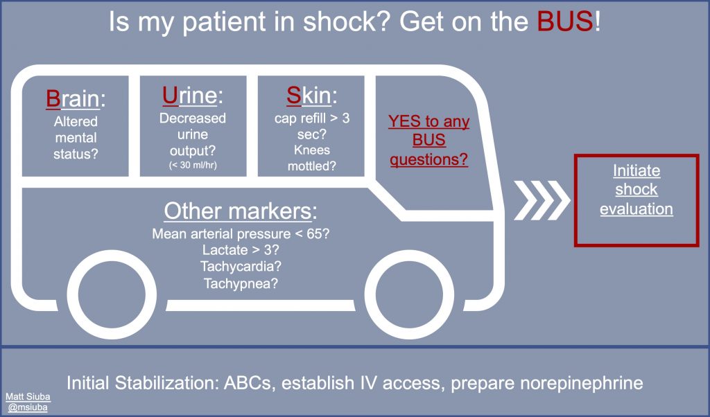
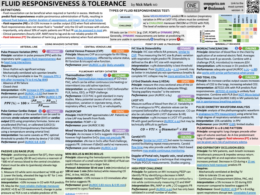
Key graphics

Courtesy of Nick Mark and ICU One Pager
Courtesy of Matt Siuba
Courtesy of Nick Mark and ICU One Pager

Definition of shock

– Shock is defined as inadequate oxygen delivery to meet the body’s needs. Decreased perfusion and oxygen delivery leads to cell injury and death

– If you define just as hypotension, you will miss people who have cryptic shock, and categorize some people with shock who don’t have it

– Cryptic shock = a patient with normal blood pressure (MAP > 65), but who still has shock based on inadequate O2 delivery

– O2 delivery is broken down in to cardiac output and arterial oxygen content

Causes of shock

Shock can be divided into three large categories:

1) A pump problem – low cardiac output. This includes cardiogenic and obstructive shock. Make sure to remember to look for tamponade and valvulopathies.

2) A pipe problem – low systemic vascular resistance. This includes distributive shock. Distributive shock is most often due to sepsis but can be due to anaphylaxis, endocrinopathies, cirrhosis, or spinal shock.

3) A tank problem – low preload. This includes hypovolemic and hemorrhagic shock. Make sure to remember about high intrathoracic pressure, which can decrease effective preload.

Examining a patient with undifferentiated shock

– See if the patient is on the “Shock BUS” by examining their brain (mental status), urine output, and skin

– Feel if their skin is warm vs cold and if it is mottled

– Feel the patient’s pulses to see if they are bounding, normal, or thready

Point of Care Ultrasound

– “Ultrasound is the new stethoscope”

– The first step is to always look at the heart and look for chamber size and function. You can then look for pericardial effusion

– Point of care ultrasound then includes looking at the lungs for signs of fluid overload, consolidation, or pneumothorax

– A complete ultrasound also involves looking at the abdomen and at the extremities for DVT

– More specific ultrasound techniques include looking at:

1) IVC exam to estimate right atrial pressure. This test is often misused. It is most helpful in states when the patient has low stroke volume and trying to figure out if they have cardiac limitation to stroke volume vs if they are hypovolemic.

2) Velocity time index as a measure of cardiac output to trend with interventions

References and links for further reading

  1. Vincent JL, De Backer D. Circulatory shock. N Engl J Med. 2013;369(18):1726-1734. doi:10.1056/NEJMra1208943
  2. Seymour CW, Liu VX, Iwashyna TJ, et al. Assessment of Clinical Criteria for Sepsis: For the Third International Consensus Definitions for Sepsis and Septic Shock (Sepsis-3). JAMA. 2016;315(8):762-774. doi:10.1001/jama.2016.0288
  3. Chukwulebe SB, Gaieski DF, Bhardwaj A, Mulugeta-Gordon L, Shofer FS, Dean AJ. Early hemodynamic assessment using NICOM in patients at risk of developing Sepsis immediately after emergency department triage. Scandinavian Journal of Trauma, Resuscitation and Emergency Medicine. 2021;29(1):23. doi:10.1186/s13049-021-00833-1
  4. Hernández G, Ospina-Tascón GA, Damiani LP, et al. Effect of a Resuscitation Strategy Targeting Peripheral Perfusion Status vs Serum Lactate Levels on 28-Day Mortality Among Patients With Septic Shock: The ANDROMEDA-SHOCK Randomized Clinical Trial. JAMA. 2019;321(7):654-664. doi:10.1001/jama.2019.0071
  5. Wang J, Zhou D, Gao Y, Wu Z, Wang X, Lv C. Effect of VTILVOT variation rate on the assessment of fluid responsiveness in septic shock patients. Medicine (Baltimore). 2020;99(47):e22702. doi:10.1097/MD.0000000000022702
  6. Sweeney DA, Wiley BM. Integrated Multiorgan Bedside Ultrasound for the Diagnosis and Management of Sepsis and Septic Shock. Semin Respir Crit Care Med. 2021;42(5):641-649. doi:10.1055/s-0041-1733896
  7. Yuan S, He H, Long Y. Interpretation of venous-to-arterial carbon dioxide difference in the resuscitation of septic shock patients. J Thorac Dis. 2019;11(Suppl 11):S1538-S1543. doi:10.21037/jtd.2019.02.79
  8. Volpicelli G, Lamorte A, Tullio M, et al. Point-of-care multiorgan ultrasonography for the evaluation of undifferentiated hypotension in the emergency department. Intensive Care Med. 2013;39(7):1290-1298. doi:10.1007/s00134-013-2919-7
  9. Perera P, Mailhot T, Riley D, Mandavia D. The RUSH exam: Rapid Ultrasound in SHock in the evaluation of the critically lll. Emerg Med Clin North Am. 2010;28(1):29-56, vii. doi:10.1016/j.emc.2009.09.010

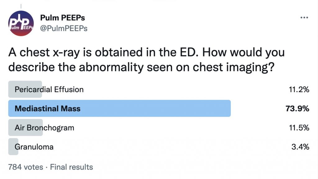
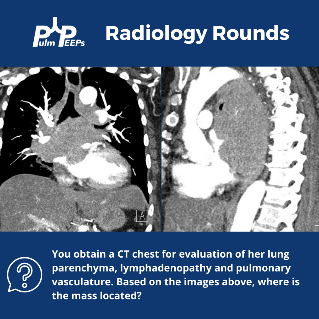
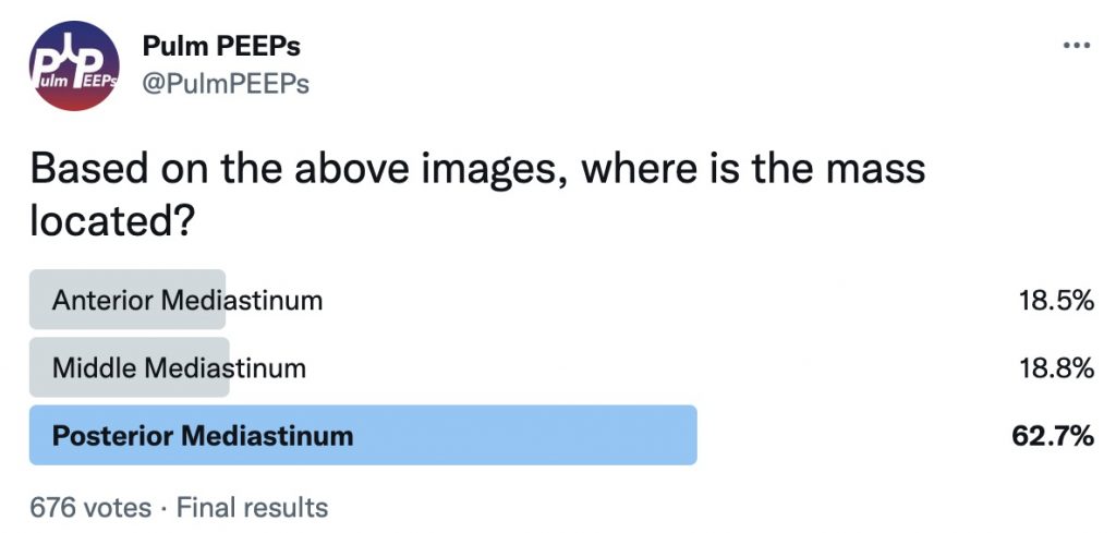
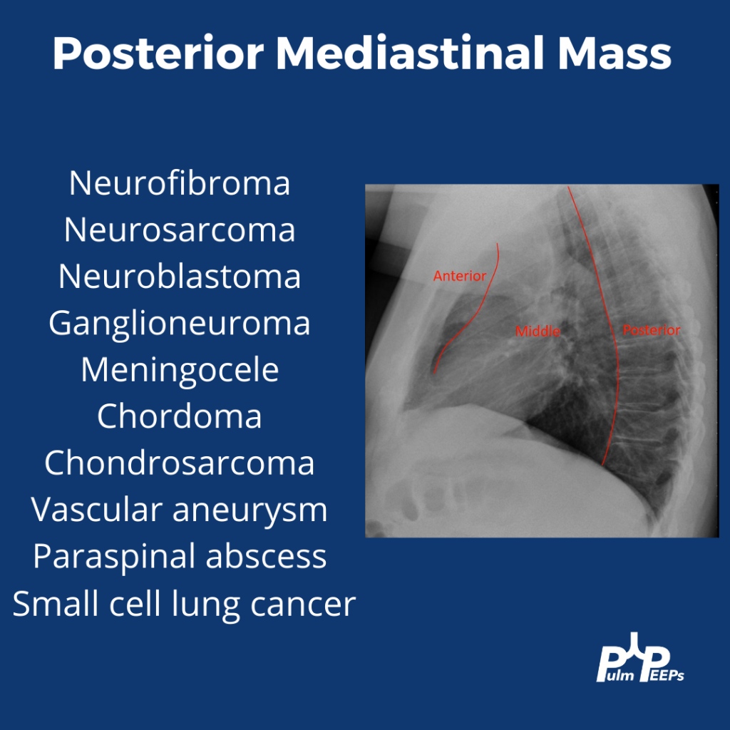
Radiology Rounds 9: Posterior Mediastinal Mass

We’re excited to bring you another Radiology Rounds today that combines pulmonary and critical care.

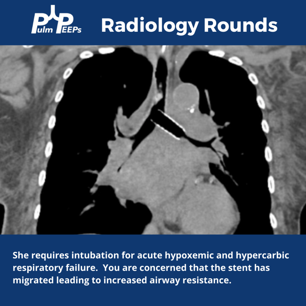
The patient is diagnosed with small cell lung cancer and requires a left bronchial stent. She develops acute hypoxemic and hypercapnic respiratory failure requiring intubation.

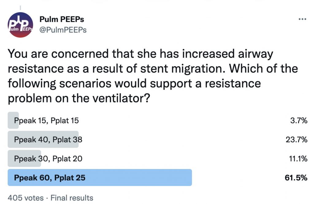
You are concerned that she has increased airway resistance as a result of stent migration. What would you expect to see on the ventilator if this is the case?

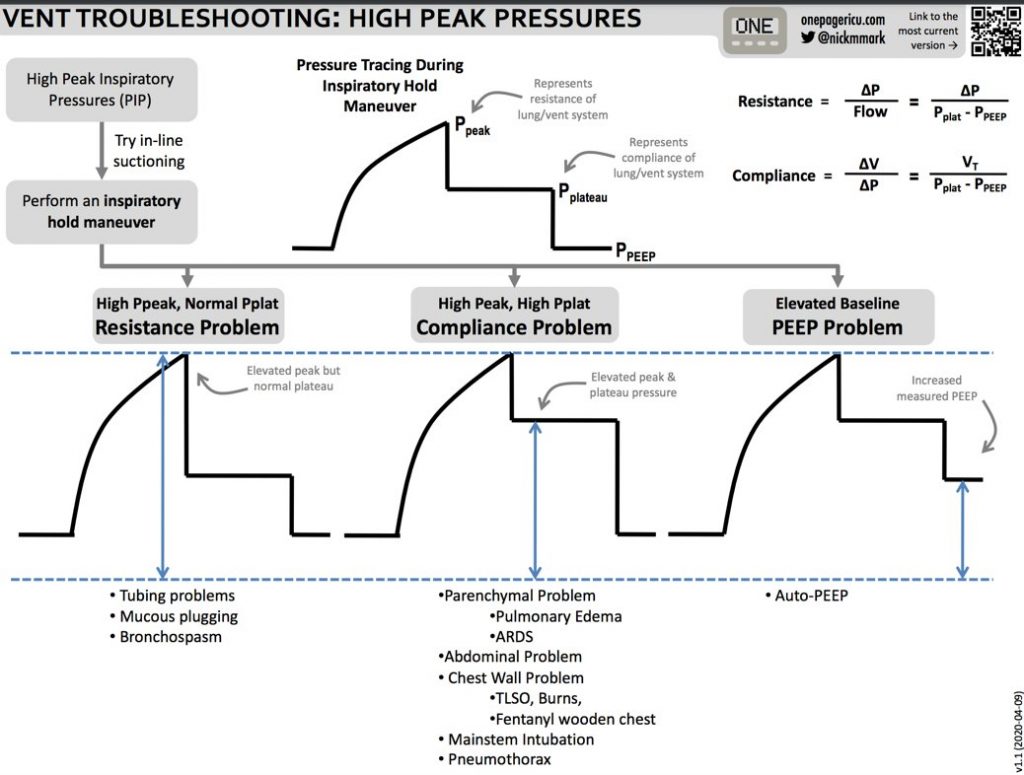
Here are some tips from ICU OnePager on interpreting high peak pressures on the ventilator

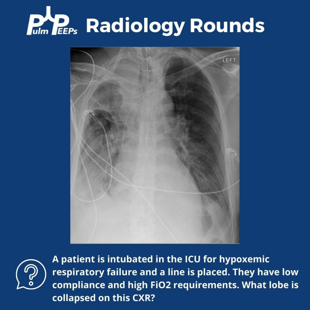
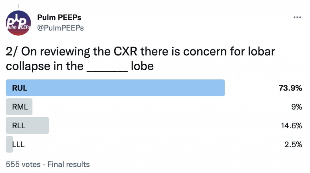
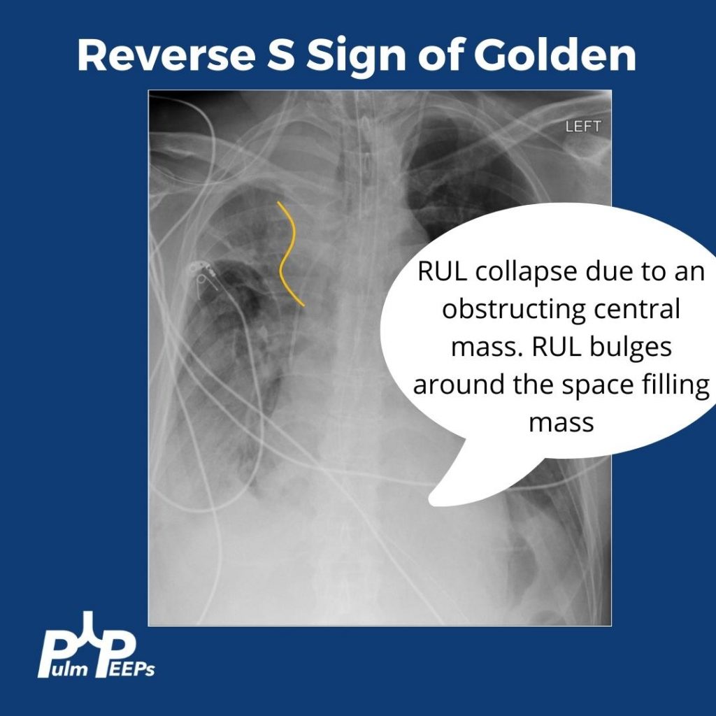
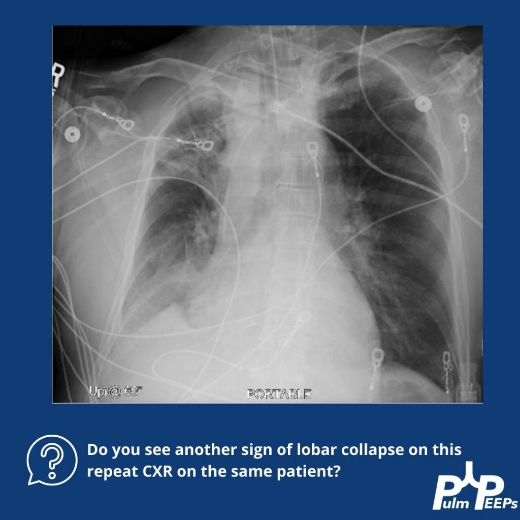
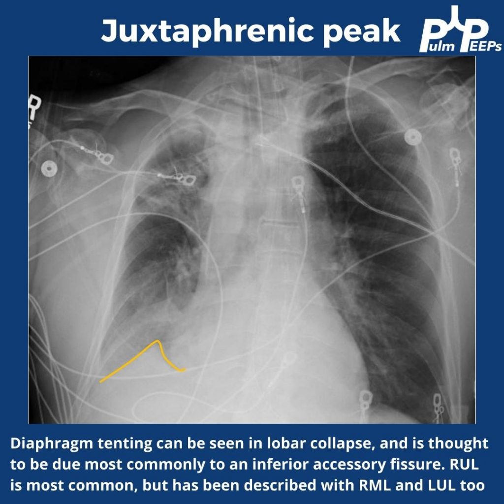
Radiology Rounds 6: Right Upper Lobe Collapse – CXR Signs

This week for radiology rounds we’re looking at some classic imaging signs of lobar collapse. Take a look, respond to our polls on Twitter, and make sure to subscribe to our podcast to get all of the Pulm PEEPs content!

References

  1. Kumaresh A, Kumar M, Dev B, Gorantla R, Sai PV, Thanasekaraan V. Back to Basics – ‘Must Know’ Classical Signs in Thoracic Radiology. J Clin Imaging Sci. 2015;5:43. doi:10.4103/2156-7514.161977
  2. Algın O, Gökalp G, Topal U. Signs in chest imaging. Diagn Interv Radiol. 2011;17(1):18-29. doi:10.4261/1305-3825.DIR.2901-09.1
  3. Gupta P. The Golden S Sign. Radiology. 2004;233(3):790-791. doi:10.1148/radiol.2333021407

7. Top Consults: Severe Asthma Exacerbation

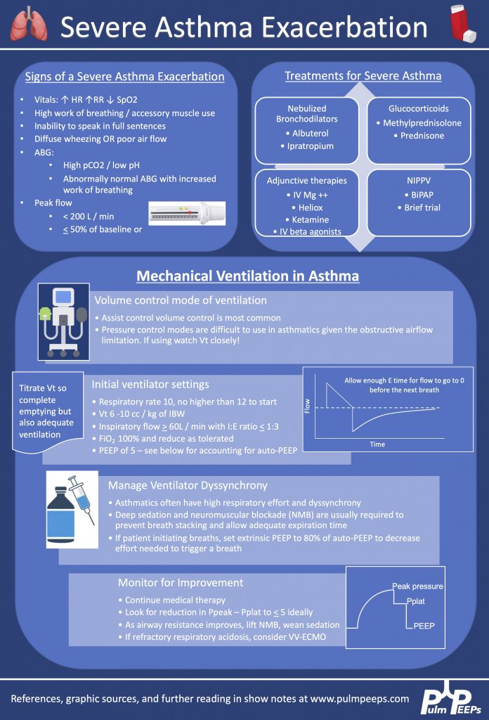
We are excited to bring you another episode in our Pulm PEEPs Top Consults series! Kristina Montemayor and David Furfaro, are joined by Sandy Zaeh to discuss the assessment and management of a patient with a severe asthma exacerbation. We’ll follow a consult patient from the emergency department to the ICU, and cover everything from the physiology of pulsus paradoxus in asthma to how to manage the ventilator in status asthmaticus. Listen today and please send any questions our way on Twitter @pulmPEEPS.

Meet Our Guests

Sandy Zaeh is an Instructor of Medicine and Pulmonary & Critical Care Medicine physician at Yale School of Medicine.

Key Learning Points

References and links for further reading

  1. Chung KF, Wenzel SE, Brozek JL, et al. International ERS/ATS guidelines on definition, evaluation and treatment of severe asthma. European Respiratory Journal. 2014;43(2):343-373. doi:10.1183/09031936.00202013
  2. Rodrigo GJ, Rodrigo C, Hall JB. Acute asthma in adults: a review. Chest. 2004;125(3):1081-1102. doi:10.1378/chest.125.3.1081
  3. Godwin HT, Fix ML, Baker O, Madsen T, Walls RM, Brown CA. Emergency Department Airway Management for Status Asthmaticus With Respiratory Failure. Respir Care. 2020;65(12):1904-1907. doi:10.4187/respcare.07723
  4. Althoff MD, Holguin F, Yang F, et al. Noninvasive Ventilation Use in Critically Ill Patients with Acute Asthma Exacerbations. Am J Respir Crit Care Med. 2020;202(11):1520-1530. doi:10.1164/rccm.201910-2021OC
  5. Brenner B, Corbridge T, Kazzi A. Intubation and Mechanical Ventilation of the Asthmatic Patient in Respiratory Failure. Proc Am Thorac Soc. 2009;6(4):371-379. doi:10.1513/pats.P09ST4
  6. Laher AE, Buchanan SK. Mechanically Ventilating the Severe Asthmatic. J Intensive Care Med. 2018;33(9):491-501. doi:10.1177/0885066617740079
  7. Leatherman J. Mechanical ventilation for severe asthma. Chest. 2015;147(6):1671-1680. doi:10.1378/chest.14-1733

6. PEEP in ARDS Roundtable

This week on Pulm PEEPs, Dave Furfaro and Kristina Montemayor are joined by experts in the field of critical care medicine and ARDS to discuss all things PEEP! Drs. Roy Brower, Sarina Sahetya, Todd Rice, and Elias Baedorf-Kassis discuss everything ranging from PEEP basics to their approach to optimizing PEEP in patients with ARDS.

Meet Our Guests

Roy Brower is a Professor of Medicine at Johns Hopkins where he served as the MICU director for over 33 years, and he has been one of the pioneers for lung-protective ventilation for patients with ARDS.

Elias Baedorf-Kassis is an Assistant Professor of Medicine at Beth Israel Deaconess Medical Center and Harvard Medical School. He is the Medical Director of Respiratory Care at BIDMC, and helps lead the VV-ECMO program.

Todd Rice is an Associate Profess of Medicine in the Division of Allergy, Pulmonary, and Critical Care Medicine at Vanderbilt University and Vice President for Clinical Trial Innovation and Operations in the Vanderbilt Institute for Clinical and Translational Research.

Sarina Sahetya is an Assistant Professor of Medicine at Johns Hopkins Hospital and does research in the diagnosis and treatment of ARDS.


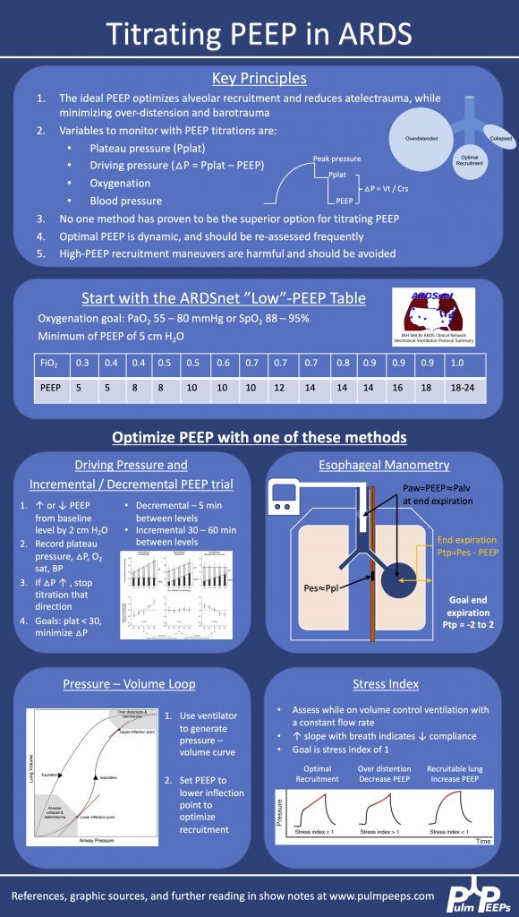
Key Learning Points

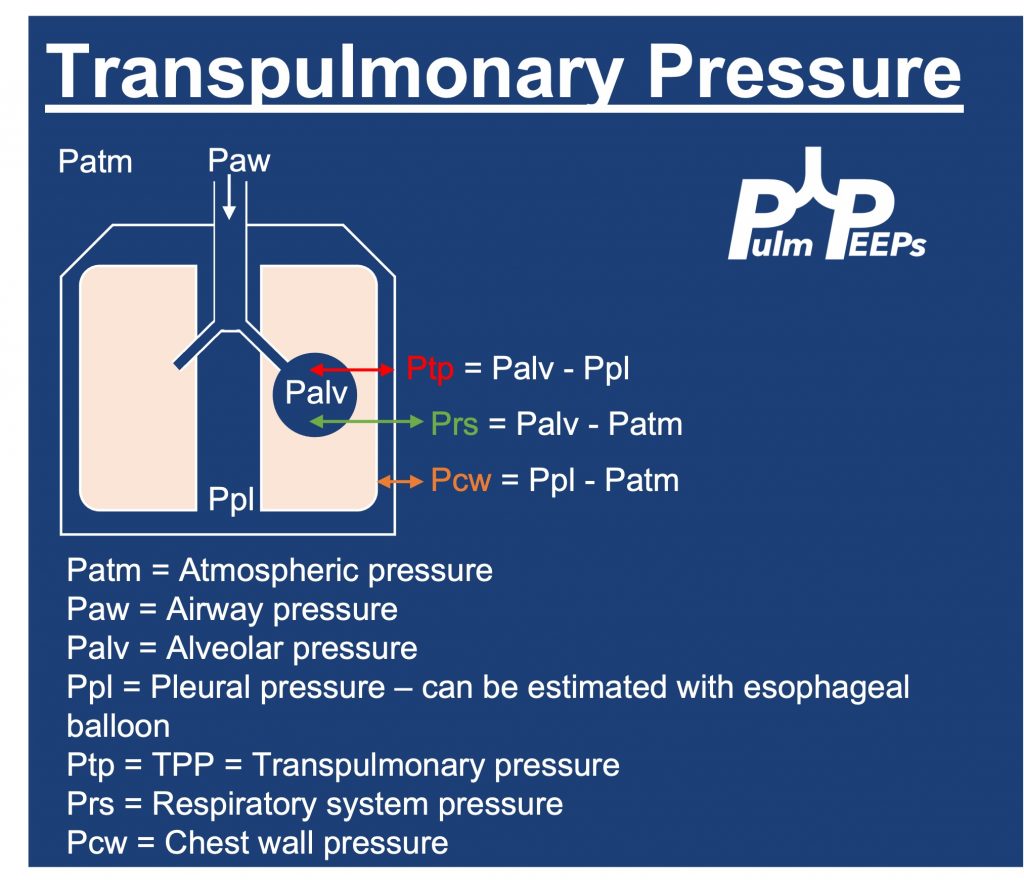
Driving Pressure figure from Amato et al. 2015. Stress index figure from Hess 2014.
  • The plateau pressure can be measured on the ventilator with an inspiratory hold maneuver
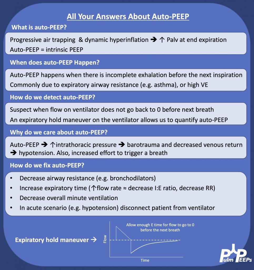
  • Extrinsic PEEP is applied by the ventiilator, while intrinsic PEEP, or auto-PEEP, occurs when there is incomplete emptying of the lungs due to inadequate time for exhalation. This often happens with obstructive lung disease. Intrinsic PEEP can be measured on the ventilator with an end-expiratory hold maneuver
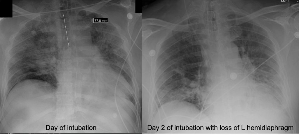
  • We utilize PEEP in all intubated patients to minimize atelectasis. When patients are supine, the heart moves back 2 cm and the diaphragm raises by 2 cm, so often the left lower lobe of the lung is compressed and there is atelectasis there. This is often seen on CXR:

References, Image Sources, and Further Reading

  1. Higher versus Lower Positive End-Expiratory Pressures in Patients with the Acute Respiratory Distress Syndrome. New England Journal of Medicine. 2004;351(4):327-336. doi:10.1056/NEJMoa032193
  2. Amato MBP, Meade MO, Slutsky AS, et al. Driving Pressure and Survival in the Acute Respiratory Distress Syndrome. New England Journal of Medicine. 2015;372(8):747-755. doi:10.1056/NEJMsa1410639
  3. Writing Group for the Alveolar Recruitment for Acute Respiratory Distress Syndrome Trial (ART) Investigators. Effect of Lung Recruitment and Titrated Positive End-Expiratory Pressure (PEEP) vs Low PEEP on Mortality in Patients With Acute Respiratory Distress Syndrome: A Randomized Clinical Trial. JAMA. 2017;318(14):1335-1345. doi:10.1001/jama.2017.14171
  4. Beitler JR, Sarge T, Banner-Goodspeed VM, et al. Effect of Titrating Positive End-Expiratory Pressure (PEEP) With an Esophageal Pressure-Guided Strategy vs an Empirical High PEEP-Fio2 Strategy on Death and Days Free From Mechanical Ventilation Among Patients With Acute Respiratory Distress Syndrome: A Randomized Clinical Trial. JAMA. 2019;321(9):846-857. doi:10.1001/jama.2019.0555
  5. LaFollette R, Hojnowski K, Norton J, DiRocco J, Carney D, Nieman G. Using pressure–volume curves to set proper PEEP in acute lung injury. Nursing in Critical Care. 2007;12(5):231-241. doi:10.1111/j.1478-5153.2007.00224.x
  6. Hess DR. Respiratory mechanics in mechanically ventilated patients. Respir Care. 2014;59(11):1773-1794. doi:10.4187/respcare.03410
  7. Sahetya SK, Hager DN, Stephens RS, Needham DM, Brower RG. PEEP Titration to Minimize Driving Pressure in Subjects With ARDS: A Prospective Physiological Study. Respir Care. 2020;65(5):583-589. doi:10.4187/respcare.07102
  8. Umbrello M, Chiumello D. Interpretation of the transpulmonary pressure in the critically ill patient. Ann Transl Med. 2018;6(19):383. doi:10.21037/atm.2018.05.31
  9. Kenny JES. ICU Physiology in 1000 Words: Driving Pressure & Stress Index. PulmCCM. Published February 13, 2016. Accessed January 1, 2022. https://pulmccm.org/review-articles/icu-physiology-in-1000-words-driving-pressure-stress-index/

Radiology Rounds 4: ARDS and Air Bronchograms

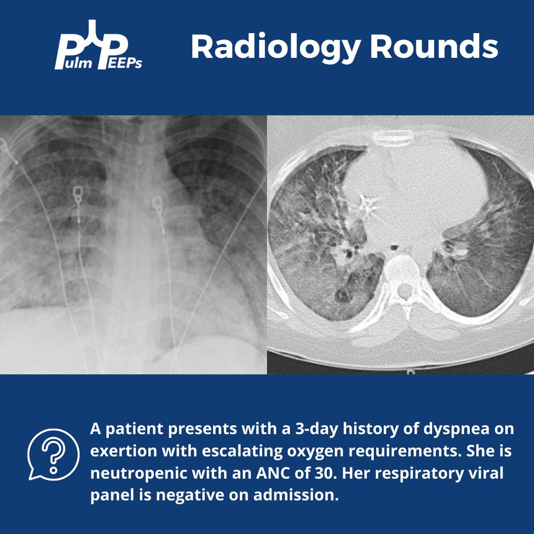
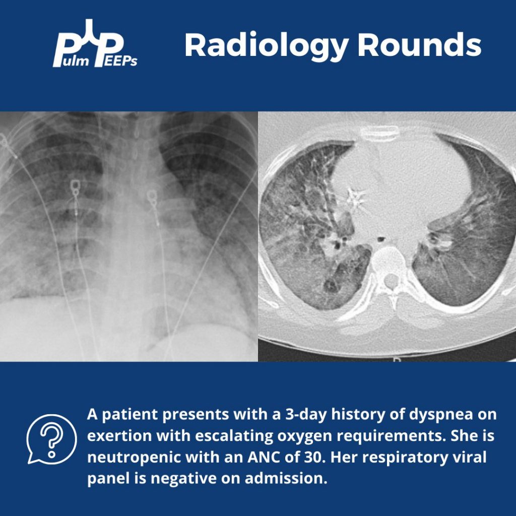
Today we’re bringing you a special edition of Radiology Rounds complete with classic imaging, and some key critical care and ventilator physiology. This case is a perfect lead-in for next week’s Pulm PEEPs Roundtable on PEEP titration, so make sure to tune in!

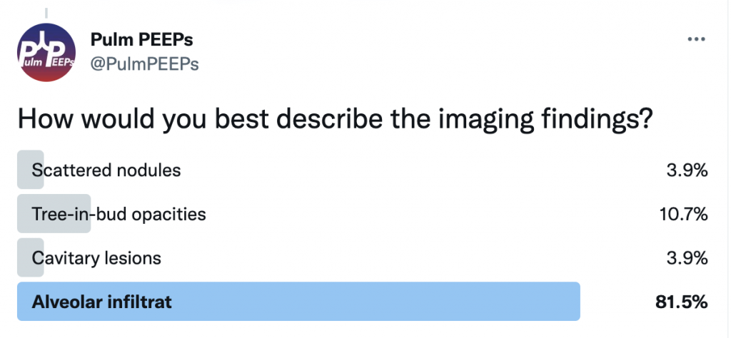
How would you best describe the imaging findings?


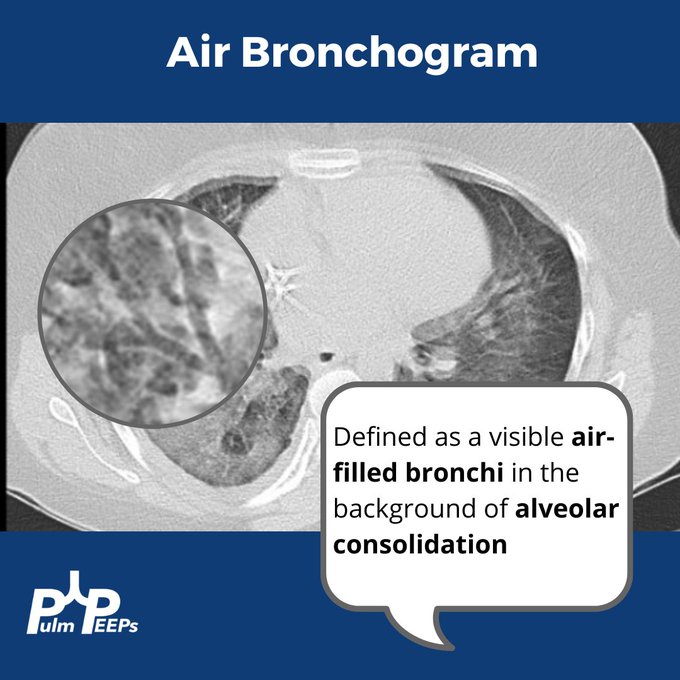
There are bilateral, diffuse alveolar infiltrates noted on imaging with evidence of an air bronchogram on the CT image.

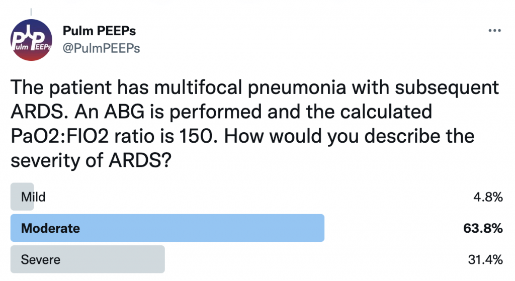
The patient develops worsening hypoxemia requiring mechanical intubation. The patient has multifocal pneumonia and requires intubation. ABG is performed and the calculated PaO2:FIO2 ratio is 150. How would you describe the severity of ARDS?


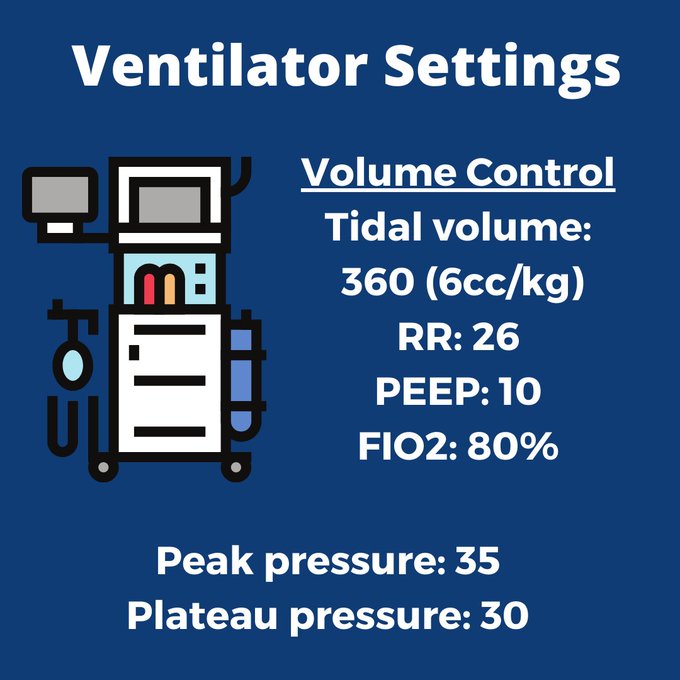
This patient has moderate ARDS based on a PaO2:FIO2 ratio that is between 100 and 200. The patient’s initial ventilator settings on volume control are:

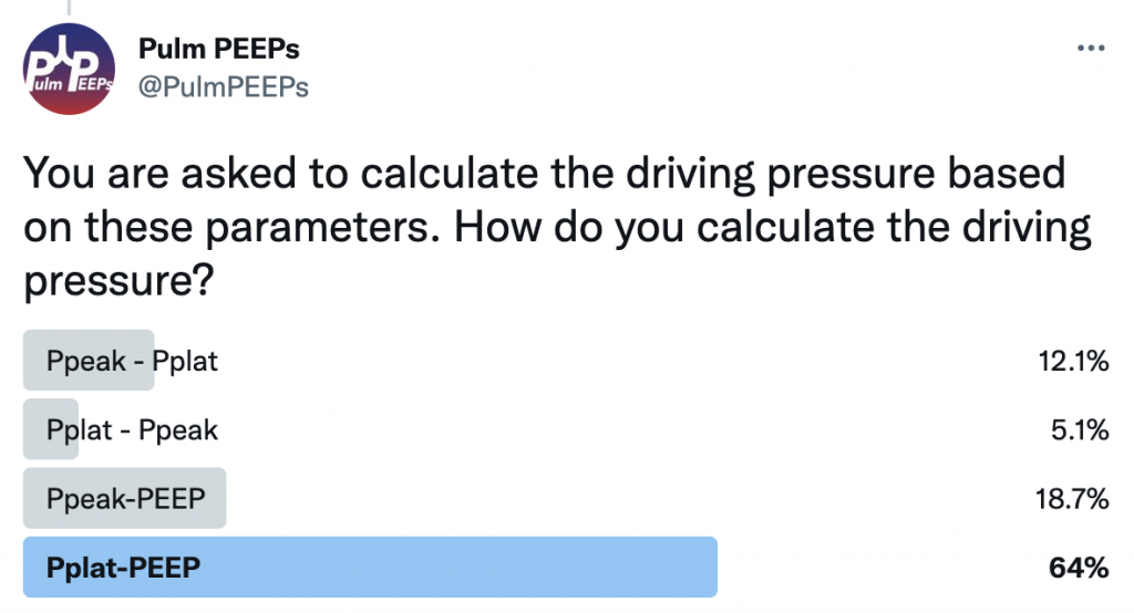
Based on these parameters, we can also calculate the driving pressure. Driving pressure is calculated by using Pplat-PEEP. In this case, Pplat (30)-PEEP (10), would give a driving pressure of 20.