125. IVC Ultrasound and Volume Responsiveness

Should this patient get more fluid? It comes up on nearly every ICU shift, and the IVC is usually the first thing we scan to answer it. In this Pulm PEEPs Pearls episode, Furf and Monty cover how to get a reliable IVC view, how to read it in intubated and spontaneously breathing patients, the cutoffs worth knowing, what the data actually show, and the pitfalls that make the IVC mislead us.

Key Learning Points

Volume Responsiveness vs. Volume Status

“Volume status,” “volume depleted” and “fluid responsive” get used interchangeably on rounds, but they are not the same thing. Fluid responsiveness is a physiologic definition: a patient is fluid responsive if a fluid bolus (usually 250 to 500 mL) increases stroke volume or cardiac output by at least 10 to 15 percent. In Frank-Starling terms, you are asking whether both ventricles are still on the steep part of the curve.

Only about half of hemodynamically unstable ICU patients are fluid responsive. This has been shown again and again, going back to the classic Michard and Teboul review. If we bolus everyone who is hypotensive, roughly half get the downsides of fluid (edema, worse oxygenation, organ congestion) with none of the upside.

Responsive does not necessarily mean needs fluid. Most healthy people are fluid responsive. A patient can even be fluid responsive and volume overloaded at the same time. Responsiveness only tells you the heart can use more preload, so the decision always sits inside the clinical picture:

  • Is there hypoperfusion?
  • Is there reason to think low cardiac output is the problem?
  • Will the patient tolerate more fluid, especially from a lung standpoint?

Why Would the IVC Tell Us Anything?

The IVC is a thin-walled, compliant vessel sitting between the abdomen and the right atrium. Its size reflects the balance between right atrial pressure on one side and intra-abdominal pressure on the other. Breathing changes the pressures around it, and how the IVC responds depends entirely on whether the patient is breathing on their own or receiving positive pressure.

  • Spontaneously breathing: inspiration makes intrathoracic pressure negative, blood is pulled into the chest, and the IVC gets smaller. This is collapsibility.
  • Passively ventilated: each positive-pressure breath raises intrathoracic and right atrial pressure, impeding venous return, so the IVC gets bigger on inspiration. This is distensibility.

The theory: a patient on the flat part of the Frank-Starling curve has a high right atrial pressure and a full, stiff IVC that barely moves. A patient on the steep part has a lower right atrial pressure and a more compliant IVC that swings with each breath. Lots of respiratory variation suggests fluid responsiveness; very little suggests fluid will not help.

Quick bedside trick: take a deep breath yourself. Your chest expands, your diaphragm drops, intrathoracic pressure falls and your IVC empties into the right atrium. Positive pressure is the reverse.

Getting the Image

You cannot do this well without practice, so scan your patients routinely, not only when the stakes are high.

  • Probe and preset: phased array or curvilinear probe, with the abdominal or cardiac preset.
  • Position: patient supine. Head position matters a little, so keep it consistent if you plan to compare scans over time.
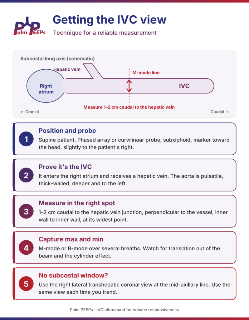
  • Find the IVC: start subxiphoid, slightly to the patient’s right, with the marker toward the head in a longitudinal (sagittal) plane. Many people start with a subcostal four-chamber view of the heart, then rotate to longitudinal to bring the IVC to the center of the screen. You want to see the IVC running through the liver into the right atrium.
  • Prove it’s the IVC: everyone has measured the aorta at some point. The IVC enters the right atrium and receives a hepatic vein. The aorta is thick-walled, pulsatile, deeper and to the patient’s left, and gives off the celiac and SMA rather than receiving hepatic veins. Fan over to find the aorta, or rotate to a transverse view to see both side by side. Color or pulsed-wave Doppler can help if you are unsure.
  • Measure in the right spot: 1 to 2 cm caudal to the hepatic vein junction (about 2 to 3 cm from the cavoatrial junction), perpendicular to the long axis of the vessel, inner wall to inner wall.
  • Capture the max and min: drop an M-mode line through that spot and record over several breaths. Many ultrasound machines have a built-in calculation package that will compute the index for you once you mark the maximum and minimum.
  • Watch for translation: the IVC moves with the diaphragm and can slide in and out of the M-mode line, which can look like collapse. Watch the B-mode clip, or measure in B-mode at a fixed anatomic landmark.
  • Avoid the cylinder effect: the IVC is a tube. A slice through its edge underestimates the diameter and can exaggerate collapse. Fan through the vessel and measure at its widest point.
  • No subcostal window? Post-op abdomen, body habitus and bowel gas are common in the ICU. Use the right lateral transhepatic coronal view at the mid-axillary line, using the liver as your window (the same view used in VExUS). Measurements are not perfectly interchangeable with the subcostal view, so use the same view each time you trend. If you cannot get confident landmarks, this may not be the right test for this patient.
Ivc 1 technique

The Intubated Patient: Distensibility

The IVC works best in mechanically ventilated patients because we control the breath. Two classic indices, both from 2004 and both in French septic shock patients, give us the numbers most people quote:

IndexFormulaCutoff suggesting responseStudy
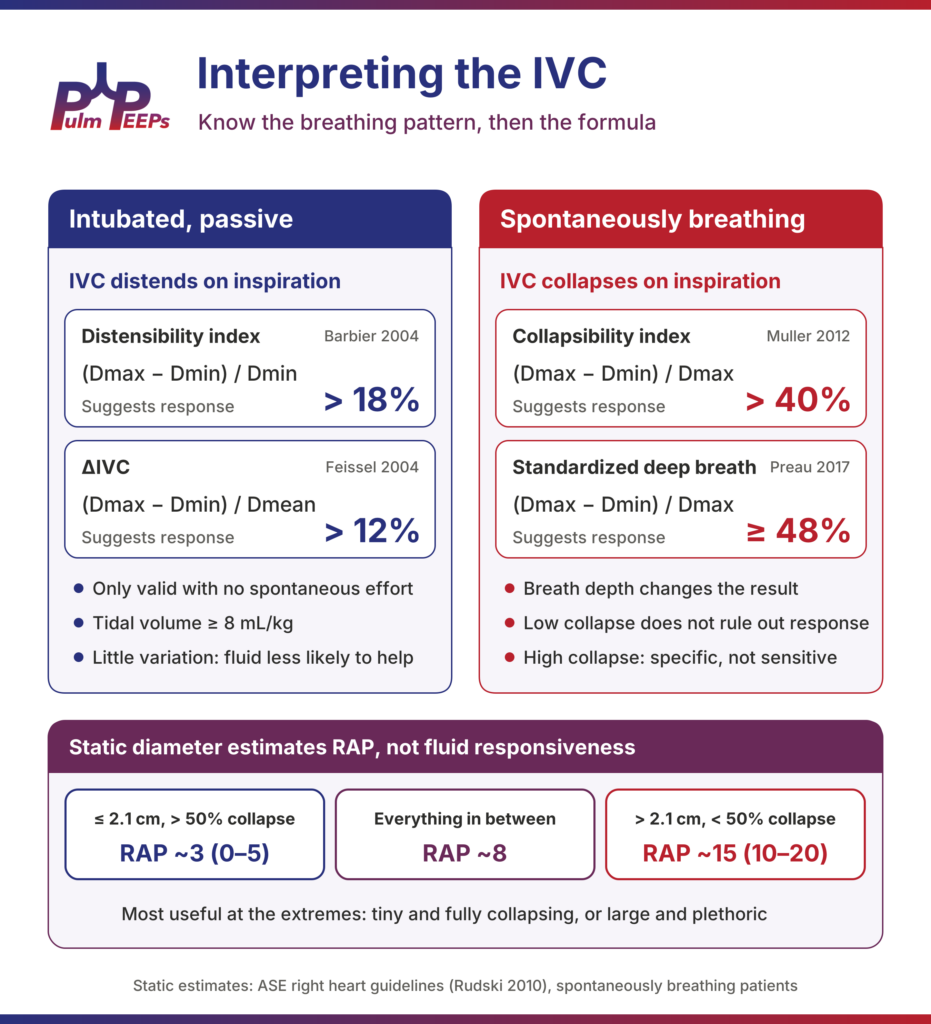
Distensibility index (dIVC)(Dmax − Dmin) / Dmin> 18%Barbier 2004, n = 23: sensitivity and specificity ~90%
ΔIVC(Dmax − Dmin) / Dmean> 12%Feissel 2004, n = 39: PPV 93%, NPV 92%

Match the formula to the cutoff. Mixing them up is one of the most common errors. The easy way to remember it: 18 percent if you divide by the minimum, 12 percent if you divide by the mean. If your machine calculates the index, check which formula it uses.

Know the conditions these were validated in. Patients were fully passive on the ventilator, in volume control, with tidal volumes of about 8 to 10 mL/kg. In today’s ICU, with lung-protective ventilation at 6 mL/kg and patients often triggering breaths, those conditions frequently do not apply. Choose your patient carefully: passive on the vent, with an adequate tidal volume.

The Spontaneously Breathing Patient: Collapsibility

In the ED or on the floor, we are looking at collapsibility (the caval index): (Dmax − Dmin) / Dmax. Here the story gets messier. We do not control the rate or the effort, and a small shallow breath versus a big deep sniff gives very different collapsibility in the same patient at the same volume status. Many septic patients are also breathing hard, which changes intrathoracic dynamics considerably.

Three studies are worth knowing:

  • Muller 2012 (n = 40, acute circulatory failure): collapsibility > 40% had a sensitivity of 70% and specificity of 80%. A value below 40% did not rule out fluid responsiveness.
  • Airapetian 2015 (n = 59): overall performance was poor (AUC 0.62), but collapsibility > 42% was very specific (97%) with a sensitivity of only 31%.
  • Preau 2017 (n = 90, septic, non-intubated): standardizing the breath, a deep inspiration followed by passive exhalation, collapsibility ≥ 48% had a sensitivity of 84%, specificity of 90% and AUC of 0.89.

The practical takeaway: near-complete collapse, over 40 to 50 percent, especially with a standardized deep breath, makes fluid responsiveness more likely. A big number means something; a small number does not tell you much. And anyone can collapse their IVC if they are breathing hard enough, so in true respiratory distress this test is of little use.

What About Just the Size of the IVC?

“The IVC is flat, give fluid” is still heard on rounds, but the static diameter answers a different question. It comes from the echo world, where it is used to estimate right atrial pressure in spontaneously breathing patients, many of them outpatients. Per the American Society of Echocardiography right heart guidelines (Rudski 2010):

IVC diameter and sniff collapseEstimated RAP (mmHg)
≤ 2.1 cm and > 50% collapse~3 (0–5)
Everything in between~8
> 2.1 cm and < 50% collapse~15 (10–20)

Estimating right atrial pressure is not predicting fluid responsiveness. Decades of ICU data show that static pressures, even a directly measured CVP, are poor predictors of fluid responsiveness. This takes us right back to the difference between volume status and volume responsiveness.

The static IVC is most useful at the extremes. A tiny, nearly virtual IVC (under about 1 cm) in a hypotensive patient makes fluid responsiveness more likely. A large, non-collapsing IVC should make you pause and think about the right heart, tamponade or volume overload. As with our FeNO episode, the extremes are informative and the middle is gray.
Ivc 2 interpretation

What Do the Data Show?

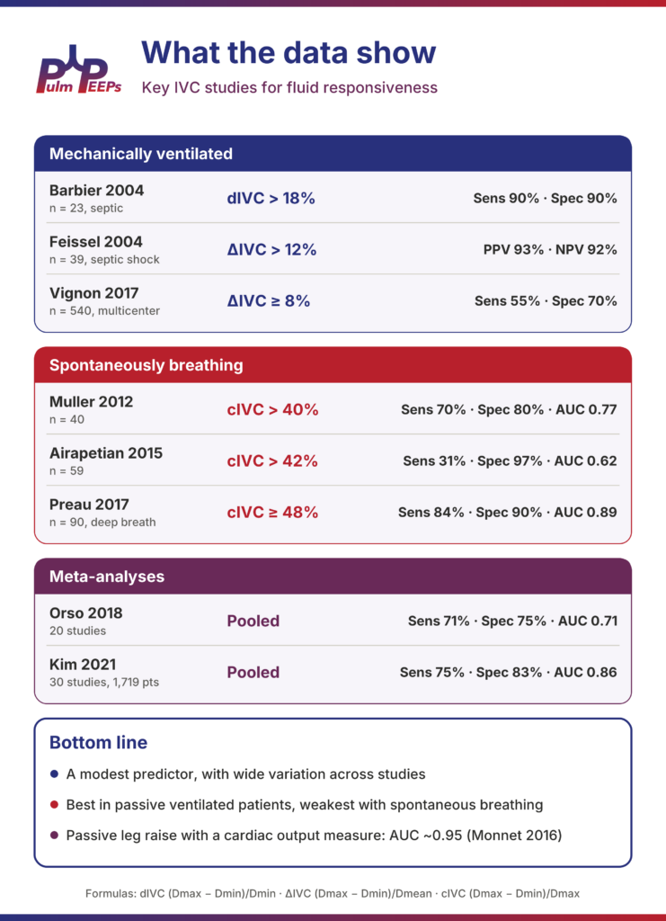
The IVC is a modest predictor of fluid responsiveness, with wide variation across studies. Performance is best in passive ventilated patients and weakest with spontaneous breathing:

StudyPopulationIndex and cutoffPerformance
Barbier 200423 ventilated, septicdIVC > 18%Sens 90%, spec 90%
Feissel 200439 ventilated, septic shockΔIVC > 12%PPV 93%, NPV 92%
Vignon 2017540 ventilated, multicenterΔIVC ≥ 8%Sens 55%, spec 70%
Muller 201240 spontaneously breathingcIVC > 40%Sens 70%, spec 80%, AUC 0.77
Airapetian 201559 spontaneously breathingcIVC > 42%Sens 31%, spec 97%, AUC 0.62
Preau 201790 spontaneously breathing, deep breathcIVC ≥ 48%Sens 84%, spec 90%, AUC 0.89
Orso (meta-analysis)20 studiesPooledSens 71%, spec 75%, AUC 0.71
Kim 2021 (meta-analysis)30 studies, 1,719 patientsPooledSens 75%, spec 83%, AUC 0.86

Note how much the large multicenter Vignon cohort pulls the ventilated numbers down compared with the small original studies. The meta-analyses are mixed and very heterogeneous: Orso and colleagues concluded the IVC is not a reliable predictor on its own, while Kim and colleagues found better pooled accuracy. Both pointed to huge variation in patient populations, formulas, cutoffs and how responsiveness was defined.

Putting it together: the IVC is fast, noninvasive and repeatable, and it is a useful piece of the puzzle. It should never be the only thing driving the decision to give fluid. When it really matters, a passive leg raise paired with a measure of cardiac output performs far better (pooled AUC ~0.95, Monnet 2016). We will cover LVOT VTI, pulse pressure variation and VExUS in future episodes.
Ivc 3 evidence

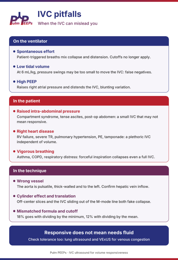
Pitfalls: When the IVC Can Mislead You

The patients we most want to answer this question for are often the ones in whom the IVC is least reliable.

On the ventilator

  • Spontaneous effort: patient-triggered breaths mix collapse and distension, and the cutoffs no longer apply.
  • Low tidal volume: at 6 mL/kg, the change in intrathoracic pressure may be too small to move the IVC, causing false negatives.
  • High PEEP: raises right atrial pressure on its own, distending the IVC and blunting variation.

In the patient

  • Raised intra-abdominal pressure: abdominal compartment syndrome, tense ascites, bowel distension or a recent abdominal surgery can compress the IVC, so a small, collapsible IVC may not mean responsive.
  • Right heart disease: RV failure, severe tricuspid regurgitation, pulmonary hypertension, PE and tamponade all give a plethoric IVC independent of volume. In tamponade or massive PE, of course, that plethoric IVC is important diagnostic information in its own right.
  • Vigorous breathing: in asthma, COPD exacerbations or any respiratory distress, forceful inspiration can collapse even a full IVC, and active exhalation distorts the measurement.

In the technique

  • Wrong vessel: the aorta is pulsatile, thick-walled and to the left. Confirm hepatic vein inflow.
  • Cylinder effect and translation: off-center slices and the IVC sliding out of the M-mode line both fake collapse.
  • Mismatched formula and cutoff: 18% goes with dividing by the minimum, 12% with dividing by the mean.

And remember: responsive does not mean needs fluid. Check tolerance too, with lung ultrasound and VExUS for venous congestion.
Ivc 4 pitfalls

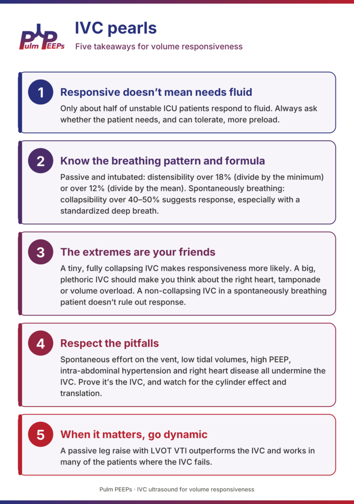
The Five Pulm PEEPs Pearls

  1. Fluid responsive doesn’t mean fluid needed. Only about half of unstable ICU patients respond to fluid, and responsiveness is only one part of the decision. Always ask whether the patient needs, and can tolerate, more preload.
  2. Know the breathing pattern and the formula. Passive and intubated: distensibility over 18% (divide by the minimum) or over 12% (divide by the mean). Spontaneously breathing: collapsibility over 40 to 50% suggests response, especially with a standardized deep breath.
  3. The extremes are your friends. A tiny, fully collapsing IVC makes responsiveness more likely. A big, plethoric IVC should make you think about the right heart, tamponade or volume overload. A non-collapsing IVC in a spontaneously breathing patient doesn’t rule out response.
  4. Respect the pitfalls. Spontaneous effort on the vent, low tidal volumes, high PEEP, intra-abdominal hypertension and right heart disease all undermine the IVC, and they are common in our sickest patients. Prove it’s the IVC, and watch for the cylinder effect and translation.
  5. When it matters, go dynamic. A passive leg raise with LVOT VTI outperforms the IVC and works in many of the patients where the IVC fails.
Ivc 5 pearls

Scan carefully, interpret in context, and as always, keep breathing!

References and Further Reading

  • Michard F, Teboul JL. “Predicting fluid responsiveness in ICU patients: a critical analysis of the evidence.” Chest. 2002;121(6):2000–2008.
  • Barbier C, Loubières Y, Schmit C, et al. “Respiratory changes in inferior vena cava diameter are helpful in predicting fluid responsiveness in ventilated septic patients.” Intensive Care Med. 2004;30(9):1740–1746.
  • Feissel M, Michard F, Faller JP, Teboul JL. “The respiratory variation in inferior vena cava diameter as a guide to fluid therapy.” Intensive Care Med. 2004;30(9):1834–1837.
  • Muller L, Bobbia X, Toumi M, et al. “Respiratory variations of inferior vena cava diameter to predict fluid responsiveness in spontaneously breathing patients with acute circulatory failure: need for a cautious use.” Crit Care. 2012;16(5):R188.
  • Airapetian N, Maizel J, Alyamani O, et al. “Does inferior vena cava respiratory variability predict fluid responsiveness in spontaneously breathing patients?” Crit Care. 2015;19:400.
  • Preau S, Bortolotti P, Colling D, et al. “Diagnostic accuracy of the inferior vena cava collapsibility to predict fluid responsiveness in spontaneously breathing patients with sepsis and acute circulatory failure.” Crit Care Med. 2017;45(3):e290–e297.
  • Vignon P, Repessé X, Bégot E, et al. “Comparison of echocardiographic indices used to predict fluid responsiveness in ventilated patients.” Am J Respir Crit Care Med. 2017;195(8):1022–1032.
  • Orso D, Paoli I, Piani T, et al. “Accuracy of ultrasonographic measurements of inferior vena cava to determine fluid responsiveness: a systematic review and meta-analysis.” J Intensive Care Med. 2020;35(4):354–363.
  • Kim DW, Chung S, Kang WS, Kim J. “Diagnostic accuracy of ultrasonographic respiratory variation in the inferior vena cava, subclavian vein, internal jugular vein, and femoral vein diameter to predict fluid responsiveness: a systematic review and meta-analysis.” Diagnostics (Basel). 2021;12(1):49.
  • Rudski LG, Lai WW, Afilalo J, et al. “Guidelines for the echocardiographic assessment of the right heart in adults.” J Am Soc Echocardiogr. 2010;23(7):685–713.
  • Monnet X, Marik P, Teboul JL. “Passive leg raising for predicting fluid responsiveness: a systematic review and meta-analysis.” Intensive Care Med. 2016;42(12):1935–1947.
122. Pulm PEEPs Pearls: Steroids in Sepsis

PPP Cover Images (1)

Today we have another Pulm PEEPs Pearls episode about a core critical care topic. Furf and Monty will be giving a high level overview of the use of steroids in sepsis including a review of the relevant literature and recent guidelines, and pragmatic bedside points.

This episode was prepared with research by Pulm PEEPs Associate Editor George Doumat.

Dustin Latimer, another Pulm PEEPs Associate Editor, assisted with audio and video editing.

Why Steroids in Sepsis?

Steroids do not treat the infection — antimicrobials are always first and remain the cornerstone. The goal is addressing critical illness–related corticosteroid insufficiency (CIRCI), where cortisol production cannot keep up with the overwhelming inflammatory demand of septic shock.

Hydrocortisone helps in two main ways:

  • Blunts the dysregulated inflammatory response — tempers the excessive vasodilation and febrile response that drive harm beyond the infection itself.
  • Restores vascular sensitivity to catecholamines — sepsis downregulates adrenergic receptors; steroids turn that responsiveness back on.

Clinical takeaway: The first thing you notice is vasopressor weaning (or a bend in the escalation curve) — not a rapid improvement in fever or white count.

Caveat: These trials predate modern sepsis phenotyping. None distinguish hyperinflammatory vs. hypoinflammatory responders — they treat all comers.

The Evidence: Four Landmark Trials

Every IM resident and critical care fellow will eventually journal-club these four. The most consistent signal across all of them is faster shock reversal and reduced vasopressor use; the mortality question remains unsettled.

Trial (Year)NRegimenKey Finding
Annane (2002)~300Hydrocortisone + fludrocortisoneMortality benefit in ACTH non-responders; criticized methodology and messy cortisol-response testing; not cleanly replicated.
CORTICUS (2008)~500Hydrocortisone aloneFaster shock reversal but no mortality benefit, regardless of cortisol responsiveness. Raised (later allayed) superinfection concern. Cornerstone for abandoning routine cort-stim testing.
ADRENAL (2018)~3,800Hydrocortisone aloneFaster vasopressor weaning; no 90-day mortality benefit.
APROCCHSS (2018)~1,200Hydrocortisone + fludrocortisoneMortality benefit at 90 days.

Bottom line: Faster shock reversal is consistent. Mortality benefit appears in 2 of 4 trials (both used fludrocortisone) but not the others. A 2026 meta-analysis showed benefit for hydrocortisone + fludrocortisone vs. placebo, but

not for hydrocortisone + fludrocortisone vs. hydrocortisone alone — suggesting hydrocortisone drives the main effect.

Who Gets Steroids, and When?

  • 2021 Surviving Sepsis: Consider steroids for norepinephrine or epinephrine ≥ 0.25 mcg/kg/min for ≥ 4 hours despite adequate resuscitation — a reasonable bedside trigger.
  • Early 2026 update: Moved away from a specific numeric trigger — consider steroids when a septic patient is not responding well to vasopressors or has escalating requirements. Make a clinical decision. (Quality of evidence: low to moderate.)
  • Go faster than the threshold when: Known/suspected adrenal insufficiency or home steroids, or florid pressor-requiring shock on arrival.

A practical escalation sequence: escalating norepinephrine → add vasopressin (per VASST) → then add steroids if requirements keep climbing.

Do NOT wait for an ACTH stimulation test. It does not reliably predict who responds and only delays treatment. Sepsis is an elevated-cortisol state but can dissociate ACTH and cortisol, and cortisol-binding globulin is depleted — the test is too messy to guide care.

What to Give: The Regimen

  • Standard dose: Hydrocortisone 200 mg/day, typically 50 mg IV Q6H. (Original trials often used continuous infusions, rarely used in the U.S.) Some start with a 100 mg bolus to gain control.
  • Higher dose: If chronically on steroids / adrenally insufficient, consider ~300 mg/day (e.g., 100 mg Q8H).
  • Fludrocortisone: Unsettled. The two mortality-benefit trials added it (50 mcg PO/NG/OG daily), but hydrocortisone already has mineralocorticoid activity and meta-analyses don’t show added benefit over hydrocortisone alone. Most clinicians omit it — adding it is reasonable and safe, just be honest about the uncertainty.

Duration & Tapering

  • Typical course: ~7 days is most common. Trial practices varied (ADRENAL ~7 days; VANISH used a taper after 6 days; some continue until pressors are off).
  • No taper needed. You do not need to taper for adrenal insufficiency after a short course — just stop. If pressors dramatically rebound, you can restart, but most patients have gained the benefit they’ll get by day 7.

Pitfalls & Safety

  • Hyperglycemia: Expected and must be managed (monitor closely; insulin drip if needed). No signal for major DKA / severe complications in the trials.
  • Superinfection / fungal infection: The most-quoted concern, but the overall literature does not show a convincing, statistically significant increase. Be disciplined about stopping on schedule.
  • Muscle weakness: Steroids can worsen critical illness myopathy; a short 7-day course likely has limited effect, but be aware.
  • Other: GI bleeding (follow general PPI prophylaxis guidance) and sodium disturbances (watch for hyper-/hyponatremia).

Two things we know: (1) steroids shorten duration of vasopressor support, and (2) they are relatively safe in sepsis. Whether they improve mortality — and in whom — remains open.

The Five Pulm PEEPs Pearls

  1. Mechanism: Steroids restore catecholamine vascular sensitivity and blunt dysregulated inflammation. The clinical target is vasopressor weaning, not infection treatment.
  2. Evidence: Faster shock reversal is the most consistent finding. Mortality benefit is seen in 2 of 4 trials but not the others — still controversial. Some patients likely benefit; we don’t yet know who.
  3. Trigger: A practical 2021 threshold is levo/epi ≥ 0.25 mcg/kg/min for ≥ 4 hours. Newer guidance drops the strict number — make a clinical decision based on poor pressor response or escalation.
  4. Dose: Hydrocortisone 200 mg/day (e.g., 50 mg Q6H). Adding fludrocortisone mirrors two trials, but meta-analyses find no benefit over hydrocortisone alone.
  5. Safety: Steroids appear safe in sepsis. Monitor and treat hyperglycemia; no marked increase in superinfection.

Annane, Djillali et al. “Effect of treatment with low doses of hydrocortisone and fludrocortisone on mortality in patients with septic shock.” JAMA vol. 288,7 (2002): 862-71. doi:10.1001/jama.288.7.862

Sprung, Charles L et al. “Hydrocortisone therapy for patients with septic shock.” The New England journal of medicine vol. 358,2 (2008): 111-24. doi:10.1056/NEJMoa071366

Venkatesh, Balasubramanian et al. “Adjunctive Glucocorticoid Therapy in Patients with Septic Shock.” The New England journal of medicine vol. 378,9 (2018): 797-808. doi:10.1056/NEJMoa1705835

Annane, Djillali et al. “Hydrocortisone plus Fludrocortisone for Adults with Septic Shock.” The New England journal of medicine vol. 378,9 (2018): 809-818. doi:10.1056/NEJMoa1705716

Sun, Alin et al. “Correction: Hydrocortisone combined with fludrocortisone for treatment of adults with septic shock: an updated meta-analysis and systematic review.” Frontiers in medicine vol. 13 1811616. 2 Mar. 2026, doi:10.3389/fmed.2026.1811616

Prescott, Hallie C et al. “Executive Summary: Surviving Sepsis Campaign: International Guidelines for Management of Sepsis and Septic Shock 2026.” Critical care medicine vol. 54,4 (2026): 715-724. doi:10.1097/CCM.0000000000007089

111. Pulm PEEPs Pearls: Methylene Blue

PPP Cover Images (1)

Furf and Monty are back today with another Pulm PEEPs Pearls episode, and discussing the use of methylene blue for patients with septic shock. They review the clinical scenarios when this comes up, the mechanism, some key data, and some take aways, all in 15 minutes! Let us know any other topics you’d like covered on the show and make sure to like, give us 5 stars, and subscribe wherever you’re listening to this podcast.

This episode was prepared in conjunction with George Doumat MD. Goerge is an internal medicine resident at UT Southwestern and joined us for a Pulm PEEPs – BMJ Thorax journal club episode. He is now acting as a Pulm PEEPs Editor for the Pulm PEEPs Pearls series.

  1. Clinical context: when does methylene blue even come up?
  • This is not a first-line sepsis drug.
  • It’s considered in catecholamine-refractory vasoplegic septic shock, typically when:
    • Norepinephrine is at high dose
    • Vasopressin is on board
    • Often a 3rd or 4th vasopressor is being used (e.g., phenylephrine, angiotensin II)
  • The phenotype is strongly vasodilatory/vasoplegic (warm, distributive shock) rather than primarily cardiogenic.
  1. Mechanism of action (why it might help)
  • Methylene blue:
    • Inhibits inducible nitric oxide synthase and guanylate cyclase.
    • Blunts excess nitric oxide and cyclic GMP–mediated vasodilation, which are key in vasoplegic sepsis.
  • Practical translation:
    • It restores vascular tone and can make the vasculature more responsive to catecholamines.
  • It’s also used in post-CPB vasoplegia (e.g., after cardiac surgery, especially in patients on ACE inhibitors) and has migrated from that world into ICU sepsis practice.
  1. Typical dosing strategy (as described in the episode)
  • Common approach:
    • 1–3 mg/kg IV bolus, then
    • Reassess hemodynamics (MAP, dynamic perfusion markers).
    • If there’s a response, consider a continuous infusion or repeat bolus.
  • Key nuance: unlike other pressors that start as drips, methylene blue is often trialed as a bolus first to see if it’s doing anything.
  1. What does the evidence suggest?

Most data are from small, single-center, heterogeneous studies, so evidence quality is low. Meta-analyses and systematic reviews (through ~2024–25) suggest:

  • Hemodynamics
    • Can increase MAP (roughly 1–10 mmHg across studies).
    • May shorten total vasopressor duration (one meta-analysis ~30 hours less, though this is not definitive).
  • Secondary physiologic effects
    • Some small improvements in PaO₂/FiO₂ (P/F) ratio in certain studies.
  • Clinical outcomes
    • Possible reduction in hospital length of stay (≈ up to 2 days in some pooled analyses).
    • Some signal toward lower short-term mortality, but:
      • Studies are small
      • Heterogeneous
      • Evidence is very low certainty
  • Bottom line:
    • There’s a repeatable signal that methylene blue:
      • Raises MAP
      • Helps reduce catecholamine requirements
    • But hard clinical outcomes (mortality, LOS, ventilator days) remain uncertain.
  1. Safety profile & important adverse effects

Things to watch for:

  • Methemoglobinemia
  • Serotonin syndrome
    • Especially in patients on SSRIs, though in life-threatening refractory shock the hosts still lean toward using it with caution.
  • Pulse oximeter artifact
    • Can distort SpO₂ readings.
  • Urine discoloration
    • Blue/green urine—benign but striking.

Notably:

  • Methylene blue is both a treatment for and a potential cause of methemoglobinemia, depending on context and dosing.
  1. Guidelines & where it fits in practice
  • Surviving Sepsis Campaign 2021:
    • Does not recommend methylene blue for routine use in septic shock.
  • No major critical care society includes it in standard septic shock bundles or protocols.
  • The hosts frame methylene blue as:
    • A rescue therapy, not guideline therapy.
    • Something to consider only in refractory vasoplegic shock, ideally with:
      • Multidisciplinary discussion (intensivist, pharmacist, etc.).
      • Clear documentation that this is off-guideline, salvage use.
  1. Practical bedside framing (“2 a.m. in the ICU”)

They emphasize three pillars of practice:

  1. Physiology – mechanism makes sense (NO / cGMP / vasodilation).
  2. Empiric evidence – small studies and meta-analyses show a signal but low-quality data.
  3. Bedside reality – at 2 a.m., with a patient in multi-pressor, refractory vasoplegic shock, you sometimes reach for imperfect tools.

So, the practical take:

  • You should NOT:
    • Use methylene blue early.
    • Treat it as part of standard sepsis care.
  • You may consider it when:
    • Shock is clearly vasoplegic and refractory.
    • Norepi + vasopressin + at least one more vasopressor are maxed.
    • Team agrees this is salvage therapy and understands the limited evidence and side effects.

103. Fellows’ Case Files: University of Virginia

Case Files

Today, we’re virtually visiting the University of Virginia for another Fellows’ Case Files. This is a fantastic case that covers ARDS, the infectious work up of an immunosuppressed patient, and the evaluation of undifferentiated shock. Please let us know what you think of the episode and always feel free to reach out with interesting cases!


John Popovich completed his residency training and chief year at UVA and has stayed on there for his pulmonary and critical care fellowship.

Tim Scialla is an associate professor of medicine at UVA. He completed his residency and fellowship at Johns Hopkins Hospital where he was also an ACS. His clinical and research focuses are advanced airways disease. He is also the program director of the PCCM fellowship.

Matt Freedman completed his residency training at Virginia Commonwealth University and is currently a second year fellow at University of Virginia.


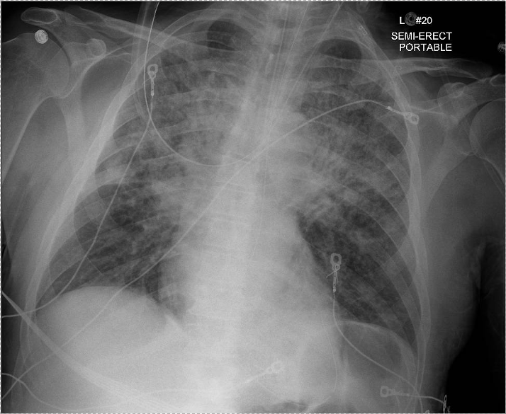
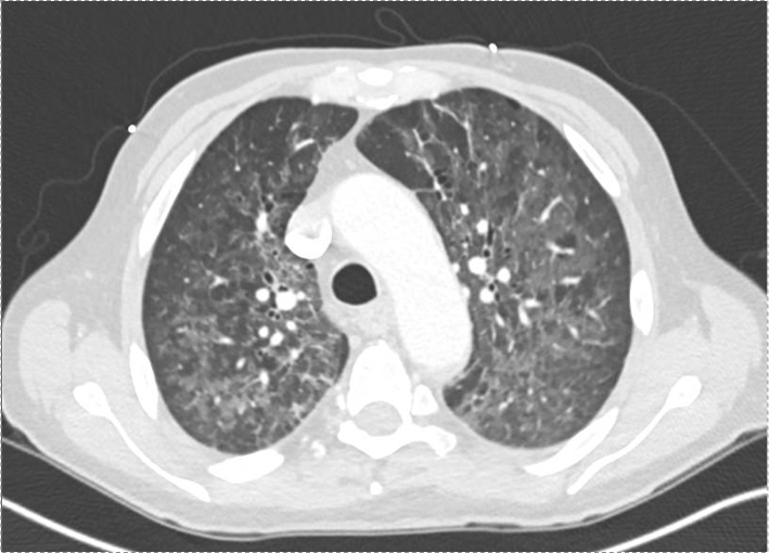
Patient: 52-year-old male with psoriasis, HIV/AIDS (CD4 count: 71), presenting with progressive shortness of breath, fever, non-productive cough, and weight loss.

Vital signs: Febrile (103°F), tachycardic (HR 110), hypoxemic on 6L O₂ (SpO₂ 90–92%).

Exam: Diffuse crackles, ill-appearing.

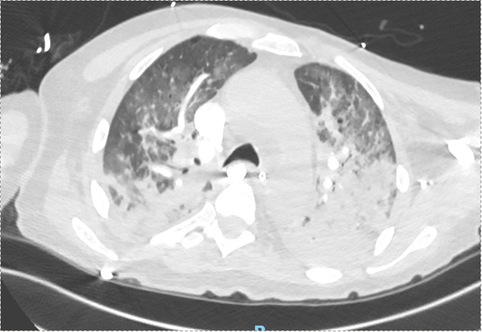
Imaging: CXR and CT showed bilateral upper lobe infiltrates, ground-glass opacities, septal thickening, and peripheral cystic changes.


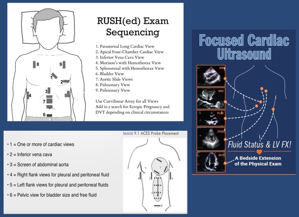
POCUS algorithms for investigating shock

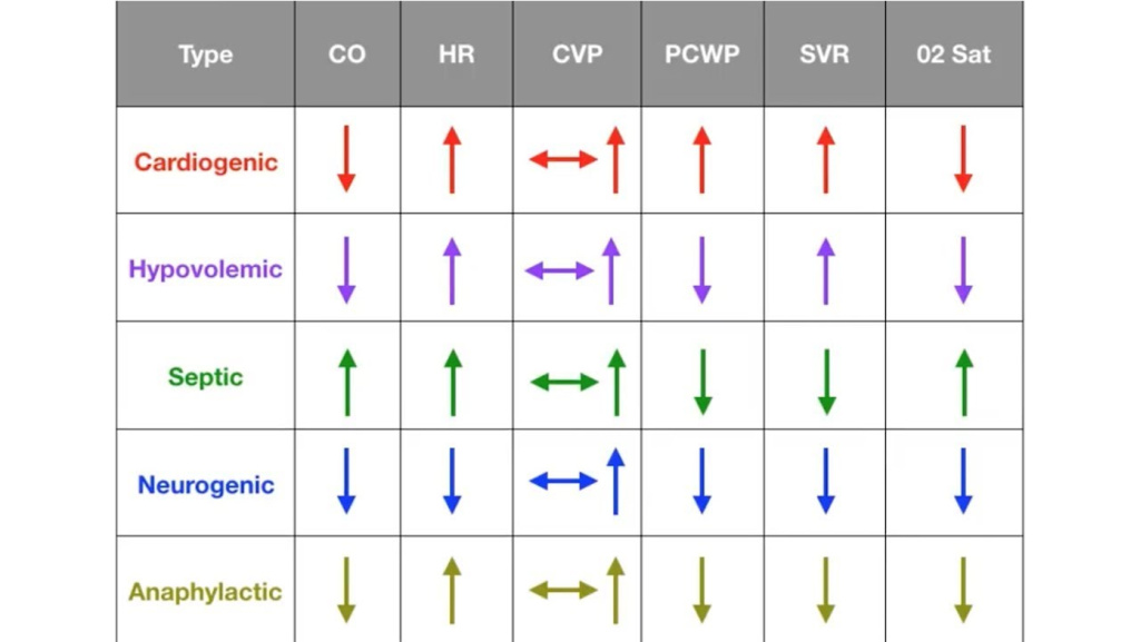
Shock physiology:


Diagnostic Reasoning in Immunocompromised Hosts

  • Framework: Anchor the differential based on type of immunosuppression.
    • HIV/AIDS → T-cell dysfunction, affecting susceptibility to PCP, TB, CMV, fungi (e.g. histo/blasto), and common CAP organisms.
  • PCP considerations:
    • PCP can occur despite prophylaxis (e.g. Bactrim), especially if adherence or resistance issues exist.
    • Classic symptoms in AIDS: acute, febrile, hypoxemic respiratory failure.

Use of Serum Markers and Imaging

  • LDH: Elevated in PCP, but non-specific. High negative predictive value when normal.
  • 1,3-β-D-glucan: Elevated in PCP and other fungal infections. Very sensitive for PCP (up to 95%).
  • Imaging: Ground-glass opacities with cystic changes support PCP diagnosis.

Role of Bronchoscopy and Diagnostic Yield

  • BAL studies to obtain:
    • DFA for PCP (rapid, high specificity, lower sensitivity)
    • PCR for PCP (higher sensitivity, slower turnaround)
    • Cultures: bacterial, fungal, mycobacterial
    • Cytology, galactomannan, histo/blasto urine antigens
  • Bronch Risk-Benefit:
    • Can change management in 40–60% of cases.
    • Complication rate: ~10–15%, most often hypoxemia.
    • Heuristic for pre-bronch ABG on non-rebreather:
      • PaO₂ >150 → likely safe
      • 100–150 → ~25% risk of intubation
      • <100 → high risk of decompensation

Steroids in PCP and Severe CAP

  • Steroids indicated in PCP with significant hypoxemia (PaO₂ <70 mmHg).
  • With new CAP guidelines (Cape Cod trial), steroids may also be considered in severe bacterial CAP.

Shock Evaluation in ICU

  • Framework: Simplify into likely causes — distributive most common, but rule out cardiogenic, obstructive, hypovolemic.
  • Physical exam + POCUS essential early.
    • POCUS: cardiac views, IVC, lung US, abdominal free fluid.
    • Low EF doesn’t exclude distributive shock.
  • PA catheter (Swan) utility:
    • Useful when physiology unclear or when tracking response to therapy is critical.
    • Swan data in this patient: low CVP and wedge, high SVR → distributive shock, not cardiogenic despite low EF.

90. Rapid Fire Journal Club: ANDROMEDA-SHOCK

Screenshot 2024 11 27 at 7.58.33 AM

We are excited to be back with a Rapid Fire Journal Club. Today’s episode is hosted by PulmPEEPs Associate Editor, Luke Hedrick, and will delve into the ANDROMEDA-SHOCK trial published in JAMA in 2019.

Jose Meade Aguilar is a second year Internal Medicine resident at Boston University Medical Campus (BUMC).

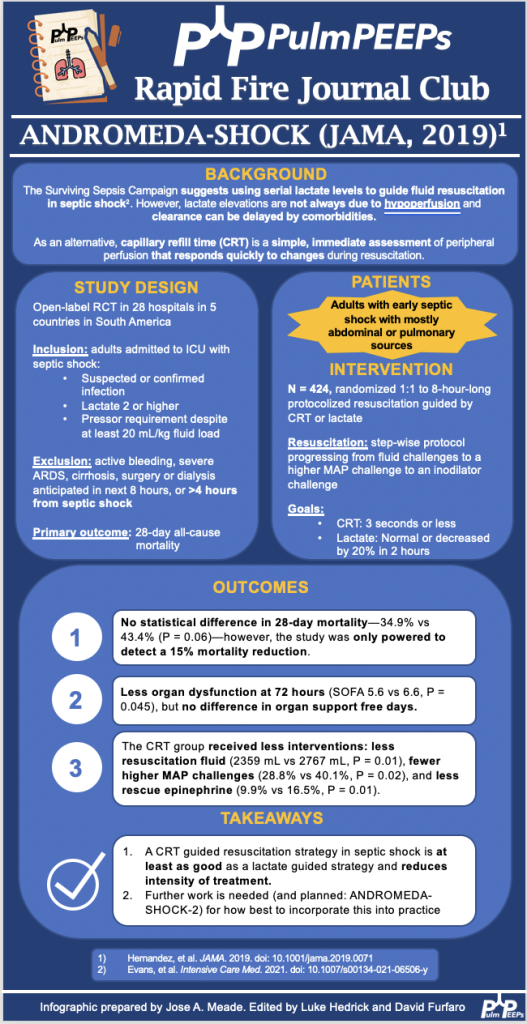
Today the discussion highlights the ANDROMEDA-SHOCK trial (JAMA, 2019) which evaluated whether resuscitation guided by capillary refill time (CRT) is superior to lactate-guided resuscitation in reducing mortality in patients with septic shock.

Hernández G, Ospina-Tascón GA, Damiani LP, Estenssoro E, Dubin A, Hurtado J, Friedman G, Castro R, Alegría L, Teboul JL, Cecconi M, Ferri G, Jibaja M, Pairumani R, Fernández P, Barahona D, Granda-Luna V, Cavalcanti AB, Bakker J; The ANDROMEDA SHOCK Investigators and the Latin America Intensive Care Network (LIVEN); Hernández G, Ospina-Tascón G, Petri Damiani L, Estenssoro E, Dubin A, Hurtado J, Friedman G, Castro R, Alegría L, Teboul JL, Cecconi M, Cecconi M, Ferri G, Jibaja M, Pairumani R, Fernández P, Barahona D, Cavalcanti AB, Bakker J, Hernández G, Alegría L, Ferri G, Rodriguez N, Holger P, Soto N, Pozo M, Bakker J, Cook D, Vincent JL, Rhodes A, Kavanagh BP, Dellinger P, Rietdijk W, Carpio D, Pavéz N, Henriquez E, Bravo S, Valenzuela ED, Vera M, Dreyse J, Oviedo V, Cid MA, Larroulet M, Petruska E, Sarabia C, Gallardo D, Sanchez JE, González H, Arancibia JM, Muñoz A, Ramirez G, Aravena F, Aquevedo A, Zambrano F, Bozinovic M, Valle F, Ramirez M, Rossel V, Muñoz P, Ceballos C, Esveile C, Carmona C, Candia E, Mendoza D, Sanchez A, Ponce D, Ponce D, Lastra J, Nahuelpán B, Fasce F, Luengo C, Medel N, Cortés C, Campassi L, Rubatto P, Horna N, Furche M, Pendino JC, Bettini L, Lovesio C, González MC, Rodruguez J, Canales H, Caminos F, Galletti C, Minoldo E, Aramburu MJ, Olmos D, Nin N, Tenzi J, Quiroga C, Lacuesta P, Gaudín A, Pais R, Silvestre A, Olivera G, Rieppi G, Berrutti D, Ochoa M, Cobos P, Vintimilla F, Ramirez V, Tobar M, García F, Picoita F, Remache N, Granda V, Paredes F, Barzallo E, Garcés P, Guerrero F, Salazar S, Torres G, Tana C, Calahorrano J, Solis F, Torres P, Herrera L, Ornes A, Peréz V, Delgado G, López A, Espinosa E, Moreira J, Salcedo B, Villacres I, Suing J, Lopez M, Gomez L, Toctaquiza G, Cadena Zapata M, Orazabal MA, Pardo Espejo R, Jimenez J, Calderón A, Paredes G, Barberán JL, Moya T, Atehortua H, Sabogal R, Ortiz G, Lara A, Sanchez F, Hernán Portilla A, Dávila H, Mora JA, Calderón LE, Alvarez I, Escobar E, Bejarano A, Bustamante LA, Aldana JL. Effect of a Resuscitation Strategy Targeting Peripheral Perfusion Status vs Serum Lactate Levels on 28-Day Mortality Among Patients With Septic Shock: The ANDROMEDA-SHOCK Randomized Clinical Trial. JAMA. 2019 Feb 19;321(7):654-664. doi: 10.1001/jama.2019.0071. PMID: 30772908; PMCID: PMC6439620.

76. Fellows’ Case Files: University of Rochester

Case Files U of Rochester

Today we’re back with another stop on our Fellows’ Case Files journey and making our way to the University of Rochester. Tune in to hear about this fascinating case and learn some key teaching points along the way.

Dr. Shiv Patel completed his IM residency and a Chief year at the California Pacific Medical Center- Van Ness Campus and is currently a second-year PCCM fellow at the University of Rochester.

Dr. Mary Anne Morgan is an Associate Professor of Medicine and the Fellowship Program Director for the PCCM Fellowship at the University of Rochester. Her clinical interests range from the care of critically ill patients in the ICU to the diagnosis and management of rare lung disease in her role as Director of the University of Rochester LAM Clinic. She loves unwrapping clinical reasoning with trainees, exploring issues around communication and teamwork in the ICU, and is excited about curriculum revitalization in the growing URMC PCCM fellowship program.

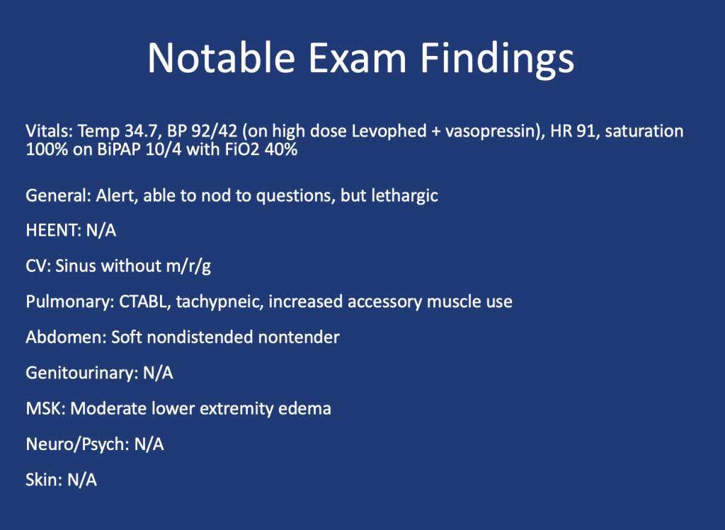
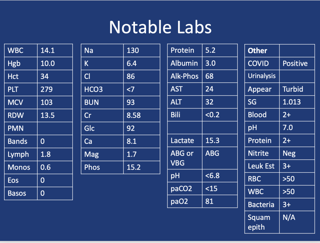
 A 75 y.o. female with a history of Hypertension, Hyperlipidemia, and Type 2 Diabetes presented for evaluation of hypoglycemia and generalized fatigue. She had felt poorly for about a week with symptoms of back pain, generalized weakness, and dyspnea, all of which acutely worsened on the day of presentation. 

She was found to be hypoglycemic with a blood glucose level in the to 40’s. Initial vital signs included a heart rate of 56, blood pressure of 70/40, respiratory rate of 30, and temperature of 28.5 degrees Celsius.

Lactic Acidosis: Type A, Type B and Type D

Type A: Typically secondary to conditions that impair oxygen delivery (respiratory failure, PE) to tissues or decrease tissue perfusion (severe anemia, shock). Patients typically present with hypotension, tachycardia, tachypnea, altered mental status, and signs of organ dysfunction.


Type B: Typically secondary to conditions that directly affect cellular metabolism or lactate clearance and characterized by the presence of hyperlactatemia without evidence of tissue hypoperfusion or hypoxia. Conditions associated include liver dysfunction (e.g., liver failure, cirrhosis), malignancies (especially hematological malignancies), medications/toxins (e.g., metformin, cyanide poisoning), inborn errors of metabolism, and mitochondrial disorders.

Type D: Less common presentation and can be seen in patients with short gut syndrome.

1.Blough B, Moreland A, Mora A Jr. Metformin-induced lactic acidosis with emphasis on the anion gap. Proc (Bayl Univ Med Cent). 2015 Jan;28(1):31-3. doi: 10.1080/08998280.2015.11929178. PMID: 25552792; PMCID: PMC4264704.

2.Callelo et al. Extracorporeal Treatment for Metformin Poisoning: Systematic Review and Recommendations From the Extracorporeal Treatments in Poisoning Workgroup. DOI: 10.1097/CCM.0000000000001002

3.Friesecke, S., Abel, P., Roser, M. et al. Outcome of severe lactic acidosis associated with metformin accumulation. Crit Care 14, R226 (2010). https://doi.org/10.1186/cc9376

4.Madias NE. Lactic acidosis. Kidney Int. 1986 Mar;29(3):752-74. doi: 10.1038/ki.1986.62. PMID: 3702227.

5. Stiller RH, Luks AM, Çoruh B. All That Raises Lactate Is Not Sepsis. ATS Sch. 2023 Jun 12;4(3):385-386. doi: 10.34197/ats-scholar.2023-0032OT.

62. Sepsis Roundtable: Best Practices and Future Directions

Case Files

We’re starting off 2024 with a bang!! Today we’re hosting another expert Roundtable discussion and we’re joined by internationally recognized experts in the field. We’ll tackle everything from teaching about sepsis, to how to incorporate guidelines into education and practice, to future research directions in the field. This is a can’t-miss discussion. Let us know what you think and other sepsis questions you have!

Meet Our Guests

Dr. Derek Angus is a Professor at the University of Pittsburgh where he holds the Mitchell P. Fink Endowed Chair in Critical Care Medicine and is the Chair of the Department of Critical Care Medicine. He is a world-renowned researcher in a range of critical care topics including sepsis, has hundreds of publications, and has led numerous NIH-funded studies.

Dr. Hallie Prescott is an Associate Professor in Pulmonary and Critical Care Medicine at the University of Michigan. She is the Co-Chair of the Surviving Sepsis Campaign Guidelines and is also an internationally recognized expert due to her research in improving sepsis outcomes. She has been recognized by both medical journals and professional societies for her outstanding contributions to the field.

Summary of Episode Discussion Topics

1. Sepsis Guidelines and Education

  • Surviving Sepsis Guidelines: Stressed as essential reading for professionals in pulmonary and critical care. They provide a structured approach to sepsis management.
  • Teaching Approaches: Transition from during-rounds teaching to focused, separate teaching sessions for trainees. Emphasizes the need to go beyond guidelines to include discussions on seminal articles, management strategies, and areas lacking robust data.

2. Clinical Skills and Decision Making in Sepsis Care

  • Early Recognition and Polypharmacy: Highlighted the need for timely sepsis identification and caution against excessive polypharmacy.
  • Mental Models in Care: Encourages building comprehensive mental models for understanding sepsis, stressing the importance of not just treating symptoms but understanding underlying causes.

3. Implementation of Sepsis Guidelines

  • Guideline Application in Bedside Care: Discusses the challenge of applying guidelines while considering patient-specific factors.
  • Fluid Resuscitation Practices: Identifies fluid resuscitation as a key area for improvement, with a shift towards more conservative approaches.
  • Overcoming Institutional Barriers: Addresses the fear of causing harm as a significant barrier to guideline implementation and emphasizes the need for balanced decision-making.

4. Advances in Sepsis Care and Prevention

  • Pre-Hospital Sepsis Management: Explores the role of early intervention in community settings and the potential of wearables for early detection.
  • Paramedic Role in Early Antibiotic Administration: Underlines the importance of starting antibiotics in the ambulance for suspected sepsis cases.

5. Recovery and Post-Discharge Care

  • Post-Discharge Initiatives: Focuses on improving handoffs from ICU to ward and from hospital to home. Highlights the importance of medication reconciliation and clear communication with primary care.
  • Challenges in Continuity of Care: Discusses the need for clear documentation and communication during patient transitions to ensure continuity of care.

6. Future Directions in Sepsis Treatment and Research

  • Phenotyping for Targeted Treatment: The potential of identifying patient subgroups through phenotyping for more effective, tailored treatments.
  • Adaptive Trial Designs: Advocates for large-scale adaptive platform trials that can test multiple interventions across diverse patient populations.

7. Personal Involvements and Perspectives

  • Experts’ Current Work: The panelists share their ongoing projects and research in sepsis care, reflecting a commitment to advancing the field through comprehensive and adaptive approaches.

References and Further Reading

  1. Evans L, Rhodes A, Alhazzani W, Antonelli M, Coopersmith CM, French C, Machado FR, Mcintyre L, Ostermann M, Prescott HC, Schorr C, Simpson S, Wiersinga WJ, Alshamsi F, Angus DC, Arabi Y, Azevedo L, Beale R, Beilman G, Belley-Cote E, Burry L, Cecconi M, Centofanti J, Coz Yataco A, De Waele J, Dellinger RP, Doi K, Du B, Estenssoro E, Ferrer R, Gomersall C, Hodgson C, Hylander Møller M, Iwashyna T, Jacob S, Kleinpell R, Klompas M, Koh Y, Kumar A, Kwizera A, Lobo S, Masur H, McGloughlin S, Mehta S, Mehta Y, Mer M, Nunnally M, Oczkowski S, Osborn T, Papathanassoglou E, Perner A, Puskarich M, Roberts J, Schweickert W, Seckel M, Sevransky J, Sprung CL, Welte T, Zimmerman J, Levy M. Surviving Sepsis Campaign: International Guidelines for Management of Sepsis and Septic Shock 2021. Crit Care Med. 2021 Nov 1;49(11):e1063-e1143. doi: 10.1097/CCM.0000000000005337. PMID: 34605781.
  2. Rudd KE, Kissoon N, Limmathurotsakul D, Bory S, Mutahunga B, Seymour CW, Angus DC, West TE. The global burden of sepsis: barriers and potential solutions. Crit Care. 2018 Sep 23;22(1):232. doi: 10.1186/s13054-018-2157-z. PMID: 30243300; PMCID: PMC6151187.
  3. Talisa VB, Yende S, Seymour CW, Angus DC. Arguing for Adaptive Clinical Trials in Sepsis. Front Immunol. 2018 Jun 28;9:1502. doi: 10.3389/fimmu.2018.01502. PMID: 30002660; PMCID: PMC6031704.
  4. Prescott HC, Angus DC. Enhancing Recovery From Sepsis: A Review. JAMA. 2018 Jan 2;319(1):62-75. doi: 10.1001/jama.2017.17687. PMID: 29297082; PMCID: PMC5839473.
  5. https://mi-hms.org/quality-initiatives/sepsis-initiative
  6. Kowalkowski M, Chou SH, McWilliams A, Lashley C, Murphy S, Rossman W, Papali A, Heffner A, Russo M, Burke L, Gibbs M, Taylor SP; Atrium Health ACORN Investigators. Structured, proactive care coordination versus usual care for Improving Morbidity during Post-Acute Care Transitions for Sepsis (IMPACTS): a pragmatic, randomized controlled trial. Trials. 2019 Nov 29;20(1):660. doi: 10.1186/s13063-019-3792-7. PMID: 31783900; PMCID: PMC6884908.
  7. Schmidt K, Worrack S, Von Korff M, Davydow D, Brunkhorst F, Ehlert U, Pausch C, Mehlhorn J, Schneider N, Scherag A, Freytag A, Reinhart K, Wensing M, Gensichen J; SMOOTH Study Group. Effect of a Primary Care Management Intervention on Mental Health-Related Quality of Life Among Survivors of Sepsis: A Randomized Clinical Trial. JAMA. 2016 Jun 28;315(24):2703-11. doi: 10.1001/jama.2016.7207. PMID: 27367877; PMCID: PMC5122319.

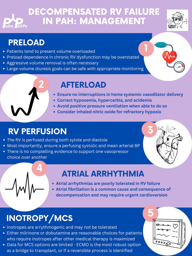
44. Decompensated Right Ventricular Failure in Pulmonary Arterial Hypertension

Pulm Peeps + CardioNerds

We are extremely excited to be hosting this episode in collaboration with CardioNerds! We have known Amit and Dan for many years, and they have been huge supporters of Pulm PEEPs, so it is an honor to address a topic we’re all interested in together.

We are joined by experts in the field today to discuss acute, decompensated right ventricle failure in patients with Pulmonary Arterial Hypertension (PAH). This topic can be quite intimidating, so we hope this will serve as a valuable guide for anyone who encounters a patient like this in the ICU.

Meet Our Guests

Leonid “Leon” Mirson is an internal medicine resident at the Johns Hopkins Hospital Osler Medical Residency and an Associate Editor here at Pulm PEEPs. He was born in Ukraine and moved to Philadelphia in early childhood with his family. He received his undergraduate degree from the University of Pittsburgh where he studied biomedical engineering and received his medical degree from the University of Pittsburgh School of Medicine. His current interests include pulmonary and critical care medicine with a focus on pulmonary hypertension as well as medical education. He is a rising PCCM fellow at the University of Pennsylvania.

Bhavya Varma completed her medical school at the University of Pittsburgh, her internal medicine residency at Johns Hopkins, and is a rising Cardiology fellow at NYU. She is interested in medical education and has done work with CardioNerds during her residency.

Mardi Gomberg-Maitland is a Professor of Medicine at George Washington University. She serves as the Medical Director of the Pulmonary Hypertension Program at George Washington Hospital. She completed her medical degree at Albert Einstein College of Medicine, completed her residency at the Weill-Cornell Medical Center, and completed her fellowship in cardiovascular diseases at Mount Sinai Medical Center. Her research focus is on understanding the epidemiology of pulmonary hypertension and the development of novel therapeutics and biomarkers. Dr. Gomberg-Maitland is internationally known for her work, she has had extensive grant funding and has published over 150 articles, abstracts, reviews, and chapters.

Rachel Damico is a pulmonologist and an Associate Professor of Medicine at Johns Hopkins Hospital, where she is also the Associate Director of the physician-scientist training program. Dr. Damico received her medical degree and doctoral degree in Molecular and Cellular biology from the University of Pennsylvania. She completed her residency in the Osler Internal Medicine training program and continued on as a PCCM fellow at Johns Hopkins. She has quickly achieved an international reputation in the field of pulmonary vascular biology and both basic and translational research, as well as clinical excellence, in Pulmonary Arterial Hypertension.

Patient Presentation

A 21-year-old woman with a past medical history notable for congenital heart disease (primum ASD and sinus venosus with multiple surgeries) complicated by severe PAH on home oxygen, sildenafil, ambrisentan, and subcutaneous treprostinil is presenting with palpitations, chest pain, and syncope. She presented as a transfer from an outside ED where she arrived in an unknown tachyarrhythmia and had undergone DCCV due to tachycardia into the 200s and hypotension. On arrival at our hospital, she denied SOB but did endorse nausea, leg swelling, and poor medication adherence. Her initial vitals were notable for a BP of 80/50, HR 110, RR 25, and saturating 91% on 5L O2.  On exam, she was uncomfortable appearing but mentating well. She had cool extremities with 1-2+ LE edema. Her JVP was 15cm H2O. She has an RV Heave and 2/6 systolic murmur. Her lungs were clear bilaterally. Her labs were notable for Cr 2.0, an anion gap metabolic acidosis (HCO3 = 11), elevated lactate (4.1), elevated troponin to 14,  and a pro-BNP of ~5000.  Her CBC was unremarkable. Her EKG demonstrated 2:1 atrial flutter at a rate of 130.

Key Learning Points

Diagnosing RV failure in patients with PH:

RV dysfunction and RV failure are two separate entities. RV dysfunction can be measured on echocardiography, but RV failure can be thought of as a clinical syndrome where there is evidence of RV dysfunction and elevated right sided filling pressures.

RV failure is a spectrum and can present with a range of manifestations from evidence of R sided volume overload and markers of organ dysfunction, all the way to frank cardiogenic shock. Most patients with RV failure are not in overt shock.

One of the first signs of impending shock in patients with RV failure is the development of new or worsening hypoxemia. Patients with decompensated RV failure approaching shock often do not present with symptoms classic for LV low flow state. Instead, hypoxia 2/2 VQ mismatching may be the first sign and they can be otherwise well appearing. Particularly because patients with PH tend to be younger, they can often appear compensated until they rapidly decompensate.

Causes of decompensation for patients with RV dysfunction and PH:

Iatrogenesis (inadvertent cessation of pulmonary vasodilators by providers, surgery if providers are not familiar with risks of anesthesia), non-adherence to pulmonary vasodilators (either due to affordability issues or other reasons), infections, arrhythmias (particularly atrial arrhythmias), and progression of underlying disease.

Patients with atrial arrhythmias (atrial flutter or atrial fibrillation) and pulmonary hypertension do not tolerate the loss of the atrial kick well as it contributes a significant amount to their RV filling and impacts their cardiac output. It is often difficult to determine if the atrial arrhythmia is a cause or effect of decompensated RV failure, but its presence is associated with a worse prognosis. Efforts should be made to re-establish normal sinus rhythm in patients with decompensated RV failure and atrial arrhythmias. 

A patient’s home PH medications should never be stopped for any reason upon admission unless on the basis of recommendations by a pulmonary hypertension provider as this is often a cause of decompensation inpatient

Interpreting findings on echocardiogram: 

Echo is a useful screening tool. When interpreting evidence of RV dysfunction, it is important to look at the global picture and not just one measurement.

RVSP, though commonly reported, may be of limited value when evaluating for decompensation. It’s a function of blood pressure, heart rate, and cardiac output. RVSP may even decline as shock worsens.

TAPSE is useful as a marker of RV dysfunction if it is reduced, but it is difficult to follow over time and only gives information about cardiac function around the annulus; it may be normal even when apical RV function is depressed. RV fractional area of change may be more useful for global RV function. It is important to pay attention to the RV size overall, the degree of TR, and the presence of effusion all of which are associated with RV dysfunction.

­Tips regarding the interpretation of invasive hemodynamics:

Cardiac output by thermodilution is the standard way to calculate PVR. Despite the degree of TR that is typically present, it is thought to be a better representation of cardiac output than the estimated Fick calculation.  

Our experts agree that routine monitoring of invasive hemodynamics for acute decompensated RV failure is likely not helpful and has significant risks. A good external volume exam or CVP off a central venous catheter + central venous saturation will likely be all you need to navigate a patient with shock secondary to RV failure. A right heart catheterization (should be only done under fluoroscopy for patients with large RVs) may be helpful if the etiology of shock is unclear. 

Management of decompensated RV failure in patients with pulmonary hypertension

Managing preload is of utmost importance, perhaps the most important tenant of management of decompensated RV failure.  The overwhelming majority of patients with PH and decompensation are volume overloaded, it is exceptionally rare that someone would be dry. Furthermore, the myth that the RV is “preload responsive” is only true in the setting of acute RV injury (eg. RV infarction) and not so in patients with acute on chronic RV dysfunction. It is important to optimize preload in someone in decompensated RV failure and it is safe to do this more rapidly than traditionally taught. Exact goals varied between our experts, but anywhere from 2-4L net negative per day is reasonable especially if the patient is hemodynamically tolerating the fluid removal. If the patient is not responding to diuretics, hemodialysis with ultrafiltration may be necessary to optimize the patient.

Afterload is the next tenant of management. Optimizing the following parameters will reduce the patient’s pulmonary vascular resistance and reduce afterload to the right ventricle.

— Avoiding hypoxic pulmonary vasoconstriction, liberalize the patient’s O2 goal 

— Avoid permissive hypercapnia and academia in this patient population

— Do not withhold a patient’s pulmonary vasodilator until discussion with the PH team. If stopped inadvertently, restart this medication immediately. For patients with malfunctioning pumps, there is a phone number on the back that you can call for rapid troubleshooting. Sildanefil can be given IV if a patient is NPO. 

— Inhaled nitric oxide can improve oxygenation and reduce afterload  

— Intubation and mechanical ventilation greatly increase PVR and are poorly tolerated. Exacting care must be taken to titrate PEEP and tidal volume, and avoid intubation when possible.

— Starting a new systemic pulmonary vasodilator in decompensated RV failure may be considered under close guidance from the pulmonary hypertension team

Management of atrial arrhythmias:

As above, patients with severe pulmonary hypertension do not tolerate loss of sinus rhythm well. If they are decompensated, every effort should be made to re-establish normal sinus rhythm. 

Management of RV perfusion:

Unlike the LV, the RV is perfused during BOTH systole and diastole. Maintaining effective coronary perfusion to the RV is essential in RV failure. For this reason, the systemic systolic pressure (as well as the mean arterial pressure) should be kept high enough to ensure that the RV is able to perfuse. There is no great body of evidence as to which pressor works best. Norepinephrine, vasopressin, and even phenylephrine are all reasonable choices to maintain appropriate perfusing blood pressure. 

Inotropy:

Patients in shock and RV failure do not always require inotropes, but if they do it’s often a sign of a grim prognosis. Either dobutamine or milrinone is reasonable, but the negative effects of these drugs (arrhythmias, tachycardia, and systemic hypotension) may limit their uses. 

Mechanical circulatory support:

Limited options are available. Balloon pumps and Impella devices have limited roles except in expert centers, and ECMO remains the standard of care. ECMO (either V-V or V-A) may have utility as a bridge to recovery if a reversible cause is identified, or a bridge to transplant if the patient is on the transplant list. 

Goals of care:

The prognosis of a patient admitted to the ICU with acute on chronic decompensated RV failure is guarded, with very high mortality rates even if not in shock

It is important for the patient’s longitudinal pulmonary hypertension provider to discuss the prognosis and goals of care ahead of time but this is not always possible. If they are admitted, early discussions regarding code status and prognosis are essential. It may be helpful to bring in the patient’s longitudinal pulmonary hypertension doctor into these discussions if possible. 

Infographic:

References and further reading:

Ventetuolo CE, Klinger JR. Management of acute right ventricular failure in the intensive care unit. Ann Am Thorac Soc. 2014 Jun;11(5):811-22. doi: 10.1513/AnnalsATS.201312-446FR. PMID: 24828526; PMCID: PMC4225807.

Arrigo M, Huber LC, Winnik S, Mikulicic F, Guidetti F, Frank M, Flammer AJ, Ruschitzka F. Right Ventricular Failure: Pathophysiology, Diagnosis and Treatment. Card Fail Rev. 2019 Nov 4;5(3):140-146. doi: 10.15420/cfr.2019.15.2. PMID: 31768270; PMCID: PMC6848943.

Kholdani CA, Fares WH. Management of Right Heart Failure in the Intensive Care Unit. Clin Chest Med. 2015 Sep;36(3):511-20. doi: 10.1016/j.ccm.2015.05.015. Epub 2015 Jun 27. PMID: 26304287.

Houston BA, Brittain EL, Tedford RJ. Right Ventricular Failure. N Engl J Med. 2023 Mar 23;388(12):1111-1125. doi: 10.1056/NEJMra2207410. PMID: 36947468.

39. Fellows’ Case Files: Indiana University

Fellows Case Files Indiana

Join us as we head to Indiana University! Listen in as we discuss another great case and hear teaching points from our amazing guests.

Meet our Guests


Parth Savsani is currently an internal medicine resident at Indiana University School of Medicine. He received his undergraduate degree from the University of Wisconsin-Madison and his medical degree from the University of Illinois College of Medicine. He enjoys medical education and was selected to be the VA chief resident next year.

Maria Srour is a Pulmonary and Critical Care Fellow at Indiana University School of Medicine. She completed her internal medicine residency at Saint Louis University where she was also a chief resident, and received her medical degree from IU. She works in global health to improve care for sepsis patients in low resources settings, and is currently pursuing her MPH.

Laura Hinkle is a Indiana University die hard and has been there from her since medical school through residency and fellowship, and is now an Assistant Professor of Clinical  Medicine and the Associate Program Director for the Pulmonary and Critical Care Medicine Fellowship. She will be taking over as the Program Director July 1, 2023. She is a dedicated educator and is the Key Clinical Educator for Pulmonary and Critical Care, and the Director of the Clinical Transitions Curriculum.  Additionally, she is working on a Master’s Degree in Education through the University of Cincinnati. 

Case Presentation

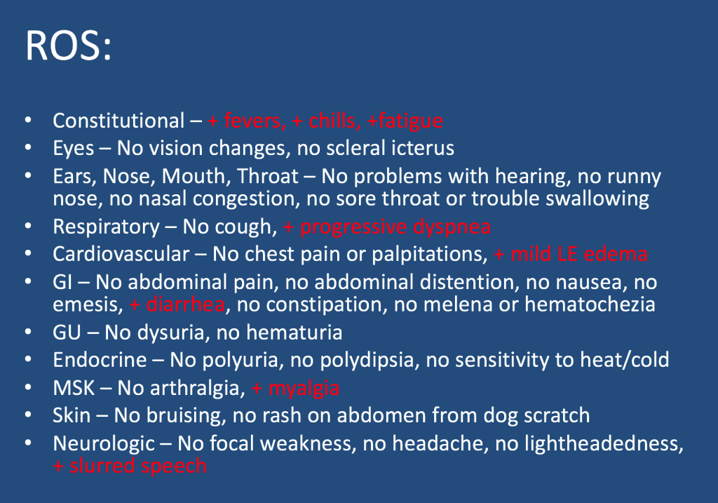
A male in her early 60s is transferred from a neighboring facility with a 1 week history of fatigue and lethargy. Three days prior to presentation he developed dyspnea and increased weakness with a near fall at home. HIs family also reported recent fevers, chills, dyspnea, and diarrhea. On his way to seek evaluation, he developed slurred speech without any other focal abnormalities.

Additional information is summarized as below:

Follow along our episode to hear the final diagnosis and key teaching points from the case!

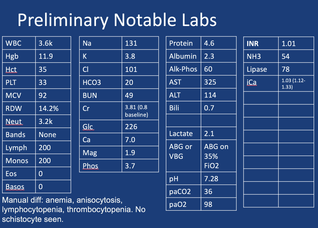
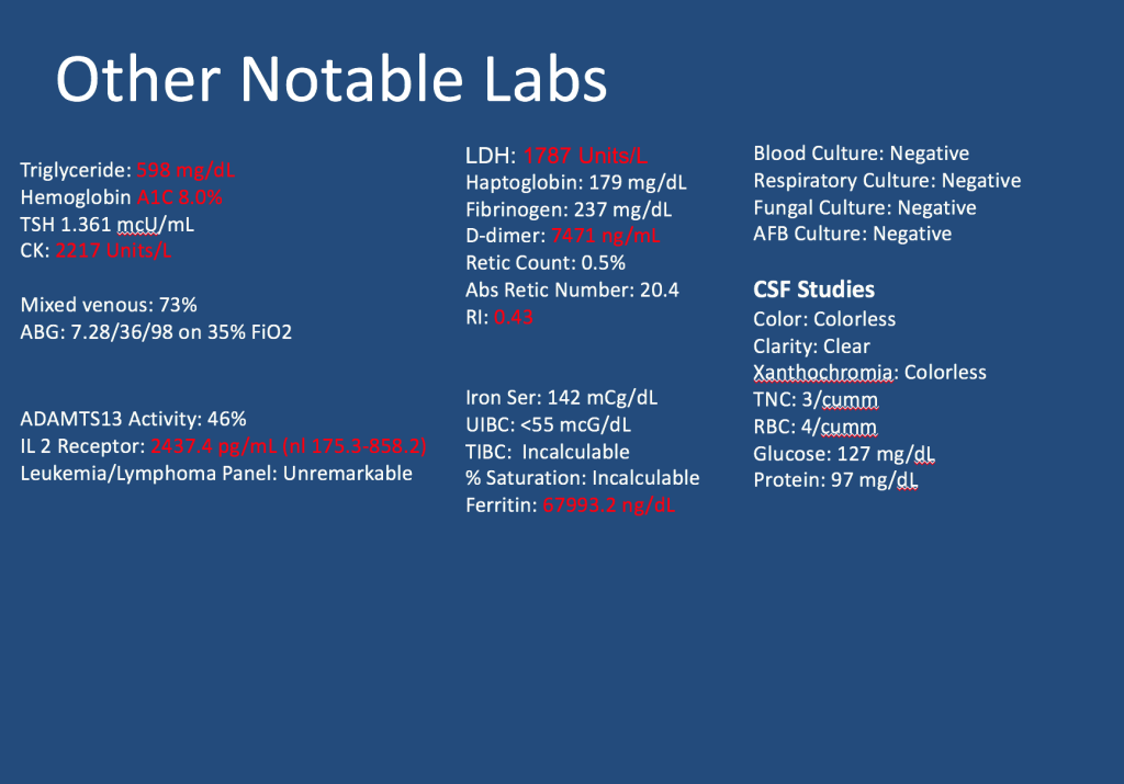
28. Fellows’ Case Files: Harvard – MGH & BIDMC

Case Files Harvard

Welcome back to our Pulm PEEPs Fellows’ Case Files series! We are joined this week by a fellow and the program director from the Harvard combined PCCM fellowship at Massachusettes General Hospital and Beth Israel Deaconess Medical Center. Listen in for a great learning case and let us know on Twitter, if you have a great case to share!

Meet our Guests

Brian Rosenberg is a third year fellow at the Harvard MGH/BI program. He completed his undergraduate degree at Harvard, received his MD  from Yale where he also got a PhD in cell biology, and then did his internal medicine residency at Columbia University Medical Center in NYC.

Asha is an Assistant Professor Medicine at Beth Israel Deaconess Medical Center and Harvard Medical School, and is the Program Director of the Harvard MGH/BI combined fellowship. She is also the Director of the Pulmonary Consult Service at BIDMC, was a Rabkin Fellow in Medical Education and has received multiple leadership and teaching awards