We are excited to bring you another #RadiologyRounds which applies some of the knowledge from our most recent episode on pneumothorax.
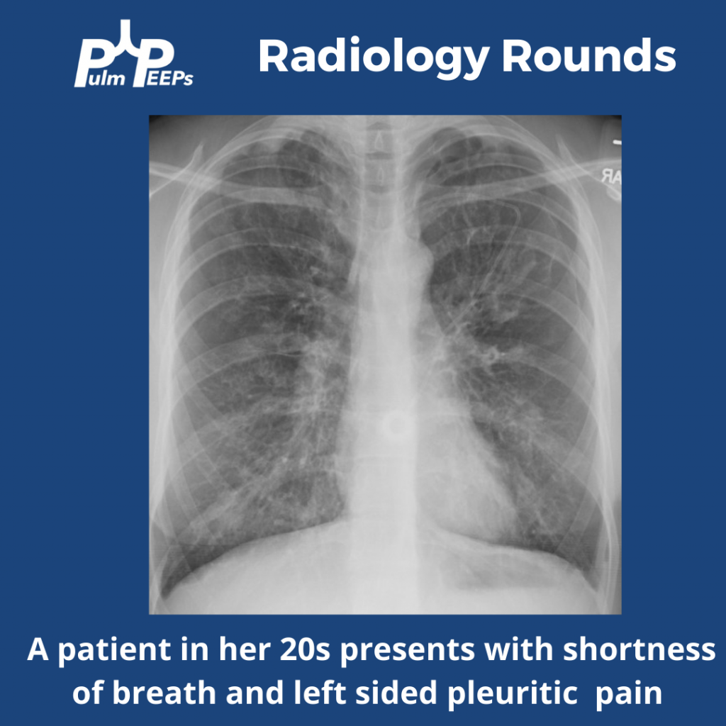
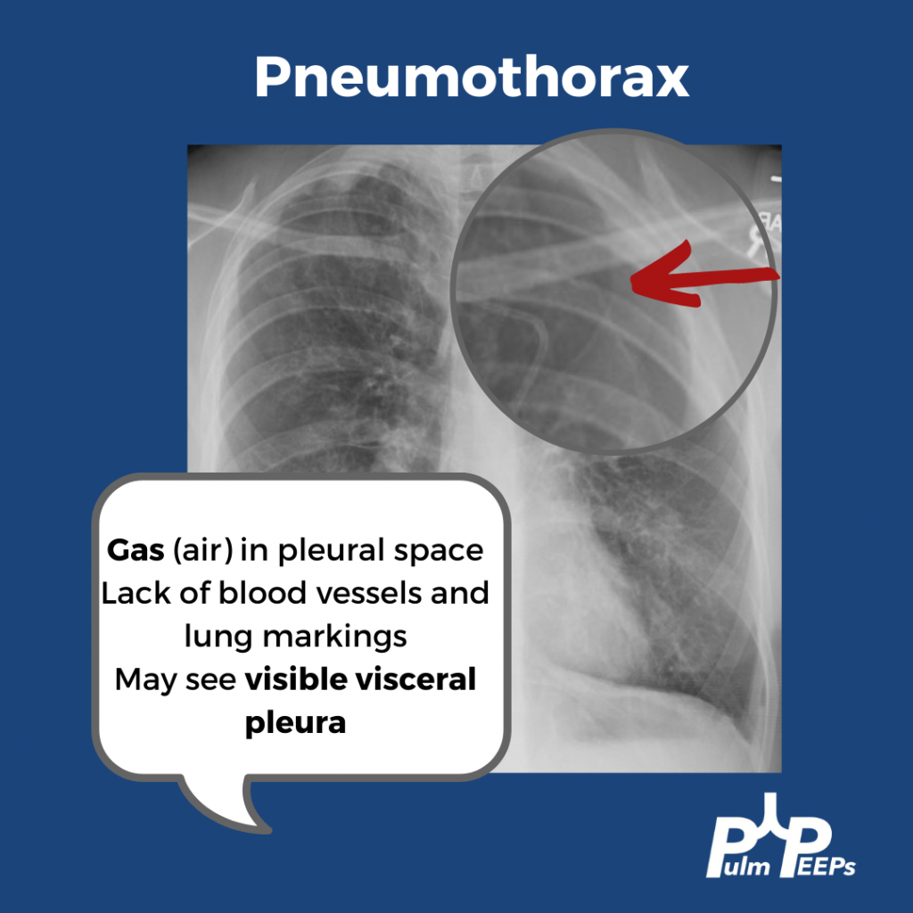
She is presenting with a 1.5 cm left pneumothorax. You can see lucency representing air in the pleural space. There are a lack of blood vessels or lung markings extending to the periphery and you can see the visceral pleura.
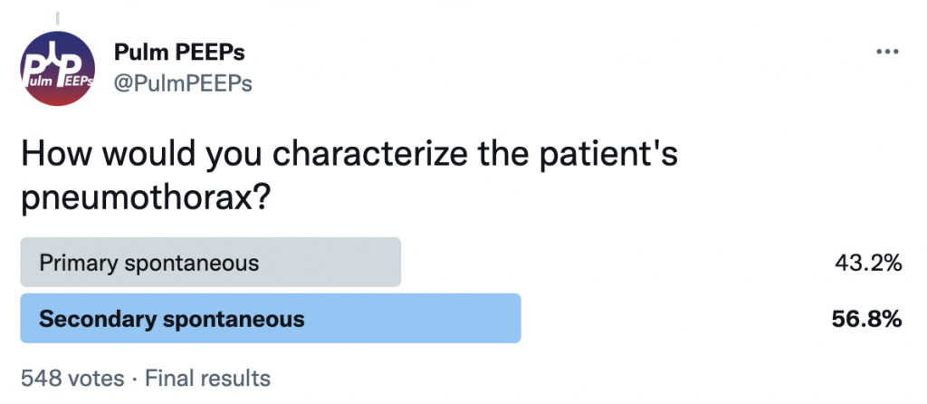
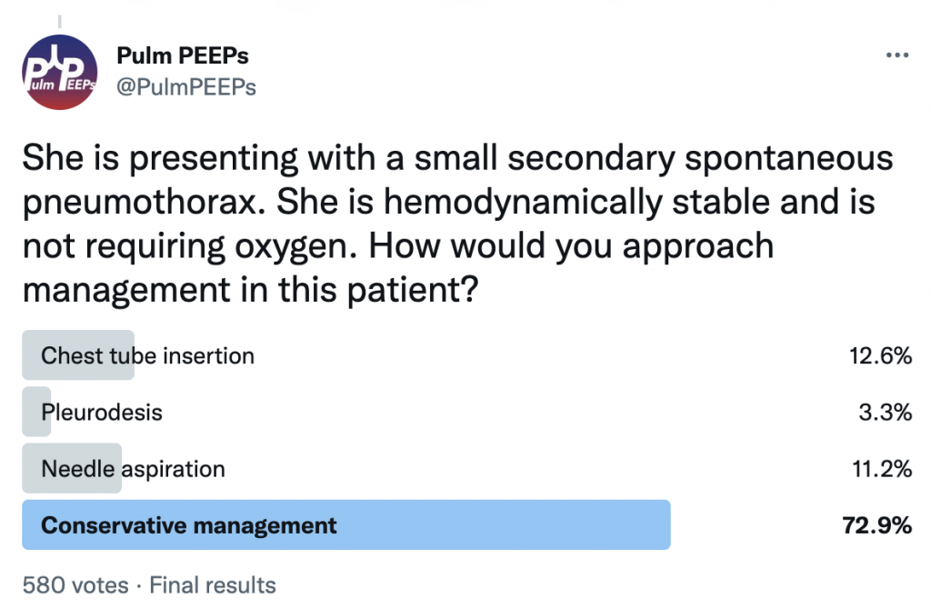
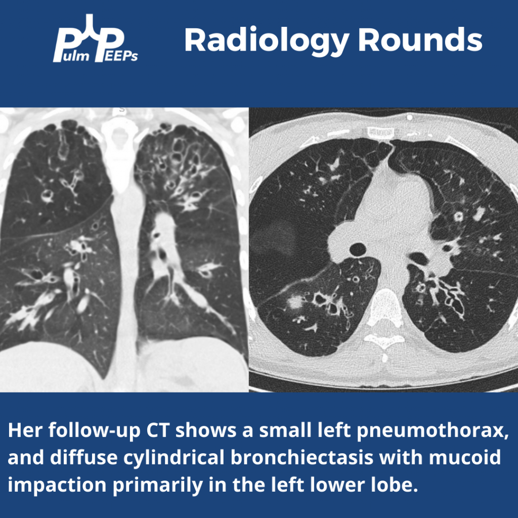
She is presenting with her first pneumothorax which is a small, spontaneous pneumothorax secondary to her underlying cystic lung disease. She was managed conservatively and followed closely outpatient with ultimate resolution of her pneumothorax.