117. Pulm PEEPs Pearls: Spontaneous Breathing Trials

This week’s Pulm PEEPs Pearls episode is all about spontaneous breathing trials (SBTs). SBTs are a standard part of the daily practice in the intensive care unit, but the exact methods vary across ICUs and institutions. Listen in to hear about the most common methods of SBTs, the physiology of each method, and what the evidence says.

This episode was prepared with research by Pulm PEEPs Associate Editor George Doumat.

Dustin Latimer, another Pulm PEEPs Associate Editor, assisted with audio and video editing.

  1. What an SBT is really testing
  • An SBT is a stress test for post-extubation work of breathing, not just a ventilator check.
  • The goal is to balance sensitivity and specificity:
    • Too hard → unnecessary failures and delayed extubation
    • Too easy → false positives and higher risk of reintubation
  1. Common SBT modalities and how they compare
  • T-piece
    • No inspiratory support and no PEEP
    • Highest work of breathing
    • Most “physiologic” but often too strict
  • Pressure support (PS) + PEEP (e.g., 5/5 or 8/5)
    • Offsets ETT resistance and provides modest assistance
    • Easier to pass than T-piece
  • CPAP (0/5)
    • No inspiratory help, but provides PEEP to counter ETT resistance
    • Sits between PS and T-piece in difficulty
  1. Evidence favors pressure-supported SBTs for most patients
  • Large meta-analysis (~6,000 patients, >40 RCTs):
    • Pressure-supported SBTs increase successful extubation (~7% absolute benefit)
    • No increase in reintubation rates
  • Trials (e.g., FAST trial):
    • Patients pass SBTs earlier
    • Leads to earlier extubation and fewer ventilator-associated risks
  • Bottom line: A 30-minute PS 5/5 SBT is evidence-based and appropriate for most stable ICU patients
  1. When a T-piece still makes sense

T-piece SBTs are useful when:

  • Cost of reintubation is high
    • Difficult airway
    • Prior failed extubation
  • Pretest probability of success is low
    • Prolonged or difficult weaning
    • Tracheostomy vs extubation decisions
  • Need to mimic physiology without positive pressure
    • In LV dysfunction or pulmonary edema even small amounts PEEP may significantly improve physiology
  • Some centers use a hybrid approach: PS SBT → short confirmatory T-piece before extubation
  1. CPAP as a middle ground
  • Rationale:
    • Allows full patient effort while compensating for ETT resistance
  • Evidence:
    • Fewer and smaller trials
    • Possible modest improvement in extubation success
    • No clear mortality or LOS benefit
  • Reasonable option based on patient physiology, institutional protocols, and clinician comfort
  1. No single “perfect” SBT mode
  • Across PS, T-piece, CPAP, and newer methods (e.g., high-flow via ETT) there are no consistent differences in mortality or length of stay
  • What matters most:
    • Daily protocolized screening
    • Thoughtful bedside clinical judgment
    • Matching SBT difficulty to patient-specific risk
  1. Institutional variation is normal—and acceptable
  • Examples:
    • PS 10/5 in postoperative surgical ICU patients
    • PS 5/0 as an intermediate difficulty option
  • Key question clinicians should ask: What does passing or failing this specific SBT tell me about this patient’s likelihood of post-extubation success?
  1. Take-home pearls
  1. SBTs are stress tests of post-extubation physiology.
  2. PS 5/5 for 30 minutes is a strong default for most ICU patients.
  3. T-piece trials are valuable when false positives are costly or physiology demands it.
  4. CPAP is reasonable but supported by less robust data.
  5. Consistency, daily screening, and judgment matter more than the exact mode.

  • Burns KEA, Khan J, Phoophiboon V, Trivedi V, Gomez-Builes JC, Giammarioli B, Lewis K, Chaudhuri D, Desai K, Friedrich JO. Spontaneous Breathing Trial Techniques for Extubating Adults and Children Who Are Critically Ill: A Systematic Review and Meta-Analysis. JAMA Netw Open. 2024 Feb 5;7(2):e2356794. doi: 10.1001/jamanetworkopen.2023.56794. PMID: 38393729; PMCID: PMC10891471.
  • Burns KEA, Sadeghirad B, Ghadimi M, Khan J, Phoophiboon V, Trivedi V, Gomez Builes C, Giammarioli B, Lewis K, Chaudhuri D, Desai K, Friedrich JO. Comparative effectiveness of alternative spontaneous breathing trial techniques: a systematic review and network meta-analysis of randomized trials. Crit Care. 2024 Jun 8;28(1):194. doi: 10.1186/s13054-024-04958-4. PMID: 38849936; PMCID: PMC11162018.
  • Subirà C, Hernández G, Vázquez A, Rodríguez-García R, González-Castro A, García C, Rubio O, Ventura L, López A, de la Torre MC, Keough E, Arauzo V, Hermosa C, Sánchez C, Tizón A, Tenza E, Laborda C, Cabañes S, Lacueva V, Del Mar Fernández M, Arnau A, Fernández R. Effect of Pressure Support vs T-Piece Ventilation Strategies During Spontaneous Breathing Trials on Successful Extubation Among Patients Receiving Mechanical Ventilation: A Randomized Clinical Trial. JAMA. 2019 Jun 11;321(22):2175-2182. doi: 10.1001/jama.2019.7234. Erratum in: JAMA. 2019 Aug 20;322(7):696. doi: 10.1001/jama.2019.11119. PMID: 31184740; PMCID: PMC6563557.
  • Burns KEA, Wong J, Rizvi L, Lafreniere-Roula M, Thorpe K, Devlin JW, Cook DJ, Seely A, Dodek PM, Tanios M, Piraino T, Gouskos A, Kiedrowski KC, Kay P, Mitchell S, Merner GW, Mayette M, D’Aragon F, Lamontagne F, Rochwerg B, Turgeon A, Sia YT, Charbonney E, Aslanian P, Criner GJ, Hyzy RC, Beitler JR, Kassis EB, Kutsogiannis DJ, Meade MO, Liebler J, Iyer-Kumar S, Tsang J, Cirone R, Shanholtz C, Hill NS; Canadian Critical Care Trials Group. Frequency of Screening and Spontaneous Breathing Trial Techniques: A Randomized Clinical Trial. JAMA. 2024 Dec 3;332(21):1808-1821. doi: 10.1001/jama.2024.20631. PMID: 39382222; PMCID: PMC11581551.
  • Mahul M, Jung B, Galia F, Molinari N, de Jong A, Coisel Y, Vaschetto R, Matecki S, Chanques G, Brochard L, Jaber S. Spontaneous breathing trial and post-extubation work of breathing in morbidly obese critically ill patients. Crit Care. 2016 Oct 27;20(1):346. doi: 10.1186/s13054-016-1457-4. PMID: 27784322; PMCID: PMC5081985.
  • Yi LJ, Tian X, Chen M, Lei JM, Xiao N, Jiménez-Herrera MF. Comparative Efficacy and Safety of Four Different Spontaneous Breathing Trials for Weaning From Mechanical Ventilation: A Systematic Review and Network Meta-Analysis. Front Med (Lausanne). 2021 Nov 22;8:731196. doi: 10.3389/fmed.2021.731196. PMID: 34881255; PMCID: PMC8647911.​

100. ATS 2025 Critical Care Assembly: The Future of Mechanical Ventilation

We are podcasting today directly from ATS 2025 in San Francisco! Every year, in collaboration with the ATS Critical Care Assembly, we highlight some of the scientific symposium programming from the conference. Today, Furf and Monty sit down with the three chairs of the scientific symposium entitled: Mechanical Ventilation of the Future: New Foundations For Ventilator Strategies.

Juliana Ferreira is an Associate Professor at the University of Sao Paulo, Brazil where she is also co-director of the pulmonary and critical care fellowship program. She is an MD, PhD, and a physician scientist with specific interests in mechanical ventilation and medical education. Finally, she serves ATS as the ATS MECOR Latin America Director.

Bhakti Patel is an Assistant Professor Medicine at the University of Chicago. She is a dedicated researcher and educator. Her research focuses on non-invasive ventilator support.

Akram Khan is an Associate Professor of Medicine at Oregon Health and Science University. Akram is a pulmonary, critical care, and sleep provider with specific clinical interests in critical illness, pulmonary vascular disease and sleep apnea. Additionally, he is an accomplished translational science researcher.

95. Clinical Pearl: Prone Positioning with Elevated Intracranial Pressure

Today we have a mini-episode / clinical pearl. We previously discussed the PROSEVA trial and the evidence for prone positioning in ARDS. In that trial, patients with elevated intracranial pressure (ICP) were excluded. We are joined now by Dr. Jon Rosenberg, a neuro intensivist, to discuss his how prone positioning can still be employed for patients with neurologic injuries and elevated ICP.


Dr. Jon Rosenberg is an assistant professor of neurology and neurosurgery at Westchester Medical Center, New York Medical College. He’s also the associate program director of the Neurocritical Care Fellowship at Westchester Medical Center and a frequent contributor to the Neurocritical Care Society podcast.


  1. Elevated Intracranial Pressure (ICP) and Proning: A Common Misconception
  • Elevated ICP is often considered a contraindication to proning, but this is more of a relative caution rather than an absolute contraindication.
  • Many neuro ICUs have successfully proned patients with elevated ICP, particularly since the COVID-19 pandemic, when critical care units had to manage both respiratory failure and neurological conditions simultaneously.
  1. Patient Selection for Proning with Elevated ICP
  • Most patients with elevated ICP can still be proned, including those with:
    • Global cerebral edema (e.g., post-anoxic brain injury, liver failure)
    • Focal lesions (e.g., traumatic brain injury, large ischemic strokes, intracerebral hemorrhage)
  • Situations where proning might be more concerning:
    • Severe hemodynamic instability (multi-pressor shock)
    • Morbid obesity (e.g., >300 lbs), where physically flipping the patient is a major challenge
  1. Theoretical Concerns with Proning in Elevated ICP
  • Loss of neurological exam access (sedation + flipped position makes pupil and motor exam difficult)
  • Jugular venous compression (especially if the head is turned to one side)
  • Cerebrospinal fluid (CSF) flow obstruction, depending on the lesion
  • Risk of increased ICP if venous outflow is impaired or head positioning is not optimized
  1. Best Practices for Proning Patients with Elevated ICP
  • Patients with invasive ICP monitors vs. without monitors:
    • If possible, placing an ICP monitor (EVD or parenchymal bolt) before proning provides better guidance.
    • Without a monitor, providers must rely on other practices like maintaining strict MAP goals and sodium targets, and indirect signs of increased ICP.
  • Positioning considerations:
    • Keep the head midline to prevent jugular venous compression.
    • If head positioning is not neutral, place the dominant/internal jugular facing upward to maintain venous drainage.
    • Maintain the head of the bed elevated even while prone (reverse Trendelenburg positioning).
  • Hemodynamic management:
    • Target a higher MAP (e.g., 70–75 mmHg, sometimes 75–80 mmHg) to ensure adequate cerebral perfusion pressure (CPP) if there is no ICP monitor
    • Avoid hypotension, as MAP – ICP = CPP, and low MAP could critically reduce cerebral perfusion.
      • A normal intracranial pressure is 7 – 15 mmHg
      • The recommended CPP is between 60 – 70 mmHg
  • Sedation & Sodium Management:
    • Consider deep sedation (RASS -5) to reduce metabolic demand and intracranial blood volume.
    • Consider keeping sodium >145 mmol/L prophylactically to mitigate brain swelling if no ICP monitor in place
  1. When to Reconsider Proning (i.e. when to supinate)
  • If a patient’s ICP spikes significantly (e.g., from 20 to 60 mmHg) despite medical management (hypertonic saline, sedation, paralysis, etc.).
  • If new signs of neurological deterioration emerge (e.g., changes in pupil exam once patient is repositioned).
  • Hemodynamic instability that is unmanageable in the prone position.
  1. Literature and Future Considerations
  • Small case series have demonstrated success in proning patients with traumatic brain injury (TBI) and aneurysmal subarachnoid hemorrhage.
  • While more formal research is needed, the neurocritical care community has begun embracing proning for neuro patients, provided that proper precautions are taken.

Bottom Line

  • Proning is not an absolute contraindication for patients with elevated ICP—it can be done safely with proper monitoring, patient selection, and precautions.
  • Having an ICP monitor makes the process more controlled and allows clinicians to adjust treatment in real time.
  • Key considerations: Maintain cerebral perfusion, optimize head positioning, monitor hemodynamics, and have a plan for reversing if ICP becomes unmanageable.


84. RFJC 14 – ARDS Series – Driving Pressure

In this podcast episode, we continue our summer series reviewing landmark ARDS studies. Today, Dave and Luke discuss the Driving Pressure trial (published in NEJM in 2015) which evaluated the impact of driving pressure on survival in patients with ARDS.

Article and Reference

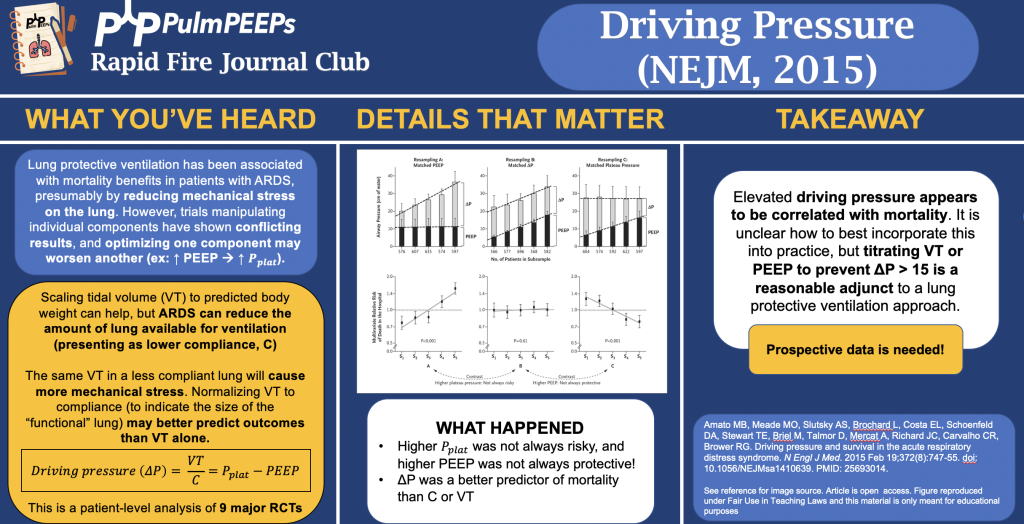
We are talking about the Driving Pressure trial today which evaluated the impact of driving pressure, as an independent variable, on survival in patients with ARDS.

Amato MB, Meade MO, Slutsky AS, Brochard L, Costa EL, Schoenfeld DA, Stewart TE, Briel M, Talmor D, Mercat A, Richard JC, Carvalho CR, Brower RG. Driving pressure and survival in the acute respiratory distress syndrome. N Engl J Med. 2015 Feb 19;372(8):747-55. doi: 10.1056/NEJMsa1410639. PMID: 25693014.

Infographic

81. RFJC 12 – ARDS Series – PROSEVA

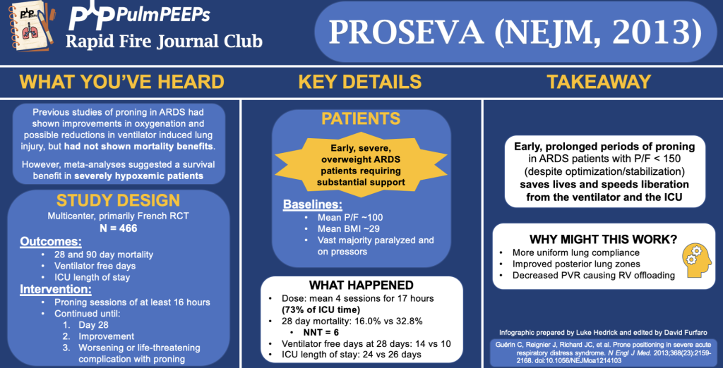
In this podcast episode, we continue our summer series reviewing landmark ARDS studies. Today, Dave and Luke discuss the PROSEVA trial (published in NEJM in 2013) which evaluated the impact of early, prolonged proning in patients with severe ARDS.

Article and Reference

We are talking about the PROSEVA trial today which evaluated the patients with severe ARDS (P/F < 150) to undergo prone-positioning sessions of at least 16 hours or to be left in the supine position.

Guérin C, Reignier J, Richard JC, Beuret P, Gacouin A, Boulain T, Mercier E, Badet M, Mercat A, Baudin O, Clavel M, Chatellier D, Jaber S, Rosselli S, Mancebo J, Sirodot M, Hilbert G, Bengler C, Richecoeur J, Gainnier M, Bayle F, Bourdin G, Leray V, Girard R, Baboi L, Ayzac L; PROSEVA Study Group. Prone positioning in severe acute respiratory distress syndrome. N Engl J Med. 2013 Jun 6;368(23):2159-68. doi: 10.1056/NEJMoa1214103. Epub 2013 May 20. PMID: 23688302.

Infographic

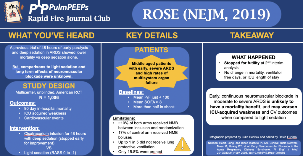
80. RFJC 11 – ARDS Series – ROSE

In this podcast episode, we continue our summer series reviewing landmark ARDS studies. Today, Dave and Luke discuss the ROSE trial (published in NEJM in 2019) which investigated use of continuous neuromuscular blockade in moderate to severe ARDS.

Article and Reference

We are talking about the ROSE trial today which was a comparison of early continuous neuromuscular blockade in patients with ARDS who were receiving mechanical ventilation.

Reference: National Heart, Lung, and Blood Institute PETAL Clinical Trials Network; Moss M, Huang DT, Brower RG, Ferguson ND, Ginde AA, Gong MN, Grissom CK, Gundel S, Hayden D, Hite RD, Hou PC, Hough CL, Iwashyna TJ, Khan A, Liu KD, Talmor D, Thompson BT, Ulysse CA, Yealy DM, Angus DC. Early Neuromuscular Blockade in the Acute Respiratory Distress Syndrome. N Engl J Med. 2019 May 23;380(21):1997-2008. doi: 10.1056/NEJMoa1901686. Epub 2019 May 19. PMID: 31112383; PMCID: PMC6741345.

Infographic

78. PREOXI Trial

Today, we’re going to be talking about pre-oxygenation methods for endotracheal intubation and the PREOXI Trial which is hot off the presses in the New England Journal of Medicine in June of 2024. This trial has potentially widespread, practice changing results and we’re lucky enough to be joined by two of the authors to discuss.


Dr. Kevin Gibbs is an Associate Professor of Medicine at Wake Forest University School of Medicine. He obtained his MD at George Washington University School of Medicine, and completed his residency and fellowship training at Johns Hopkins. He is an active researcher in critical care, ARDS, mechanical ventilation, and pragmatic trial design.

Dr. Jon Casey is an Assistant Professor of Medicine for the Division of Allergy, Pulmonary, and Critical Care Medicine at Vanderbilt University Medical Center. He obtained his MD from the University of Louisville School of Medicine, and completed his residency training at Brigham and Women’s Hospital before going to Vanderbilt for fellowship training. He is a physician scientist and also has his Masters of Science in Clinical Investigation. His research is focused on comparative effectiveness of ICU treatments and he also has a focus on pragmatic trials. He is supported with NIH funding and is active in the American Thoracic Society Critical Care Assembly.

Summarized Key Points


  • Significance of the Problem: Tracheal intubation in emergency and ICU settings is common, with significant risks such as hypoxemia (10-20% incidence) and cardiac arrest (2% incidence) associated with the procedure. This makes effective pre-oxygenation crucial.

  • Methods of Pre-oxygenation: Common methods include face mask oxygen (e.g., non-rebreather, bag-mask devices) and more advanced techniques like non-invasive ventilation (used in about 15% of cases globally). Each method has pros (e.g., simplicity, no risk of aspiration for face masks; 100% oxygen delivery, positive pressure for non-invasive ventilation) and cons (e.g., potential for gastric insufflation with non-invasive ventilation).

  • Study Design: The study discussed in the podcast is a pragmatic trial aiming to optimize pre-oxygenation strategies to prevent peri-intubation hypoxemia. Eligibility criteria were broad, encompassing most patients undergoing tracheal intubation in the ED or ICU, with exclusions mainly for safety reasons.

  • Primary Outcome: The primary outcome of the trial was hypoxemia, defined as oxygen saturation < 85%. This threshold was chosen because it signifies a critical point on the oxygen dissociation curve, where patients are at higher risk of further desaturation and adverse outcomes.

  • Secondary Outcomes: Secondary exploratory outcomes included more severe levels of hypoxemia (oxygen saturation < 80% and < 70%), aiming to capture varying degrees of oxygenation failure during intubation. Rates of cardiac arrest during intubation were an additional outcome.

  • Intervention Comparison:

    • The trial compared two methods of pre-oxygenation: non-invasive ventilation (NIV) and oxygen mask (face mask)

    • Both methods aimed to provide at least three minutes of pre-oxygenation before intubation.

    • NIV group specifics: Expiratory pressure of 5 cm H2O, Inspiratory pressure of 10 cm H2O, respiratory rate of 10 breaths per minute, and 100% oxygen delivery

    • Oxygen mask group specifics: Non-rebreather or bag mask device with at least 15 liters per minute oxygen flow.

    • Nasal cannulas and HFNC could be used in both groups.


  • Logistics and Equipment Use:

    • The trial allowed flexibility in using available equipment (invasive ventilator capable of NIPPV vs. dedicated BiPAP machine).

    • Sites were encouraged to use the same ventilator for both pre-oxygenation and subsequent ventilation to streamline workflow and reduce logistical challenges.


  • Primary and Secondary Outcomes:

    • Results showed a significant reduction in hypoxemia incidents in the NIV group compared to the oxygen mask group.

    • There was also a reduction in severe hypoxemia and a notable decrease in cardiac arrest incidents in the NIV group.


  • Aspiration Safety:

    • There was no statistical difference in aspiration-related outcomes between the NIV and oxygen mask groups, indicating that NIV did not increase the risk of aspiration.


  • Conclusions:

    • The trial concluded that NIV for pre-oxygenation significantly reduced the incidence of hypoxemia and possibly cardiac arrest during tracheal intubation.

    • It also dispelled concerns about increased aspiration risk with NIPPV as pre-oxygenation, suggesting it can be safely used in clinical practice.


Gibbs KW, Semler MW, Driver BE, Seitz KP, Stempek SB, Taylor C, Resnick-Ault D, White HD, Gandotra S, Doerschug KC, Mohamed A, Prekker ME, Khan A, Gaillard JP, Andrea L, Aggarwal NR, Brainard JC, Barnett LH, Halliday SJ, Blinder V, Dagan A, Whitson MR, Schauer SG, Walker JE Jr, Barker AB, Palakshappa JA, Muhs A, Wozniak JM, Kramer PJ, Withers C, Ghamande SA, Russell DW, Schwartz A, Moskowitz A, Hansen SJ, Allada G, Goranson JK, Fein DG, Sottile PD, Kelly N, Alwood SM, Long MT, Malhotra R, Shapiro NI, Page DB, Long BJ, Thomas CB, Trent SA, Janz DR, Rice TW, Self WH, Bebarta VS, Lloyd BD, Rhoads J, Womack K, Imhoff B, Ginde AA, Casey JD; PREOXI Investigators and the Pragmatic Critical Care Research Group. Noninvasive Ventilation for Preoxygenation during Emergency Intubation. N Engl J Med. 2024 Jun 20;390(23):2165-2177. doi: 10.1056/NEJMoa2313680. Epub 2024 Jun 13. PMID: 38869091.

77. RFJC 9 – ARDS Series – ARMA

This episode is launching our 2024 Rapid Fire Journal Club summer series on ARDS! This summer we will be talking about landmark ARDS trials that have defined the literature and shaped patient care. Journal clubs often focus on new trials, and so learners may have a less thorough understanding of the baseline literature that defines many of our ICU practices. The goal of this series is to provide a quick, but in-depth look at these papers so that learners understand the modern landscape of ARDS.

Today, we’re kicking this initiative off by looking at the ARMA or ARDSNet Trial published in the NEJM in 2000. Enjoy!

Article and Reference

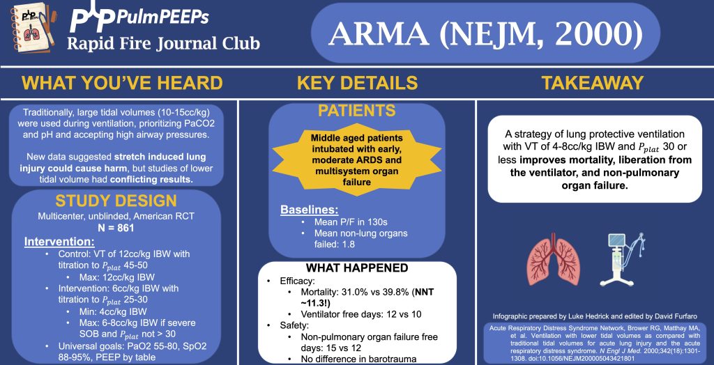
We’re talking about the ARMA trial today which examined “Ventilation with Lower Tidal Volumes as Compared with Traditional Tidal Volumes for Acute Lung Injury and the Acute Respiratory Distress Syndrome.”

Reference: Acute Respiratory Distress Syndrome Network; Brower RG, Matthay MA, Morris A, Schoenfeld D, Thompson BT, Wheeler A. Ventilation with lower tidal volumes as compared with traditional tidal volumes for acute lung injury and the acute respiratory distress syndrome. N Engl J Med. 2000 May 4;342(18):1301-8. doi: 10.1056/NEJM200005043421801. PMID: 10793162.

Infographic

Correction:

We mention a step-wise titration of tidal volume in the control group to achieve Pplats of 45-50. To clarify, there was no adjustment of Vt in the traditional Vt group unless Pplat > 50. If Vt had been decreased in the traditional Vt group because Pplat was > 50, it would not be subsequently increased back to 12 unless Pplat < 45 (to avoid a cycle of corrections and re-adjustments). Similarly in the lower Vt group, there was no adjustment (“titration”) of Vt unless Pplat > 30, and there was a similar protocol in place not to increase the Vt again unless the Pplat was < 25.

Radiology Rounds – 4/11/2023

For “#RadiologyRounds” today, we won’t be looking at any imaging, but we’ll be looking at some vent wave forms and examining dysynchrony! This is a re-booted @david_furfaro Tweetorial. I hope you enjoy and this is an open invitation for all dysynchrony waveforms!

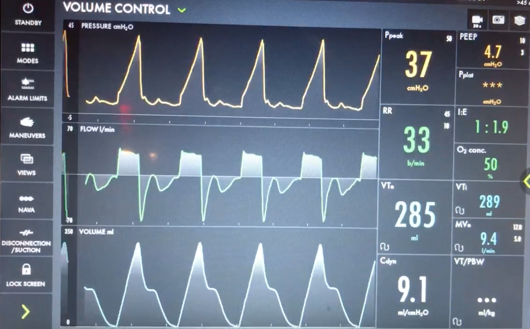
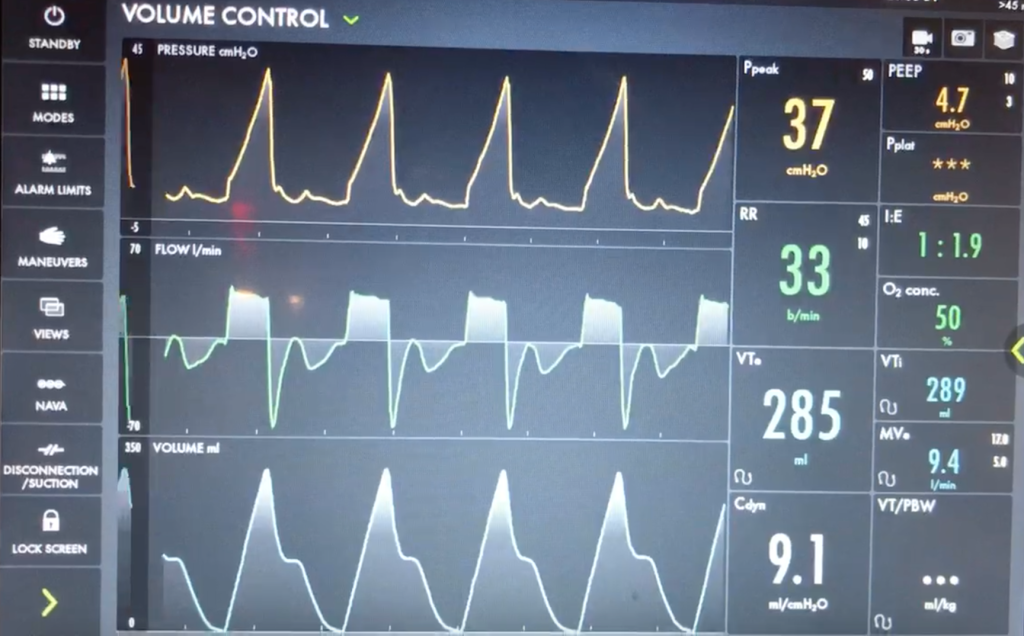
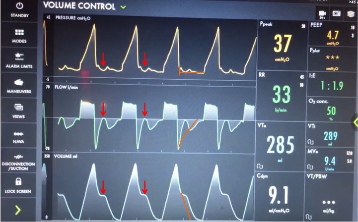
A 40s M is intubated for ARDS. In order to maintain lung protective ventilation, he was on high does of propofol, fentanyl and midazolam. His sedation is being weaned slightly now, and the RN calls for vent dysynchrony. His ventilator looks like this

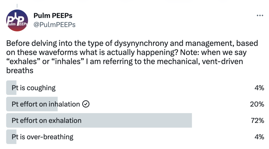
Before delving into the type of dysynynchrony and management, based on these waveforms what is actually happening? Note: when we say “exhales” or “inhales” I am referring to the mechanical, vent-driven breaths

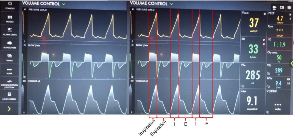
Let’s take a look at the waveforms and identify the phase of breathing. This is VC with a square flow waveform, so as a set volume is delivered, the flow is at a constant rate, and the pressure is measured. Expiration is about 2x as long as inspiration (determined by I time or flow rate)

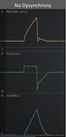
With a passive patient (no dysynchrony) after inspiration, the volume curve should show a smooth decrease with exhalation, the pressure curve should flatten to the set PEEP, and the flow will be negative and gradually return to 0 as the patient exhales

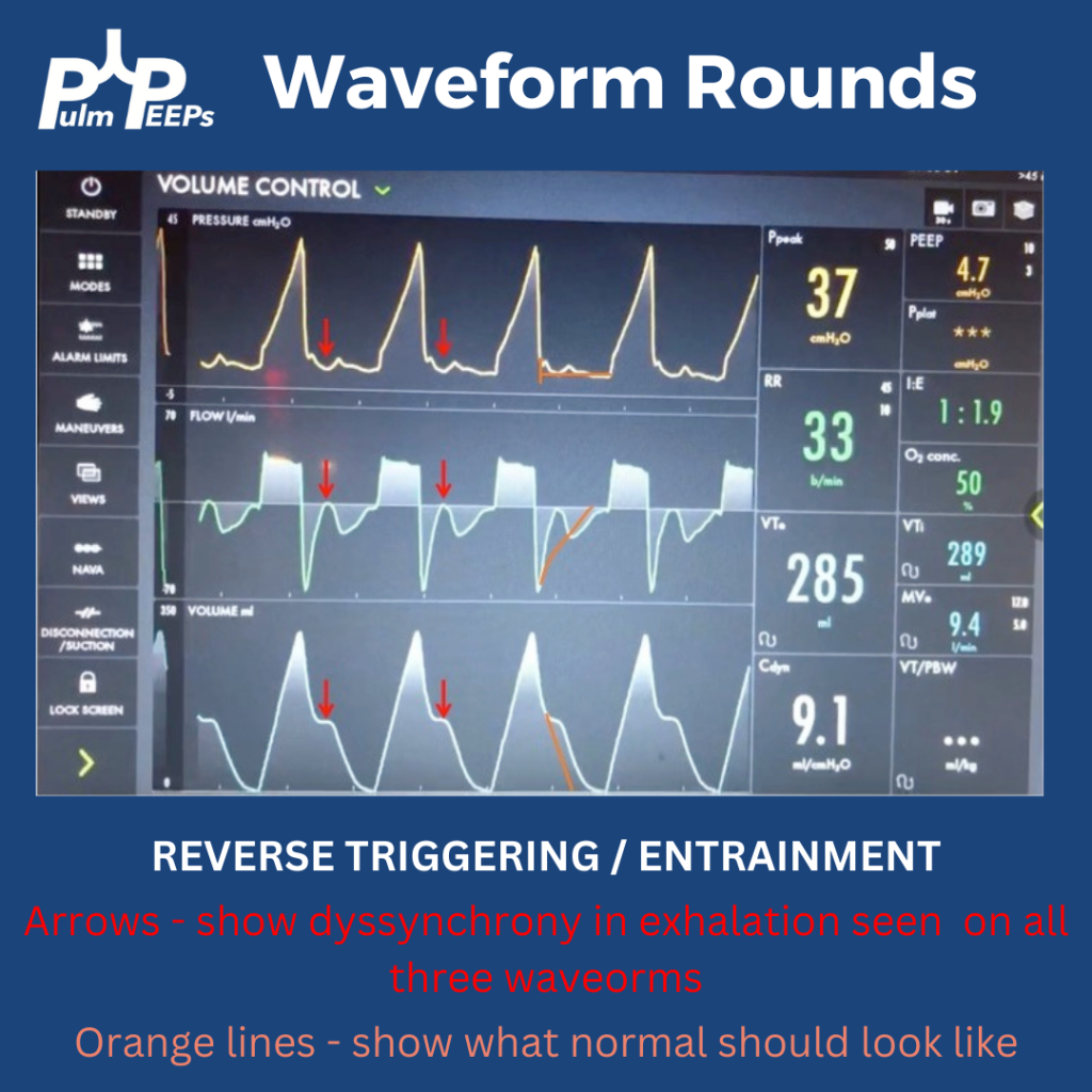
In our case, you can see simultaneous dysynchrony in all three waveforms during exhalation (red arrows) vs normal (orange lines). There is a pressure negative deflection; the flow quickly rises to 0 before returning to negative; and the volume curve plateaus as exhalation pauses

Putting this together, it means that during expiration there is an inspiratory effort. The patient is trying to inhale, which causes a negative pressure deflection, and a pause in air flowing out of the lungs. If this effort triggered a breath, there could be breath stacking. Notice that the same pattern occurs after every breath, and clinically we said the patient was still heavily sedated. This combination of findings is a type of dysynchrony called REVERSE TRIGGERING or ENTRAINMENT.

You could consider whether this dysynchrony was an ineffective effort, but the trigger sensitivity was low, and the pattern and consistent timing after inspiration is more consistent with reverse triggering.

The change in pressure and flow of a ventilator-initiated, mandatory breath stimulates an inspiratory effort from the patient. Theories differ on if this is mediated by the diaphragm or central respiratory center. This can start during the ventilator-delivered breath, or afterwards in exhalation, as with our patient.

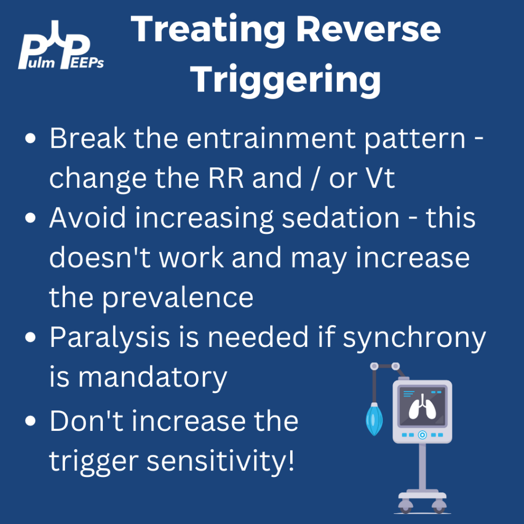
Reverse triggering often occurs in heavily sedated patients, and is defined by a stable, repetitive pattern (i.e. it is not voluntary, but reflex mediated). It can also occur in anoxic brain injury. Treating it involves breaking the pattern and avoiding harmful therapies. It can even be induced in healthy patients but this is much less clinically relevant, and is rare.

DO NOT just increase the trigger sensitivity of the vent. This can stop breath stacking but does not prevent dysynchrony and it can cause harmful changes in transpulmonary pressure. For this patient, sedation was lightened slightly, and the respiratory rate decreased and the pattern of reverse triggering ultimately broke without the need for paralysis.

Radiology Rounds – 1/31/23

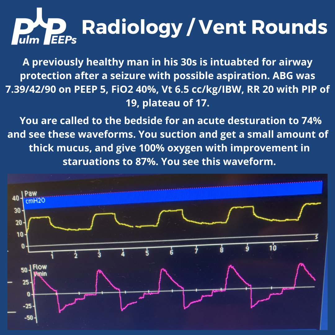
For today’s #RadiologyRounds we have a combined Radiology and Ventilator imaging rounds! You’re in the ICU caring for a young patient on a ventilator when you are called to the bedside for a desaturation.

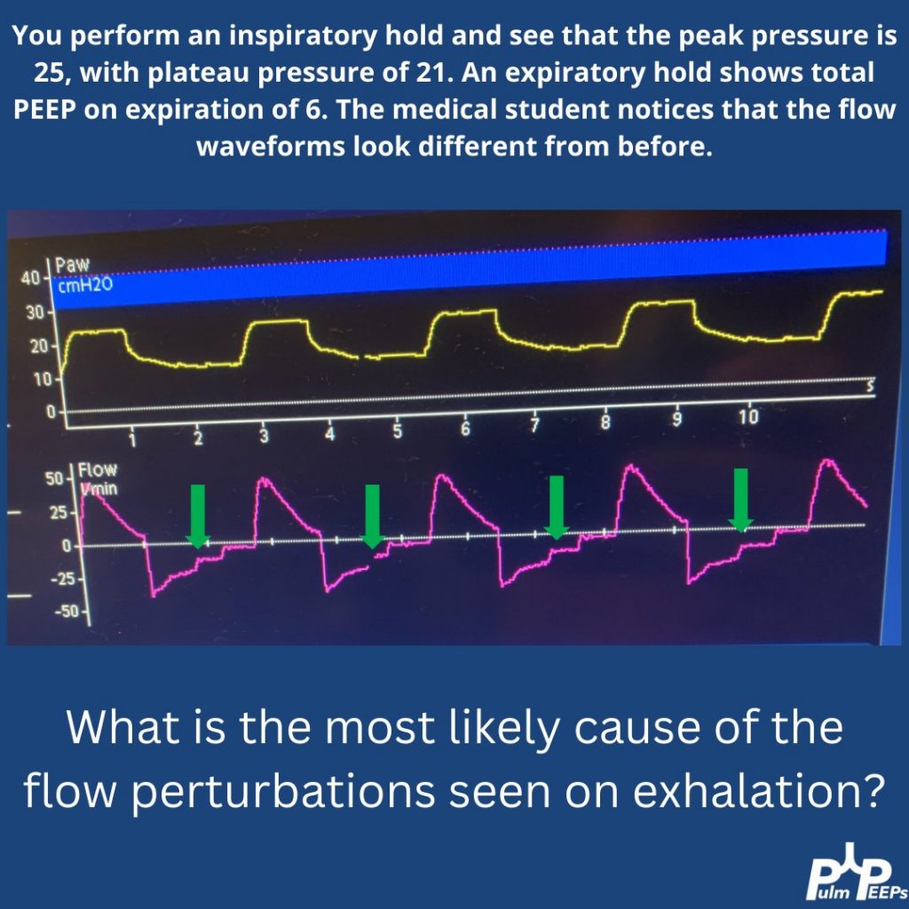
You perform an inspiratory hold and see that the PIP, plateau, and difference between peak and plateau have all increased. On exam you hear bilateral mechanical breath sounds anteriorly. You order a CXR and the student asks a question about the waveforms

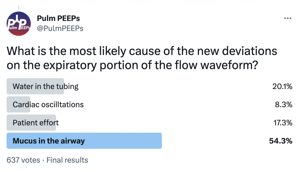
There are pressure deviations corresponding to the flow deviations.

There is no clear patient effort The fact that the PIP and plat have changed makes water in the tubing or cardiac oscillations less likely.

You think this is mucus, with a plug ball-valving in a bronchus

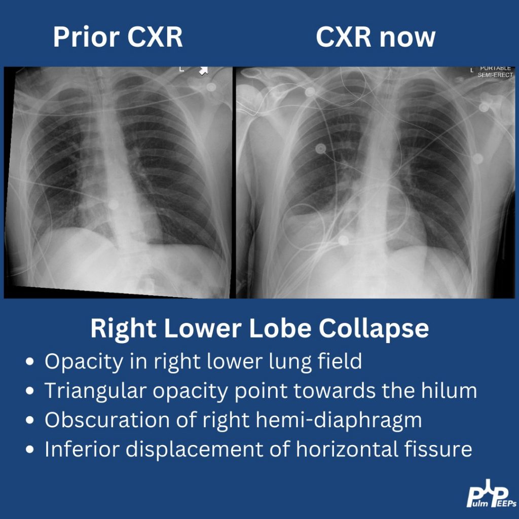
The CXR arrives and shows right lower lobe collapse.

A bedside bronchoscopy is performed with large mucus plugs suctioned out of the RLL and RML. Afterward, the patient’s oxygenation is improved, the flow deviations resolve, and the plateau pressure drops to 19