119. Guideline Series: Pulmonary Embolism

We are unbelievably excited this week to be reviewing the hot-off-the-presses 2026 Multi-Society (AHA/ACC/ACCP/ACEP/CHEST/SCAI/SHM/SIR/SVM/SVN) Pulmonary Embolism Guidelines with lead author Dr. Mark A. Creager. We will talk about key updates in these guidelines compared to prior practice, including the new risk classification model, and provide an overview from diagnosis to follow-up. Given the clinical importance and prevalence of pulmonary embolism, these guidelines are certainly going to shape practice going forward, so this episode is a can’t miss!

Watch the full video of this episode with graphics and helpful teaching visuals on our YouTube channel: https://www.youtube.com/@pulmpeeps

Dr. Mark Creager is a Professor of Medicine at Dartmouth Hitchcock Medical Center where he specializes in Cardiovascular Medicine with an emphasis on venous thromboembolic disease. He served as the lead author of the 2026 Pulmonary Embolism Guidelines.

Creager MA, Barnes GD, Giri J, Mukherjee D, Jones WS, Burnett AE, Carman T, Casanegra AI, Castellucci LA, Clark SM, Cushman M, de Wit K, Eaves JM, Fang MC, Goldberg JB, Henkin S, Johnston-Cox H, Kadavath S, Kadian-Dodov D, Keeling WB, Klein AJP, Li J, McDaniel MC, Moores LK, Piazza G, Prenger KS, Pugliese SC, Ranade M, Rosovsky RP, Russo F, Secemsky EA, Sista AK, Tefera L, Weinberg I, Westafer LM, Young MN. 2026 AHA/ACC/ACCP/ACEP/CHEST/SCAI/SHM/SIR/SVM/SVN Guideline for the Evaluation and Management of Acute Pulmonary Embolism in Adults: A Report of the American College of Cardiology/American Heart Association Joint Committee on Clinical Practice Guidelines. J Am Coll Cardiol. 2026 Feb 19:S0735-1097(25)10161-7. doi: 10.1016/j.jacc.2025.11.005. Epub ahead of print. PMID: 41712898.

Why these guidelines matter:

This is the first joint AHA/ACC clinical practice guideline specifically on acute PE, bringing together a truly multidisciplinary writing committee (cardiology, pulmonology, hematology, emergency medicine, interventional radiology, surgery, and others). Prior guidelines existed from individual societies, but nothing this comprehensive had been updated in roughly five to six years.

New PE clinical categories (A through E):

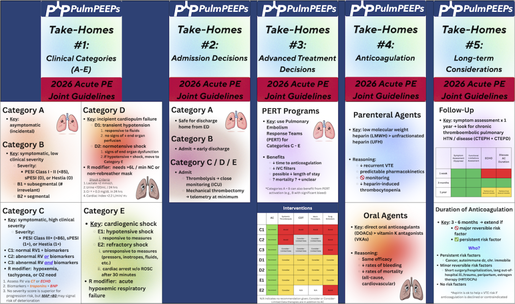
One of the most impactful changes is replacing the old “massive/submassive” and “low/intermediate/high risk” labels with five categories that form a severity continuum. Category A is subclinical (incidental PE found on imaging in asymptomatic patients). Category B covers symptomatic but low-severity patients. Category C is where much of the clinical complexity lives — symptomatic, hemodynamically stable patients subdivided into C1, C2, and C3 based on RV function and biomarkers. Category D represents incipient cardiopulmonary failure (transient hypotension, normotensive shock with end-organ dysfunction). Category E is frank cardiopulmonary failure, with E2 being the sickest — refractory or recurrent cardiac arrest. Respiratory modifiers (hypoxia requiring supplemental oxygen) layer onto C, D, and E.

Diagnostic approach:

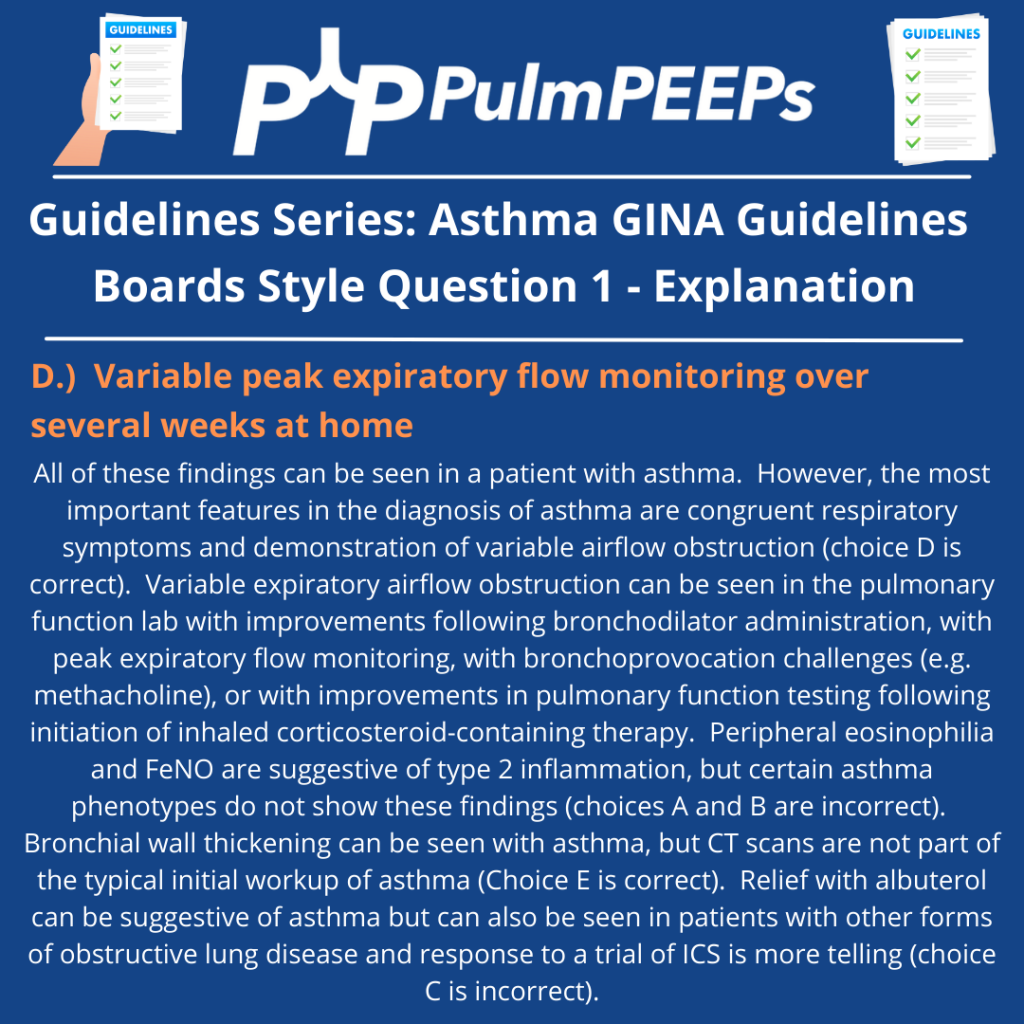
Clinical evaluation comes first — history, exam, and validated decision tools (Wells score, revised Geneva, PERC). If clinical probability is low and D-dimer is normal, imaging can be safely avoided. If either is concerning, imaging is warranted. CTPA remains the preferred imaging modality due to superior sensitivity, specificity, wide availability, and ability to assess clot burden and alternative diagnoses. VQ scanning is still appropriate when CTPA is contraindicated, and VQ SPECT offers better reproducibility and specificity than traditional planar VQ if available. Echocardiography is not a diagnostic test for PE but is important for risk stratification — RV size, TAPSE, and tissue Doppler measures all contribute prognostic information.

Anticoagulation updates:

Anticoagulation remains the cornerstone of treatment. For patients potentially needing advanced therapies (C3, D, E), parenteral anticoagulation is started first. A notable recommendation: low molecular weight heparin is generally preferred over unfractionated heparin, based on evidence showing more effective VTE risk reduction, more predictable pharmacokinetics, no need for routine monitoring, lower rates of heparin-induced thrombocytopenia, and no increase in major bleeding. The committee acknowledged this may create discomfort for clinicians accustomed to unfractionated heparin’s easy reversibility, but the difficulty of achieving and maintaining therapeutic levels with UFH was a significant concern.

Advanced therapies:

Catheter-based thrombolysis, mechanical thrombectomy, systemic thrombolysis, and surgical embolectomy all received mostly class 2B recommendations (“can consider”) for C3 and D categories, reflecting that current evidence shows improvement in short-term surrogate measures (RV/LV ratio, hemodynamics) but lacks definitive hard outcome data on mortality. For category E1 patients, recommendations are stronger (class 2A). Multiple trials are expected soon — HI-PEITHO, PEERLESS-2, PE-TRACT, PERSEVERE, TORPEDO, and PROG — that should substantially inform future updates.

PERT teams:

Pulmonary embolism response teams are encouraged, particularly for C3, D, and E patients. They’ve been shown to reduce length of stay. For institutions without PERT capability, establishing consultation networks with larger centers is recommended.

Post-PE follow-up:

Patients shouldn’t be “left in the wilderness” after discharge. The guidelines recommend communication within the first week to ensure understanding of diagnosis and treatment, an in-person visit at or before three months to assess for persistent symptoms and discuss anticoagulation duration, ongoing surveillance for chronic thromboembolic pulmonary disease, and periodic reassessment for those on extended anticoagulation.

Infographics
116. Guidelines Series: Pulmonary Hypertension – Risk Stratification and Treatment Goals

On this week’s episode, we’re continuing our Guidelines Series exploring the 2022 ESC/ERS Guidelines for the diagnosis and treatment of Pulmonary Hypertension. If you missed our first episode in the series, give it a listen to hear about the most recent recommendations regarding Pulmonary Hypertension definitions, screening, and diagnostics. Today, we’re talking about the next steps after diagnosis. Specifically, we’ll be discussing risk stratification, establishing treatment goals, and metrics for re-evaluation. We’ll additionally introduce the mainstays of pharmacologic therapy for Pulmonary Hypertension.

Rupali Sood  grew up in Las Vegas, Nevada and made her way over to Baltimore for medical school at Johns Hopkins. She then completed her internal medicine residency training at Massachusetts General Hospital before returning back to Johns Hopkins, where she is currently a pulmonary and critical care medicine fellow. Rupali’s interests include interstitial lung disease, particularly as related to oncologic drugs, and bedside medical education.

Tom Di Vitantonio  is originally from New Jersey and attended medical school at Rutgers, New Jersey Medical School in Newark. He then completed his internal medicine residency at Weill Cornell, where he also served as a chief resident. He currently is a pulmonary and critical care medicine fellow at Johns Hopkins, and he’s passionate about caring for critically ill patients, how we approach the management of pulmonary embolism, and also about medical education of trainees to help them be more confident and patient centered.

1) Episode Roadmap

  • How to set treatment goals, assess symptom burden, and risk-stratify patients with suspected/confirmed pulmonary arterial hypertension (PAH).
  • What tools to use to re-evaluate patients on treatment
  • Intro to major PAH medication classes and how they map to pathways.

2) Case-based diagnostic reasoning

Patient: 37-year-old woman with exertional dyspnea, mild edema, abnormal echo, telangiectasias + epistaxis → raises suspicion for HHT (hereditary hemorrhagic telangiectasia) and/or early connective tissue disease.

  • Key reasoning move: start broad (Groups 2–5) and narrow using history/exam/testing.
  • In a young patient without obvious left heart or lung disease, think more about Group 1 PAH (idiopathic/heritable/associated).

HHT teaching point: HHT can cause PH in more than one way:

  • More common: high-output PH from AVMs (often hepatic/pulmonary)
  • Rare (1–2% mentioned): true PAH phenotype (vascular remodeling; associated with ALK1 in some patients), behaving like Group 1 PAH.

3) Functional class assessment

WHO Functional Class:

  • Class I: no symptoms with ordinary activity, only with exertion
  • Class II: symptoms with ordinary activity
  • Class III: symptoms with less-than-ordinary activity (can’t do usual chores/shopping without dyspnea)
  • Class IV: symptoms at rest

Practical bedside tip they give:

  • Ask if the patient can walk at their own pace or keep up with a similar-age peer/partner. If not, think Class II (or worse).

4) Risk stratification at diagnosis: why, how, and which tools

Big principle: treatment choices are driven by risk, and the goal is to move patients to low-risk quickly.

ESC/ERS approach at diagnosis (as described):

  • Use a 3-strata model predicting 1-year mortality:
    • Low: <5%
    • Intermediate: 5–20%
    • High: >20%

ESC/ERS risk assessment variables (10 domains discussed):

  • Clinical progression, signs of right heart failure, syncope
  • WHO FC
  • Biomarkers (NT-proBNP)
  • Exercise capacity (6MWD)
  • Hemodynamics
  • Imaging (echo; sometimes cardiac MRI)
  • CPET (peak VO₂; VE/VCO₂ slope)

They note: even if you don’t have everything, the calculator can still be useful with ≥3 variables.

REVEAL 2.0:

  • Builds on similar core variables but adds further patient context (demographics, renal function, BP, DLCO, etc.)

Case result: both tools put her in intermediate risk (ESC/ERS ~1.6; REVEAL 2.0 score 8), underscoring that mild symptoms can still equal meaningful mortality risk.

5) Treatment goals and follow-up philosophy

What they explicitly prioritize:

  • Help patients feel better, live longer, and stay out of the hospital
  • Use risk tools to communicate prognosis and to track improvement
  • Reassess frequently (they mention ~every 3 months early on) until low risk is achieved
  • “Time-to-low-risk” is an important treatment goal

Also emphasized:

  • The diagnosis is psychologically heavy; patients need clear counseling, reassurance about the plan, and connection to support groups.

6) Medication classes for the treatment of PAH

Nitric oxide–cGMP pathway

  • PDE5 inhibitors: sildenafil, tadalafil
  • Soluble guanylate cyclase stimulator: riociguat
  • Important safety point: don’t combine PDE5 inhibitors with riociguat (risk of significant hypotension/hemodynamic effects)

Endothelin receptor antagonists (ERAs)

  • “-sentan” drugs: bosentan (less used due to side effects/interactions), ambrisentan, macitentan
  • Teratogenicity emphasized
  • Hepatotoxicity that requires LFT monitoring
  • Can cause fluid retention and peripheral edema

Prostacyclin pathway

  • Prostacyclin analogs/agonists:
    • Epoprostenol (potent; short half-life; IV administration)
    • Treprostinil (IV/SubQ/oral/inhaled options)
    • Selexipag (oral prostacyclin receptor agonist)

7) Sotatercept (post-guidelines)

They note sotatercept wasn’t in 2022 ESC/ERS but is now “a game changer” in practice:

  • Mechanism: ligand trap affecting TGF-β signaling / remodeling biology
  • Positioned as potentially more disease-modifying than pure vasodilators
  • Still evolving: where to place it earlier vs later in regimens is an active question in the field

8) How risk category maps to initial treatment intensity

General approach they outline:

  • High risk at diagnosis: parenteral prostacyclin (IV/SubQ) strongly favored, often aggressive early
  • Intermediate risk: at least dual oral therapy (typically PDE5i + ERA); escalate if not achieving low risk
  • Low risk: at least one oral agent; many still use dual oral depending on etiology/trajectory

For the case: intermediate-risk → start dual oral therapy (they mention tadalafil + ambrisentan as a typical choice), reassess in ~3 months; add a third agent (e.g., selexipag/prostacyclin pathway) if not low risk.

Humbert M, Kovacs G, Hoeper MM, Badagliacca R, Berger RMF, Brida M, Carlsen J, Coats AJS, Escribano-Subias P, Ferrari P, Ferreira DS, Ghofrani HA, Giannakoulas G, Kiely DG, Mayer E, Meszaros G, Nagavci B, Olsson KM, Pepke-Zaba J, Quint JK, Rådegran G, Simonneau G, Sitbon O, Tonia T, Toshner M, Vachiery JL, Vonk Noordegraaf A, Delcroix M, Rosenkranz S; ESC/ERS Scientific Document Group. 2022 ESC/ERS Guidelines for the diagnosis and treatment of pulmonary hypertension. Eur Heart J. 2022 Oct 11;43(38):3618-3731. doi: 10.1093/eurheartj/ehac237. Erratum in: Eur Heart J. 2023 Apr 17;44(15):1312. doi: 10.1093/eurheartj/ehad005. PMID: 36017548.

Condon DF, Nickel NP, Anderson R, Mirza S, de Jesus Perez VA. The 6th World Symposium on Pulmonary Hypertension: what’s old is new. F1000Res. 2019 Jun 19;8:F1000 Faculty Rev-888. doi: 10.12688/f1000research.18811.1. PMID: 31249672; PMCID: PMC6584967.

Maron BA. Revised Definition of Pulmonary Hypertension and Approach to Management: A Clinical Primer. J Am Heart Assoc. 2023 Apr 18;12(8):e029024. doi: 10.1161/JAHA.122.029024. Epub 2023 Apr 7. PMID: 37026538; PMCID: PMC10227272.

Hoeper MM, Badesch DB, Ghofrani HA, Gibbs JSR, Gomberg-Maitland M, McLaughlin VV, Preston IR, Souza R, Waxman AB, Grünig E, Kopeć G, Meyer G, Olsson KM, Rosenkranz S, Xu Y, Miller B, Fowler M, Butler J, Koglin J, de Oliveira Pena J, Humbert M; STELLAR Trial Investigators. Phase 3 Trial of Sotatercept for Treatment of Pulmonary Arterial Hypertension. N Engl J Med. 2023 Apr 20;388(16):1478-1490. doi: 10.1056/NEJMoa2213558. Epub 2023 Mar 6. PMID: 36877098.

Ruopp NF, Cockrill BA. Diagnosis and Treatment of Pulmonary Arterial Hypertension: A Review. JAMA. 2022 Apr 12;327(14):1379-1391. doi: 10.1001/jama.2022.4402. Erratum in: JAMA. 2022 Sep 6;328(9):892. doi: 10.1001/jama.2022.13696. PMID: 35412560.

112. Guidelines Series: Pulmonary Hypertension – Definitions, Screening, and Diagnosis

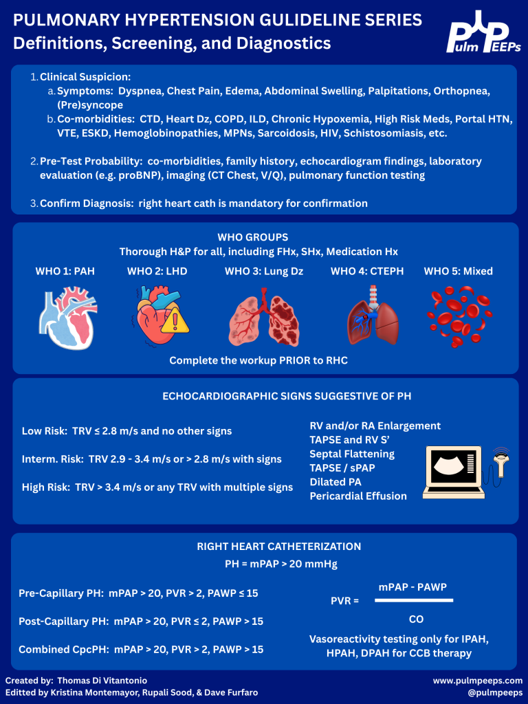
Today we’re kicking off another segment in our Guidelines Series, and doing a deep dive into the 2022 ESC/ERS Guidelines for the diagnosis and treatment of pulmonary hypertension. Over a series of episodes we’ll talk about the most recent updates to definitions around pulmonary hypertension, recognizing and diagnosing Group 1 – 5 pulmonary hypertension, risk stratification, and treatments. In this first episode, we will review the most recent definitions, including changes to the definitions that were new in 2022. We’ll then talk about recognizing and diagnosing pulmonary hypertension with tips and insights along the way.


Rupali Sood  grew up in Las Vegas, Nevada and made her way over to Baltimore for medical school at Johns Hopkins. She then completed her internal medicine residency training at Massachusetts General Hospital before returning back to Johns Hopkins, where she is currently a pulmonary and critical care medicine fellow alongside Tom. Rupali’s interests include interstitial lung disease, particularly as related to oncologic drugs. And she also loves bedside medical education.

Tom Di Vitantonio  is originally from New Jersey and attended medical school at Rutgers, New Jersey Medical School in Newark. He then completed his internal medicine residency at Weill Cornell, where he also served as a chief resident. He currently is a pulmonary and critical care medicine fellow at Johns Hopkins, and he’s passionate about caring for critically ill patients, how we approach the management of pulmonary embolism, and also about medical education of trainees to help them be more confident and patient centered in the care they have going forward.

  1. Why to have a high index of suspicion for pulmonary hypertension (PH)
  • PH often presents subtly with slowly progressive dyspnea on exertion, fatigue, lightheadedness, exertional chest pain, or syncope.
  • There’s often a delay of 1–2+ years from symptom onset to diagnosis, which is associated with worse mortality.
  • Early recognition and treatment, especially for pulmonary arterial hypertension (PAH, WHO group 1), can significantly change outcomes.


  1. When to suspect PH

Think PH when:

  • Dyspnea is out of proportion to:
    • CT parenchymal findings (relatively normal lungs)
    • Spirometry (normal FEV₁/FVC, volumes)
  • There are subtle but progressive symptoms over months:
    • Reduced exercise tolerance
    • No obvious alternative explanation (e.g., no overt HF, CAD, big ILD, etc.)
  • Physical exam may show (often late):
    • Elevated JVP, V waves (TR)
    • Peripheral edema, hepatomegaly, ascites
    • Loud P2, RV heave

In the case: a woman with systemic sclerosis + slowly progressive exertional dyspnea + relatively normal CT parenchyma and spirometry → high suspicion.


  1. WHO classification: 5 PH groups (big picture + why it matters)

Used for pathophysiology, prognosis, and treatment choices:

  1. Group 1 – PAH
    • Idiopathic, heritable (e.g., BMPR2), drug-induced (e.g., dasatinib)
    • Connective tissue disease (esp. systemic sclerosis)
    • Portal hypertension (portopulmonary HTN)
    • HIV, HHT, congenital heart disease/shunts
    • Rare: PVOD, PCH
  2. Group 2 – PH due to left heart disease
    • HFrEF, HFpEF, valvular disease
    • Most common cause worldwide.
  3. Group 3 – PH due to lung disease/hypoxia
    • COPD, ILD, combined pulmonary fibrosis–emphysema
    • OSA/obesity hypoventilation, chronic hypoxemia
  4. Group 4 – CTEPH
    • Chronic thromboembolic pulmonary hypertension
  5. Group 5 – Multifactorial/unclear
    • Sarcoidosis, myeloproliferative disorders, CKD, sickle cell, etc.

Patients can span multiple groups (e.g., systemic sclerosis: group 1 and/or group 3; sickle cell: many mechanisms).


  1. Initial workup & refining pre-test probability

Once you suspect PH, you’re trying to answer:

  1. Does this patient likely have PH?
  2. If yes, what group(s) are most likely?

Core non-invasive tests:

  • NT-proBNP (preferred over BNP)
    • Surrogate of RV strain and prognosis.
    • Normal value makes significant RV failure less likely.
  • Oxygenation & exercise
    • Resting SpO₂ plus ambulatory sats; consider 6-minute walk test.
    • Exertional desaturation is common and clinically meaningful.
  • CXR & ECG
    • Low yield but may show RV enlargement, right axis deviation, etc.
  • Pulmonary function tests
    • Full set: spirometry, volumes, DLCO.
    • Clue: isolated or disproportionately low DLCO with relatively preserved FVC suggests pulmonary vascular disease.
  • Imaging
    • High-res CT chest – parenchymal disease (ILD, emphysema).
    • V/Q scan – best screening test for CTEPH; better than CT angiography for chronic disease.
  • Sleep testing / overnight oximetry
    • When OSA/nocturnal hypoxemia suspected.


  1. Echo: estimating PH probability (not diagnosis)

TTE is the key screening tool but does not diagnose PH.

Main elements:

  1. Peak tricuspid regurgitant (TR) velocity
    • Used to estimate pulmonary artery systolic pressure (PASP).
    • Categories:
      • Low probability: TR velocity < 2.8 m/s, no other PH signs.
      • Intermediate: 2.9–3.4 m/s ± other PH signs.
      • High: > 3.4 m/s.

The presence and severity of TR ≠ TR velocity. You can have severe TR without PH.

  1. “Other signs” of PH/RV dysfunction on echo:
    • RV enlargement or systolic dysfunction (qualitative, TAPSE < ~1.7 cm, S′ ↓)
    • RA enlargement
    • Septal flattening (D-shaped LV; systolic = pressure overload, diastolic + systolic = volume + pressure)
    • Dilated PA
    • Pericardial effusion

Interpretation pattern:

  • Low pre-test probability + TR v < 2.8 + no other signs → PH unlikely.
  • Intermediate TR v (2.9–3.4) + high pre-test probability and/or other PH signs → consider RHC.
  • High TR v (>3.4) or clearly abnormal RV → strongly consider RHC if it would change management.

Also:

  • Echo is great to follow RV size/function and PASP over time once PH is diagnosed and treated.

Case echo:

  • TR velocity 3.1 m/s + mild RA enlargement + moderate RV enlargement + TAPSE 1.6 cm → intermediate probability, consistent with PH and RV involvement.


  1. Right heart cath (RHC): gold standard & updated definitions

You cannot definitively diagnose or classify PH without RHC.

Key directly measured values:

  • RA, RV, PA pressures
  • Pulmonary capillary wedge pressure (PCWP/PAWP) ≈ LVEDP
  • Oxygen saturations in chambers/vessels
  • Cardiac output (thermodilution)

Key derived values:

  • Cardiac output (Fick)
  • Pulmonary vascular resistance (PVR)

Updated hemodynamic definitions:

  1. Pulmonary hypertension (PH)
    • mPAP ≥ 20 mm Hg (lowered from ≥ 25).
  2. Pre-capillary PH (think PAH, group 1; also groups 3, 4, some 5):
    • mPAP ≥ 20
    • PAWP ≤ 15
    • PVR > 2 Wood units (new lower threshold)
  3. Isolated post-capillary PH (IpcPH) (group 2)
    • mPAP ≥ 20
    • PAWP > 15
    • PVR ≤ 2
  4. Combined pre- and post-capillary PH (CpcPH)
    • mPAP ≥ 20
    • PAWP > 15
    • PVR > 2

Rationale for the changes:

  • Normal mPAP in healthy people is < ~19; 20 is about 2 SD above normal.
  • Patients with mPAP 20–24 (esp. systemic sclerosis) already have worse outcomes than those < 20.
  • Lowering PVR cutoff from 3 → 2 WU better aligns with these new thresholds and catches earlier precapillary disease.

Practical interpretation:

  • You use mPAP + PAWP + PVR to:
    • Confirm PH.
    • Distinguish pre- vs post-capillary.
    • Identify mixed disease.
  • Echo tells you probability; RHC tells you what type and how severe.


  1. Vasoreactivity testing (acute vasodilator testing)
  • Only indicated in:
    • Idiopathic (IPAH)
    • Heritable PAH
    • Drug-induced PAH
      Not routine for all PH patients.
  • Performed in the cath lab with short-acting vasodilator (e.g., inhaled NO).

Positive test:

  • ↓ mPAP ≥ 10 mm Hg
  • To an absolute mPAP ≤ 40 mm Hg
  • No fall in cardiac output

Why it matters:

  • Identifies a small subset who can be treated with high-dose calcium channel blockers long-term and often have better prognosis.
  • Does not predict response to other PAH therapies (ERA, PDE5i, prostacyclin, etc.).


  1. Screening high-risk populations

Some groups warrant systematic screening because of high PAH risk.

  1. a) Systemic sclerosis / systemic sclerosis spectrum
  • Annual screening if:
    • Disease duration ≥ 3 years
    • FVC ≥ 40% predicted
    • DLCO < 60% predicted
  • DETECT algorithm (2-step):
    • Step 1: uses labs and simple tests (FVC/DLCO ratio, NT-proBNP, autoantibodies, right axis deviation on ECG, telangiectasias).
    • If positive → Step 2: adds echo (TR velocity, RA size).
    • If high risk after Step 2 → RHC.
  • Goal: catch early PAH before symptoms are severe.
  1. b) Other high-risk groups

Annual screening (usually with echo ± NT-proBNP, PFTs) for:

  • Known heritable PAH mutations (e.g., BMPR2)
  • Portal hypertension (esp. considering liver transplant or TIPS)
  • HIV

Always layer this on top of clinical symptoms and progression.


  1. Big practical takeaways (what to apply on Monday)
  1. Don’t label “pulmonary hypertension” off CT or echo alone.
    • Enlarged PA on CT or elevated PASP on echo ≠ diagnosis.
    • RHC is required.
  2. Think PH early when:
    • Dyspnea is out of proportion to imaging and spirometry.
    • There is a relevant risk factor (systemic sclerosis, portal HTN, HIV, prior PE, congenital heart disease, etc.).
  3. Use the WHO groups to structure your differential and workup:
    • Group 1 vs 2 vs 3 vs 4 vs 5 → drives what tests you order and what treatments you eventually consider.
  4. Echo = probability. RHC = truth.
    • Echo gives you low / intermediate / high PH probability.
    • RHC gives you pre- vs post-capillary, PVR, and hemodynamics needed for therapy.
  5. Know the new numbers:
    • mPAP ≥ 20 = PH
    • PAWP cutoff = 15
    • PVR > 2 WU = precapillary component
  6. Don’t forget NT-proBNP, DLCO, V/Q scan, and high-risk screening (especially in systemic sclerosis and BMPR2 carriers).


Humbert M, Kovacs G, Hoeper MM, Badagliacca R, Berger RMF, Brida M, Carlsen J, Coats AJS, Escribano-Subias P, Ferrari P, Ferreira DS, Ghofrani HA, Giannakoulas G, Kiely DG, Mayer E, Meszaros G, Nagavci B, Olsson KM, Pepke-Zaba J, Quint JK, Rådegran G, Simonneau G, Sitbon O, Tonia T, Toshner M, Vachiery JL, Vonk Noordegraaf A, Delcroix M, Rosenkranz S; ESC/ERS Scientific Document Group. 2022 ESC/ERS Guidelines for the diagnosis and treatment of pulmonary hypertension. Eur Heart J. 2022 Oct 11;43(38):3618-3731. doi: 10.1093/eurheartj/ehac237. Erratum in: Eur Heart J. 2023 Apr 17;44(15):1312. doi: 10.1093/eurheartj/ehad005. PMID: 36017548.

Condon DF, Nickel NP, Anderson R, Mirza S, de Jesus Perez VA. The 6th World Symposium on Pulmonary Hypertension: what’s old is new. F1000Res. 2019 Jun 19;8:F1000 Faculty Rev-888. doi: 10.12688/f1000research.18811.1. PMID: 31249672; PMCID: PMC6584967.

Maron BA. Revised Definition of Pulmonary Hypertension and Approach to Management: A Clinical Primer. J Am Heart Assoc. 2023 Apr 18;12(8):e029024. doi: 10.1161/JAHA.122.029024. Epub 2023 Apr 7. PMID: 37026538; PMCID: PMC10227272.

109. Guidelines Series: GINA Guidelines – Special Considerations in Asthma Care

In this episode, we’re concluding our review of the Global Initiative for Asthma (GINA) guidelines on asthma today with a cased based episode on special considerations in asthma care. We’ve covered asthma diagnosis and phenotyping, the approach to therapy inhaler and oral medical therapy, and biologic therapy. On today’s episode we’re talking about complex cases that are at the edges of the guidelines, or may be in future guidelines. To help us with this exciting topic we’re joined by an expert in the field. Enjoy! 

Dr. Meredith McCormack is a Professor of Medicine at Johns Hopkins, where she leads multiple NIH funded endeavors at understanding lung health and disease. She is the Division Director for Pulmonary and Critical Care Medicine, while also directing the Asthma Precision Medicine Center of Excellence, and the BREATHE Center, which focuses on understanding the effects of the environment on lung health and disease through research and community engagement.  She is an internationally recognized expert in asthma management and is a dedicated member of the faculty who is committed to the trainees.

Rupali Sood  grew up in Las Vegas, Nevada and made her way over to Baltimore for medical school at Johns Hopkins. She then completed her internal medicine residency training at Massachusetts General Hospital before returning back to Johns Hopkins, where she is currently a second year pulmonary and critical care medicine fellow alongside Tom. Rupali’s interests include interstitial lung disease, particularly as related to oncologic drugs. And she also loves bedside medical education.

Tom Di Vitantonio  is originally from New Jersey and attended medical school at Rutgers, New Jersey Medical School in Newark. He then completed his internal medicine residency at Weill Cornell, where he also served as a chief resident. He currently is a second year pulmonary and critical care medicine fellow at Johns Hopkins, and he’s passionate about caring for critically ill patients, how we approach the management of pulmonary embolism, and also about medical education of trainees to help them be more confident and patient centered in the care they have going forward.

Episode themes

  • Built on GINA 2024: final capstone focusing on evolving topics + case-based application.

  • Three focal areas: (1) obesity/metabolic health (GLP-1s, metformin), (2) dual biologics vs switching, (3) de-escalating inhalers while on biologics.

  • Emphasis throughout on personalized care, shared decision-making, and multidisciplinary collaboration.
  • Obesity & metabolic health in asthma
  • Obesity affects mechanics, inflammation, and treatment response; tackling metabolic dysfunction can improve asthma control.

  • GLP-1 receptor agonists may provide additive benefit beyond weight loss for some patients (early clinical signals; trials ongoing).

  • Metformin is being studied as a potential adjunct targeting metabolic-inflammatory pathways.

  • Practical approach: screen/counsel on weight, activity, and metabolic disease; partner with primary care/endocrine/sleep clinics; consider GLP-1/other agents when indicated for comorbidities, with potential asthma “bonus.”

  • Biologics: switching vs dual therapy
  • Consider switching/adding when control is not achieved or sustained on a biologic despite adherence.

  • Upstream vs downstream targets:

    • Upstream: anti-TSLP (e.g., tezepelumab) may help when multiple pathways/biomarkers (e.g., high IgE + eos) suggest broader blockade.Downstream: IL-5/IL-4/13/IgE agents selected to match phenotype/endotypes.

  • Comorbidities can drive choice:

    • Nasal polyps or upper airway syndromes: there are biologic options that improve upper airway symptoms in addition to asthmaAtopic dermatitis: agents with dual indications can be life-changing.

  • Logistics matter: injection burden/needle phobia and dosing cadence (e.g., every 2 vs 4–8 weeks) can determine real-world success.
  • De-escalating inhalers on biologics
  • Don’t step down immediately. Ask patients to maintain their full regimen for ~3 months after starting a biologic to gauge true benefit.Set expectations early and share a step-down plan to prevent unsupervised discontinuation.Typical order (individualize):
    1. Remove non-essential add-ons first (e.g., antihistamines, leukotriene modifiers).Reduce ICS dose gradually (high → medium → low).Keep ICS/LABA combination among the last therapies to taper

  • Targets while stepping down: “normal” lung function when feasible, minimal/no day or night symptoms, full activity, no exacerbations.

  • When patients don’t respond to biologics
  • Re-check the fundamentals:
    • Adherence/technique for inhalers and biologic.Biomarkers behaving as expected (e.g., eosinophils falling on anti-IL-5).Revisit the diagnosis and contributors/mimics (e.g., vocal cord dysfunction, upper-airway disease).
    • Consider moving upstream (e.g., to TSLP) if a downstream agent underperforms.

    Communication & practical pearls

    • Use visual aids to verify what patients actually take and how (e.g., Asthma & Allergy Network inhaler pictogram).
    • Needle issues are common; home vs clinic administration and family support can make or break adherence.
    • Biologics are transformative for the right patient—consider them early in steroid-dependent or poorly controlled severe asthma.
    • Think longitudinally: plan for monitoring, comorbidity management, and timely adjustments.


    98. Guidelines Series: GINA Guidelines – Biologics for Treatment of Asthma

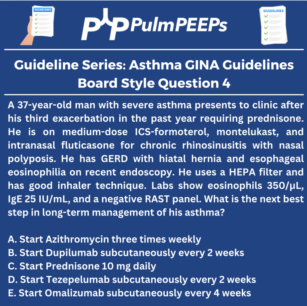
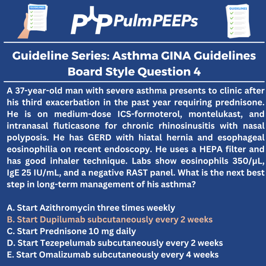
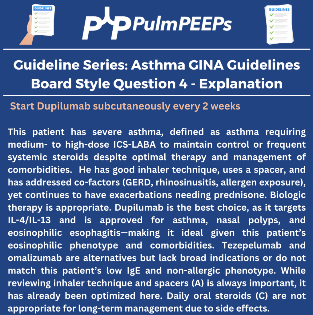
    Today, we continue our review of the Global Initiative for Asthma (GINA) guidelines on asthma. We’ve covered asthma diagnosis and phenotyping, and the initial approach to therapy. On today’s episode we’re talking about biologic therapies for asthma and will cover everything from when to consider starting them, which to choose, and what to monitor for after a patient is started. To help us with this exciting topic we’re joined by an expert in the field. We again have a great infographic prepared along with the episode, and a boards-style question for your review.


    Megan Conroy is an Assistant Professor of Medicine at The Ohio State University, and is also the associate program director for curriculum and quality in the Pulmonary and Critical Care Medicine Fellowship. Megan’s clinical area of expertise involves asthma and biologic therapies and she was recently recognized for her work in this area as the 2024 CHEST Airway Disorders Network Rising Star Award. 

    Rupali Sood  grew up in Las Vegas, Nevada and made her way over to Baltimore for medical school at Johns Hopkins. She then completed her internal medicine residency training at Massachusetts General Hospital before returning back to Johns Hopkins, where she is currently a second year pulmonary and critical care medicine fellow alongside Tom. Rupali’s interests include interstitial lung disease, particularly as related to oncologic drugs. And she also loves bedside medical education.

    Tom Di Vitantonio  is originally from New Jersey and attended medical school at Rutgers, New Jersey Medical School in Newark. He then completed his internal medicine residency at Weill Cornell, where he also served as a chief resident. He currently is a second year pulmonary and critical care medicine fellow at Johns Hopkins, and he’s passionate about caring for critically ill patients, how we approach the management of pulmonary embolism, and also about medical education of trainees to help them be more confident and patient centered in the care they have going forward.


    Mauer Y, Taliercio RM. Managing adult asthma: The 2019 GINA guidelines. Cleve Clin J Med. 2020 Aug 31;87(9):569-575. doi: 10.3949/ccjm.87a.19136. PMID: 32868307.

    Viswanathan RK, Busse WW. Biologic Therapy and Asthma. Semin Respir Crit Care Med. 2018 Feb;39(1):100-114. doi: 10.1055/s-0037-1606218. Epub 2018 Feb 10. PMID: 29427990.

    Brusselle GG, Koppelman GH. Biologic Therapies for Severe Asthma. N Engl J Med. 2022 Jan 13;386(2):157-171. doi: 10.1056/NEJMra2032506. PMID: 35020986.

    96. Guidelines Series: GINA Guidelines – Asthma Treatment and Management

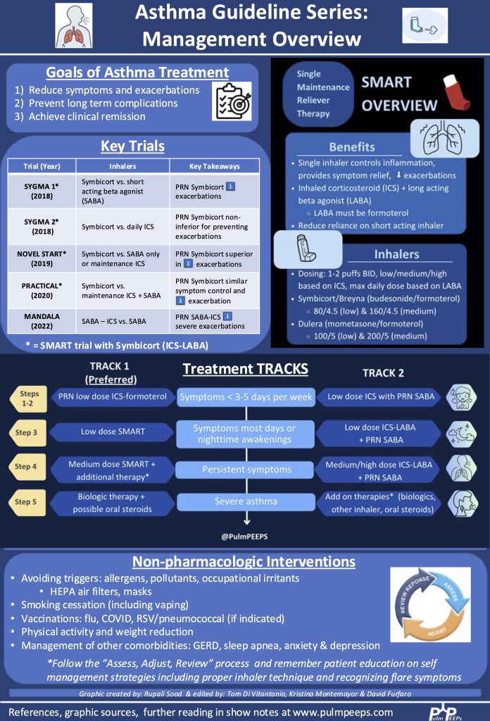
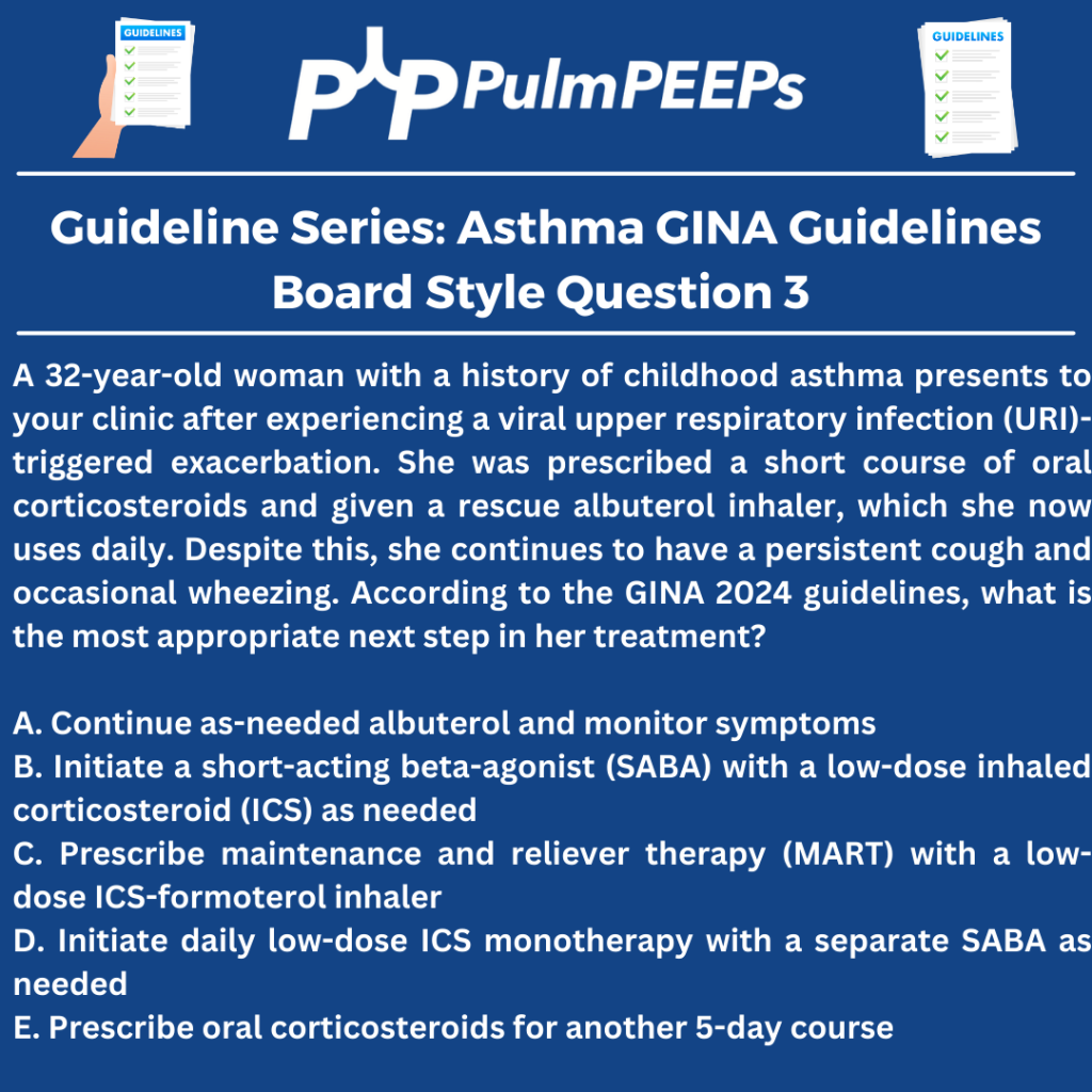
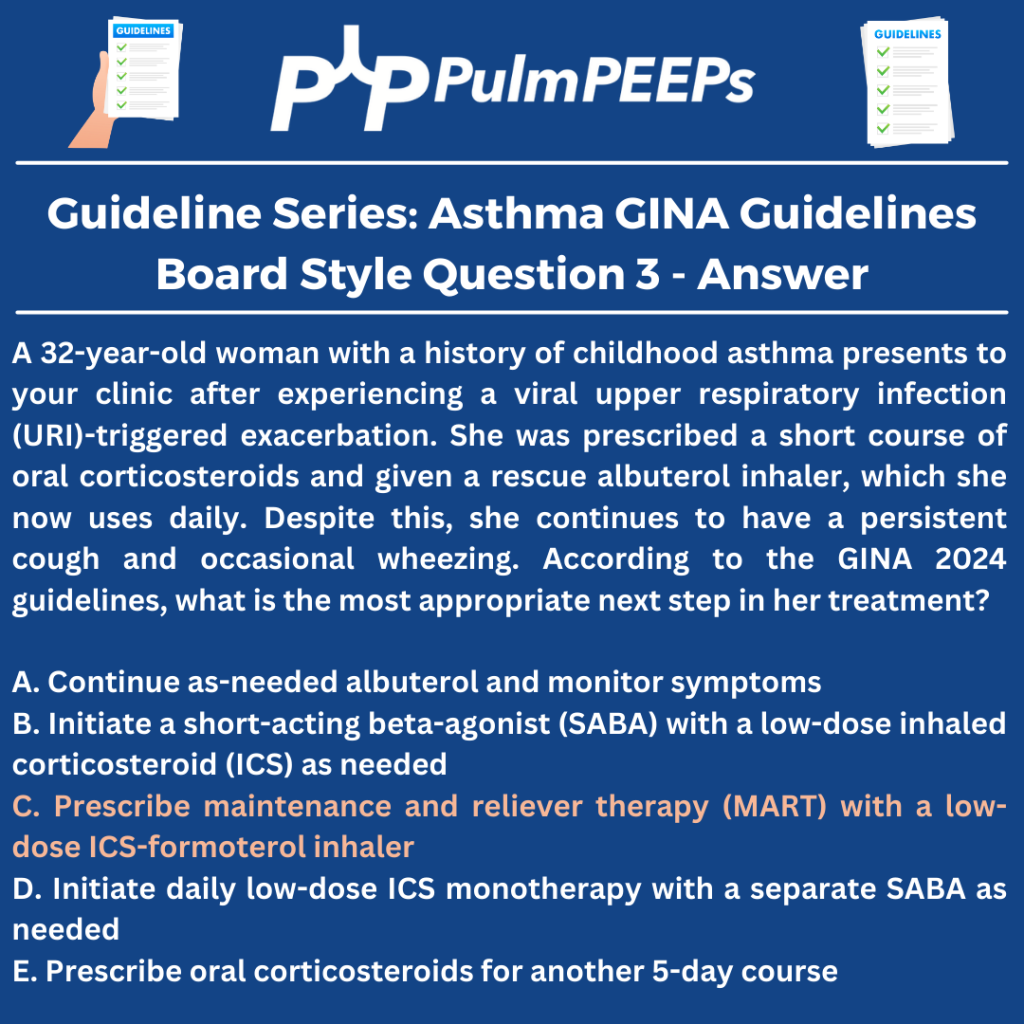
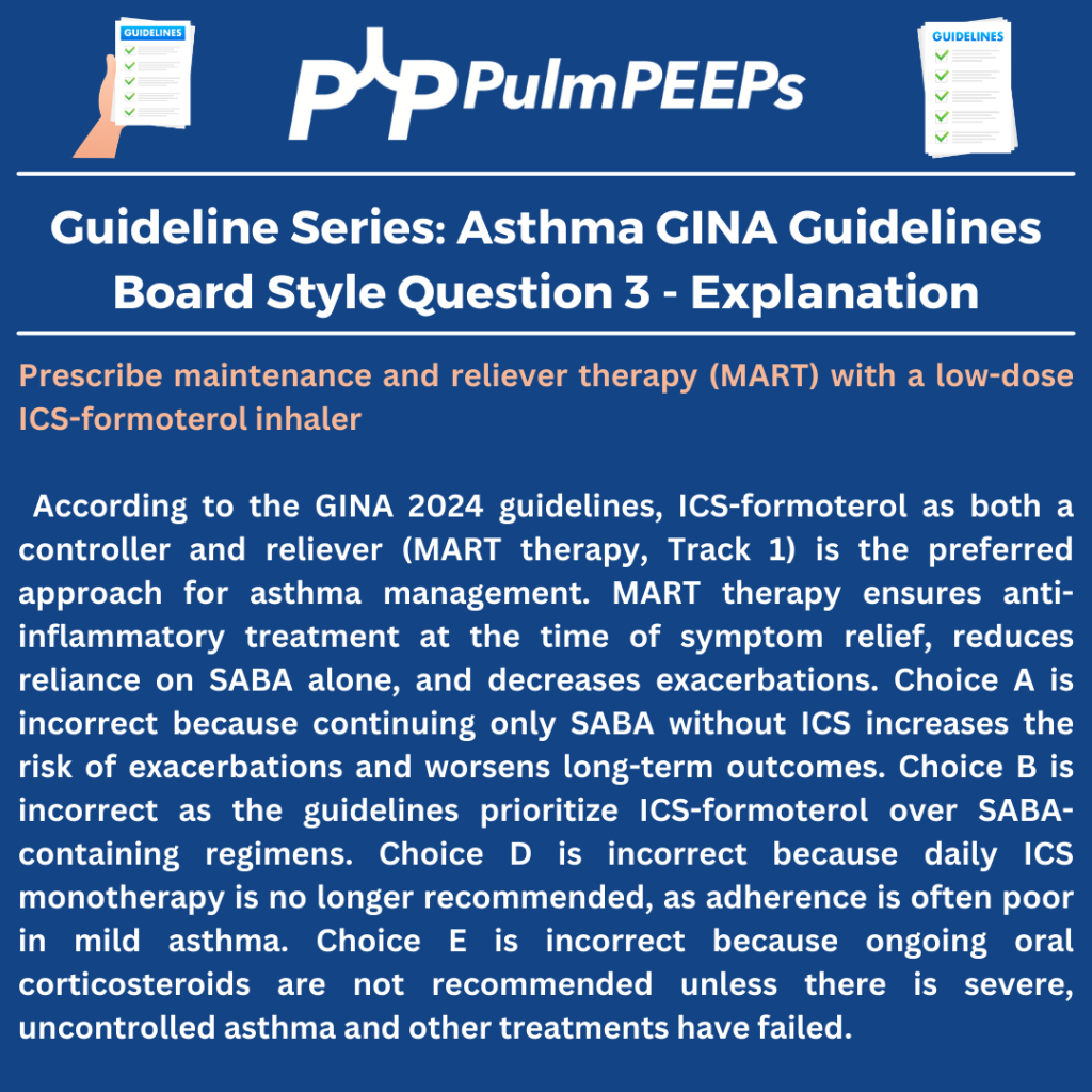
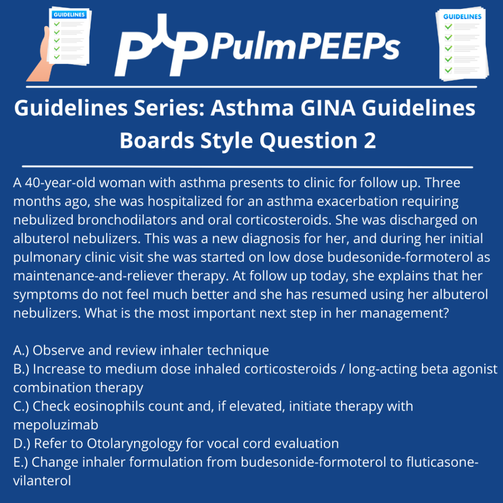
    We’re back with our second episode in our guideline initiative, and continuing our review of the Global Initiative for Asthma (GINA) guidelines on asthma. In our first episode of this series, we talked about making the diagnosis of asthma, the importance of appropriate phenotyping, and doing an initial assessment of asthma severity. Today, we’re discussing the initial management of asthma and discussing but pharmacologic and non-pharmacologic treatments. We have a great infographic prepared along with the episode, and a boards-style question for your review.

    Rupali Sood  grew up in Las Vegas, Nevada and made her way over to Baltimore for medical school at Johns Hopkins. She then completed her internal medicine residency training at Massachusetts General Hospital before returning back to Johns Hopkins, where she is currently a second year pulmonary and critical care medicine fellow alongside Tom. Rupali’s interests include interstitial lung disease, particularly as related to oncologic drugs. And she also loves bedside medical education.

    Tom Di Vitantonio  is originally from New Jersey and attended medical school at Rutgers, New Jersey Medical School in Newark. He then completed his internal medicine residency at Weill Cornell, where he also served as a chief resident. He currently is a second year pulmonary and critical care medicine fellow at Johns Hopkins, and he’s passionate about caring for critically ill patients, how we approach the management of pulmonary embolism, and also about medical education of trainees to help them be more confident and patient centered in the care they have going forward.

    1. Introduction to Asthma Guidelines
    • The podcast continues a guideline series on asthma, focusing on the Global Initiative for Asthma (GINA) 2024 guidelines.
    • Emphasizes practical applications for clinicians managing asthma in different settings.
    1. Importance of Evidence-Based Asthma Management
    • Asthma treatment must be systematic and personalized, considering recent clinical evidence.
    • Previous reliance on short-acting beta agonists (SABAs) as rescue inhalers has shifted towards inhaled corticosteroid (ICS)-containing therapies.
    • Over-reliance on SABAs is linked to increased exacerbations, airway inflammation, and poor long-term outcomes.
    1. Stepwise Approach to Asthma Management (GINA 2024)
    • The Track 1 approach (preferred) centers around ICS-formoterol as both maintenance and reliever therapy (MART).
    • Track 2 (alternative approach) includes daily ICS or ICS-LABA with a separate SABA as a reliever.

    Stepwise Therapy

    • Step 1-2 (Mild asthma): Low-dose ICS-formoterol as needed for symptom relief.
    • Step 3 (Moderate asthma): Low-dose maintenance ICS-formoterol (MART therapy).
    • Step 4 (Persistent symptoms): Medium-dose ICS-formoterol (MART) with additional inhaler adjustments.
    • Step 5 (Severe asthma): Consider biologic therapies, phenotyping, and additional controllers.
    1. MART Therapy as a Game-Changer
    • Maintenance and Reliever Therapy (MART):
      • Uses a single inhaler for both daily maintenance and symptom relief.
      • Reduces overuse of SABAs.
      • Provides real-time up-titration of ICS during exacerbations.
      • Leads to better adherence and control.
    • Supporting Evidence from Trials:
      • SIGMA 1 & 2, Novel Start, Practical (2018-2019): Showed ICS-formoterol reduces exacerbations and steroid exposure compared to SABAs.
      • MANDALA (2022): Showed ICS-SABA improves outcomes over SABA alone, though not a true MART study.
    1. Practical Considerations in Asthma Management
    • Patient adherence is critical—educate on proper inhaler use and symptom monitoring.
    • Insurance and cost barriers may require prescribing alternative inhalers.
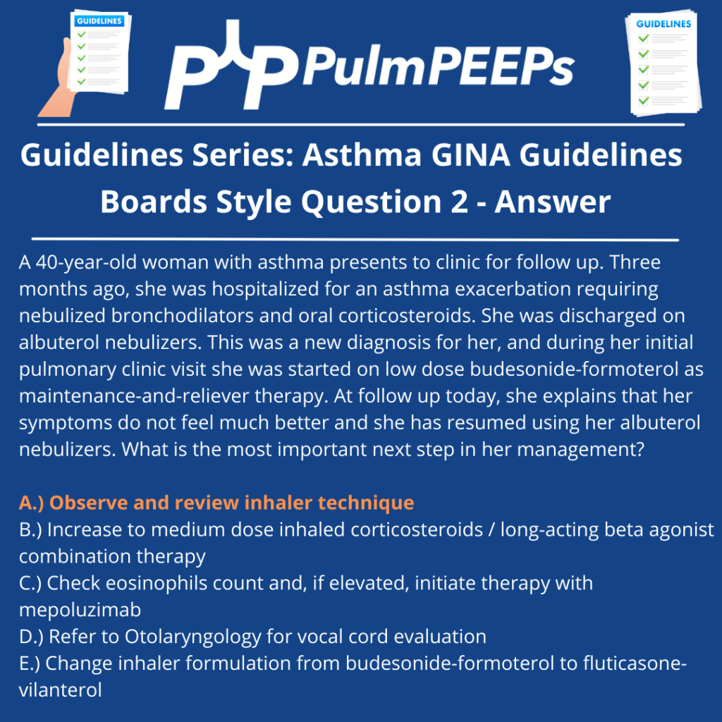
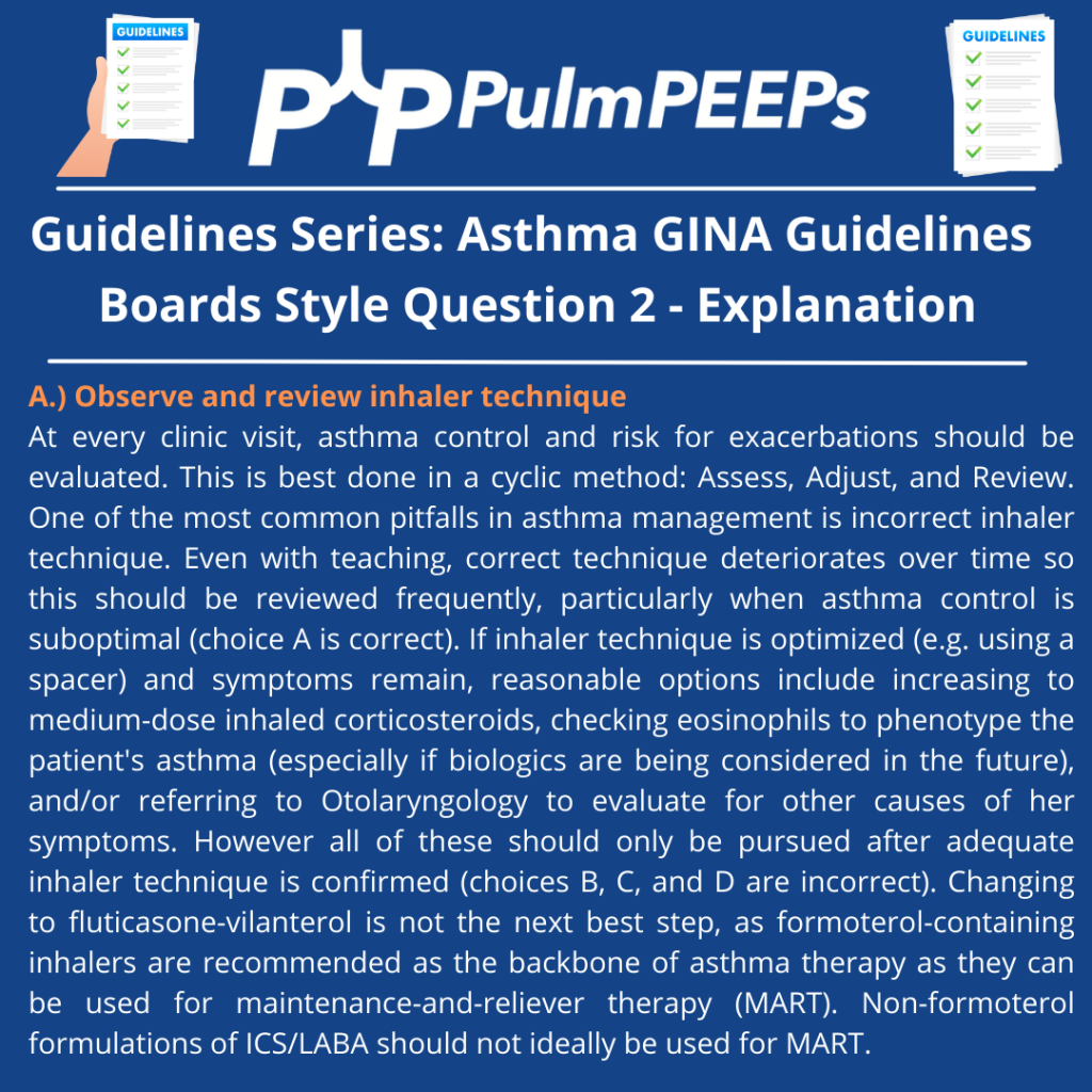
    • Review and adjust treatment regularly using the “Assess, Adjust, Review” framework.
    • Avoid high-dose ICS without exploring additional controller therapies like LAMAs, leukotriene receptor antagonists (Montelukast), and azithromycin.
    1. Non-Pharmacologic Interventions
    • Smoking cessation (including vaping/marijuana).
    • Weight management and physical activity.
    • Avoiding triggers (allergens, occupational exposures, pollution).
    • Air purifiers and HEPA filters.
    • Vaccinations (flu, COVID-19) to prevent viral exacerbations.
    • Managing comorbidities (GERD, sleep apnea, anxiety/depression).
    1. Case Discussion & Real-World Application
    • Patient with recurrent asthma symptoms post-viral illness.
    • Started on low-dose ICS-formoterol as needed.
    • Symptoms persisted, leading to maintenance ICS-formoterol (MART therapy).
    • Regular follow-up to monitor and adjust therapy.
    1. Looking Ahead
    • Next episode will focus on biologic therapies for severe asthma.
    • Emphasis on ongoing education, practical application, and patient-centered care.


    Mauer Y, Taliercio RM. Managing adult asthma: The 2019 GINA guidelines. Cleve Clin J Med. 2020 Aug 31;87(9):569-575. doi: 10.3949/ccjm.87a.19136. PMID: 32868307.

    Matera MG, Rinaldi B, Annibale R, De Novellis V, Cazzola M. The pharmacological management of asthma in adults: 2023 update. Expert Opin Pharmacother. 2024 Mar;25(4):383-393. doi: 10.1080/14656566.2024.2332627. Epub 2024 Mar 20. PMID: 38497368.

    Arismendi E, Ribo P, García A, Torrego A, Bobolea I, Casas-Saucedo R, Palomino R, Picado C, Muñoz-Cano R, Valero A. Asthma Control According to GINA 2023: Does Changing the Criteria Improve Asthma Control? J Clin Med. 2024 Nov 6;13(22):6646. doi: 10.3390/jcm13226646. PMID: 39597790; PMCID: PMC11594371.

    http://ginasthma.org/2023-gina-main-report/

    https://www.uptodate.com/contents/an-overview-of-asthma-management-in-children-and-adults

    https://onlinelibrary.wiley.com/doi/full/10.1111%2Fresp.14782

    Dubin S, Patak P, Jung D. Update on Asthma Management Guidelines. Mo Med. 2024 Sep-Oct;121(5):364-367. PMID: 39421468; PMCID: PMC11482852.


    93. Guidelines Series: GINA Guidelines – Asthma Diagnosis and Assessment

    Today we are launching a new Pulm PEEPs initiative! We are going to be reviewing some of the major guidelines that are available in pulmonary and critical care. We are starting by diving into the Global Initiative for Asthma (GINA) guidelines on asthma. The goal of this initiative is to breakdown the guidelines into digestible and helpful discussions, and to talk about key issues that are pointed out by the guideline authors. Our first episode will be the start of the GINA guidelines and we’re discussing the initial diagnosis and evaluation of patients with asthma.

    Rupali Sood  grew up in Las Vegas, Nevada and made her way over to Baltimore for medical school at Johns Hopkins. She then completed her internal medicine residency training at Massachusetts General Hospital before returning back to Johns Hopkins, where she is currently a second year pulmonary and critical care medicine fellow alongside Tom. Rupali’s interests include interstitial lung disease, particularly as related to oncologic drugs. And she also loves bedside medical education.

    Tom Di Vitantonio  is originally from New Jersey and attended medical school at Rutgers, New Jersey Medical School in Newark. He then completed his internal medicine residency at Weill Cornell, where he also served as a chief resident. He currently is a second year pulmonary and critical care medicine fellow at Johns Hopkins, and he’s passionate about caring for critically ill patients, how we approach the management of pulmonary embolism, and also about medical education of trainees to help them be more confident and patient centered in the care they have going forward.

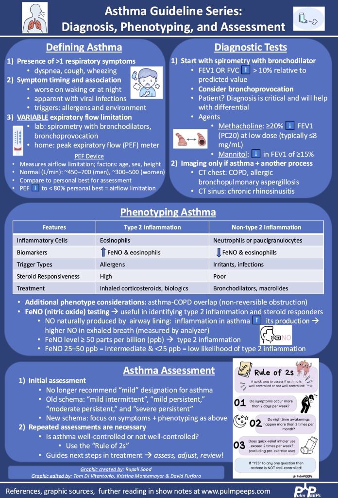
    Understanding Asthma & the GINA Guidelines

    • Asthma is a heterogeneous disease characterized by recurring respiratory symptoms (breathlessness, wheezing, cough, chest tightness) with variable airflow limitation.
    • The 2023 & 2024 Global Initiative for Asthma (GINA) guidelines emphasize phenotyping asthma to improve diagnosis and treatment.
    • Asthma differs from other obstructive lung diseases due to reversible airway obstruction, which can be demonstrated through diagnostic testing.

    Diagnosing Asthma

    • Clinical history is crucial, particularly identifying symptom triggers (cold air, exercise, allergens).
    • Spirometry is the standard diagnostic tool, looking for an increase in FEV1 or FVC ≥12% and 200 mL after bronchodilator use.
    • Alternative tests include:
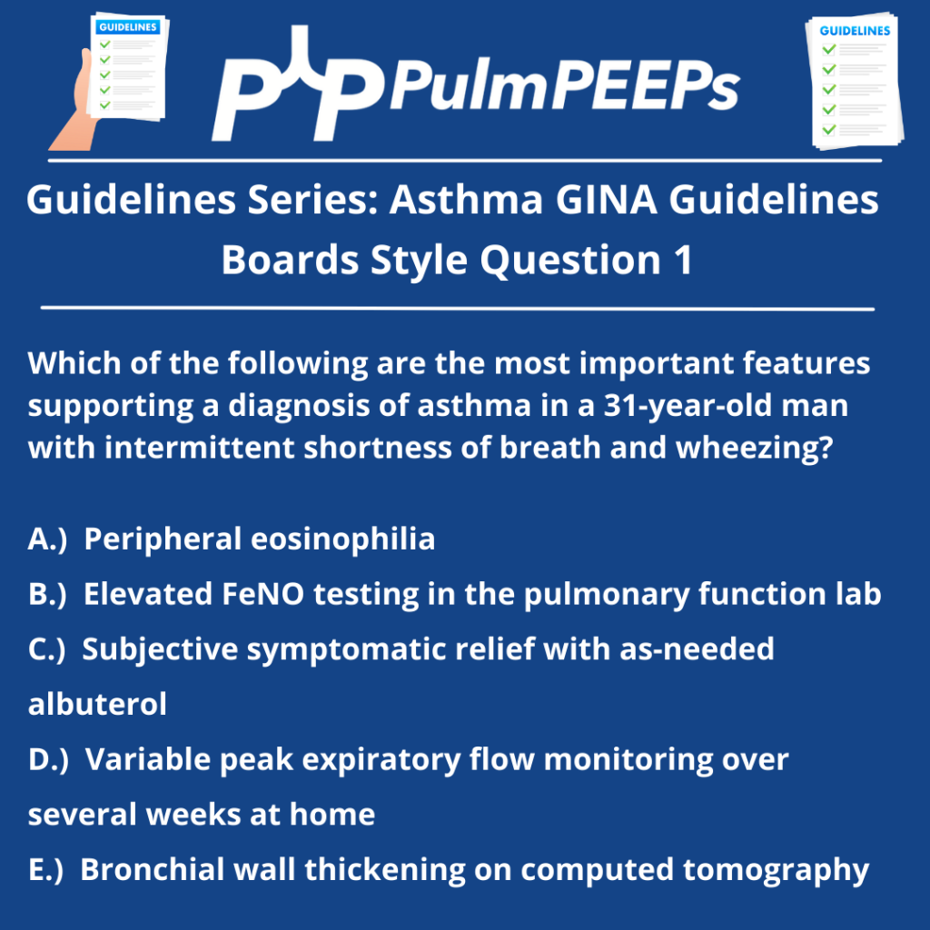
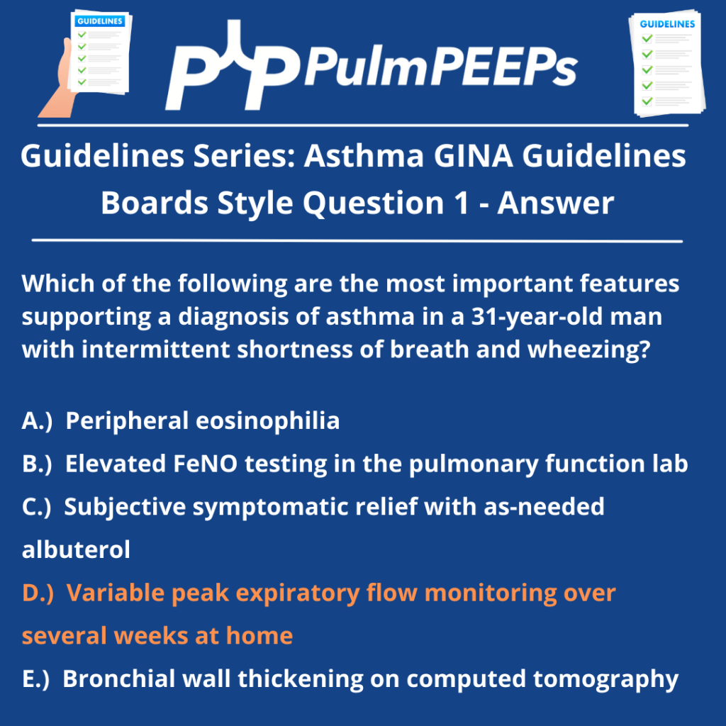
      • Peak expiratory flow monitoring over time.
      • Bronchoprovocation tests (e.g., methacholine challenge) to assess airway hyperresponsiveness.
      • Fractional exhaled nitric oxide (FENO) and blood eosinophils as markers of type 2 inflammation.

    Asthma Phenotypes & Precision Medicine

    • Different asthma phenotypes guide personalized treatment approaches:
      • Type 2  inflammation: Characterized by eosinophilic inflammation, high FeNO, good steroid responsiveness, and potential for biologic therapy.
      • Non-Type 2 inflammation: Associated with neutrophilic inflammation, poor steroid responsiveness, and potential benefit from macrolides or bronchodilators.
    • Asthma-COPD overlap requires a distinct treatment approach due to persistent obstruction.

    Imaging & Adjunctive Tests

    • Imaging is not routinely needed in asthma but can be useful for:
      • Bronchiectasis (suspected allergic bronchopulmonary aspergillosis – ABPA).
      • Asthma-COPD overlap (CT chest for emphysema).
      • Chronic sinusitis or nasal polyps (CT sinus imaging).

    Assessing Asthma Control

    • Asthma is not a one-time diagnosis; continuous reassessment is crucial.
    • Asthma control is assessed at every visit, considering:
      • Symptom frequency
      • Exacerbations
      • Inhaler technique
      • Comorbidities
    • Rule of Twos: Asthma is not well-controlled if:
      • Symptoms occur >2 times per week.
      • Nighttime awakenings >2 times per month.
      • Rescue inhaler use >2 times per week (excluding pre-exercise use).
    • Peak flow meters are valuable for self-monitoring and guiding asthma action plans.

    Conclusion

    • Asthma assessment is a continuous process, incorporating history, spirometry, biomarkers, and patient-reported symptoms.
    • Future episodes will cover asthma treatment, including biologics and inhaler therapy.
    • Infographics and questions will accompany this series for further learning.

    Mauer Y, Taliercio RM. Managing adult asthma: The 2019 GINA guidelines. Cleve Clin J Med. 2020 Aug 31;87(9):569-575. doi: 10.3949/ccjm.87a.19136. PMID: 32868307.

    Matera MG, Rinaldi B, Annibale R, De Novellis V, Cazzola M. The pharmacological management of asthma in adults: 2023 update. Expert Opin Pharmacother. 2024 Mar;25(4):383-393. doi: 10.1080/14656566.2024.2332627. Epub 2024 Mar 20. PMID: 38497368.

    Arismendi E, Ribo P, García A, Torrego A, Bobolea I, Casas-Saucedo R, Palomino R, Picado C, Muñoz-Cano R, Valero A. Asthma Control According to GINA 2023: Does Changing the Criteria Improve Asthma Control? J Clin Med. 2024 Nov 6;13(22):6646. doi: 10.3390/jcm13226646. PMID: 39597790; PMCID: PMC11594371.

    http://ginasthma.org/2023-gina-main-report/

    19. Severe COPD and Lung Volume Reduction

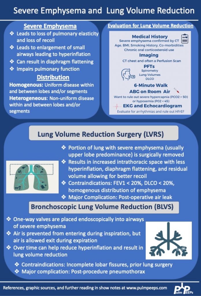
    We are extremely excited for the third and final installment in our Pulm PEEPs and ATS Clinical Problems Assembly collaborative series on COPD. Today, we are joined by Drs. Jessica Bon, Michael Lester, and Niru Putcha to discuss severe COPD management and the role of lung volume reduction procedures. If you missed the first two parts of our series, make sure to check out episode 1 on COPD diagnosis and initial management, and episode 2 on COPD exacerbations.

    Meet our Guests

    Jessica Bon is an Associate Professor of Medicine at the University of Pittsburgh School of Medicine where she is also the Program Director for the Pulmonary and Critical Care Medicine Fellowship. Her research and clinical interests focus on lung disease progression in COPD and she manages patients with difficult-to-treat and severe COPD and evaluates patients for lung volume reduction surgery. Jessica was the chair of the ATS Clinical Problems Assembly Programming Committee from 2021 – 2022.

    Michael Lester is an Assistant Professor of Medicine at Vanderbilt University Medical Center. Michael’s interests span both pulmonary and critical care medicine. He specializes in patients with advanced COPD and evaluation for bronchoscopic lung volume reduction surgery.

    Niru Putcha is an Associate Professor of Medicine at Johns Hopkins School of Medicine and is an integral member and mentor in the Obstructive Lung Disease Group. Her research and clinical interests focus on the role of comorbidities on clinical outcomes in individuals with COPD. She also manages patients with difficult-to-treat and severe COPD and evaluates patients for lung volume reduction surgery. Niru is also the new chair of the ATS  Clinical Problems Assembly Programming Committee.

    Key Learning Points

    Patients with advanced COPD should also be considered for lung transplantation. We will have an episode on lung transplant coming up soon!

    References

    1. Criner GJ, Sternberg AL. A Clinician’s Guide to the Use of Lung Volume Reduction Surgery. Proc Am Thorac Soc. 2008;5(4):461-467. doi:10.1513/pats.200709-151ET
    2. A Randomized Trial Comparing Lung-Volume–Reduction Surgery with Medical Therapy for Severe Emphysema. New England Journal of Medicine. 2003;348(21):2059-2073. doi:10.1056/NEJMoa030287
    3. Valipour A, Slebos DJ, Herth F, et al. Endobronchial Valve Therapy in Patients with Homogeneous Emphysema. Results from the IMPACT Study. Am J Respir Crit Care Med. 2016;194(9):1073-1082. doi:10.1164/rccm.201607-1383OC
    4. Sciurba FC, Ernst A, Herth FJF, et al. A Randomized Study of Endobronchial Valves for Advanced Emphysema. New England Journal of Medicine. 2010;363(13):1233-1244. doi:10.1056/NEJMoa0900928
    5. Klooster K, Slebos DJ. Endobronchial Valves for the Treatment of Advanced Emphysema. Chest. 2021;159(5):1833-1842. doi:10.1016/j.chest.2020.12.007
    6. Choi M, Lee WS, Lee M, et al. Effectiveness of bronchoscopic lung volume reduction using unilateral endobronchial valve: a systematic review and meta-analysis. Int J Chron Obstruct Pulmon Dis. 2015;10:703-710. doi:10.2147/COPD.S75314

    15. COPD Exacerbations

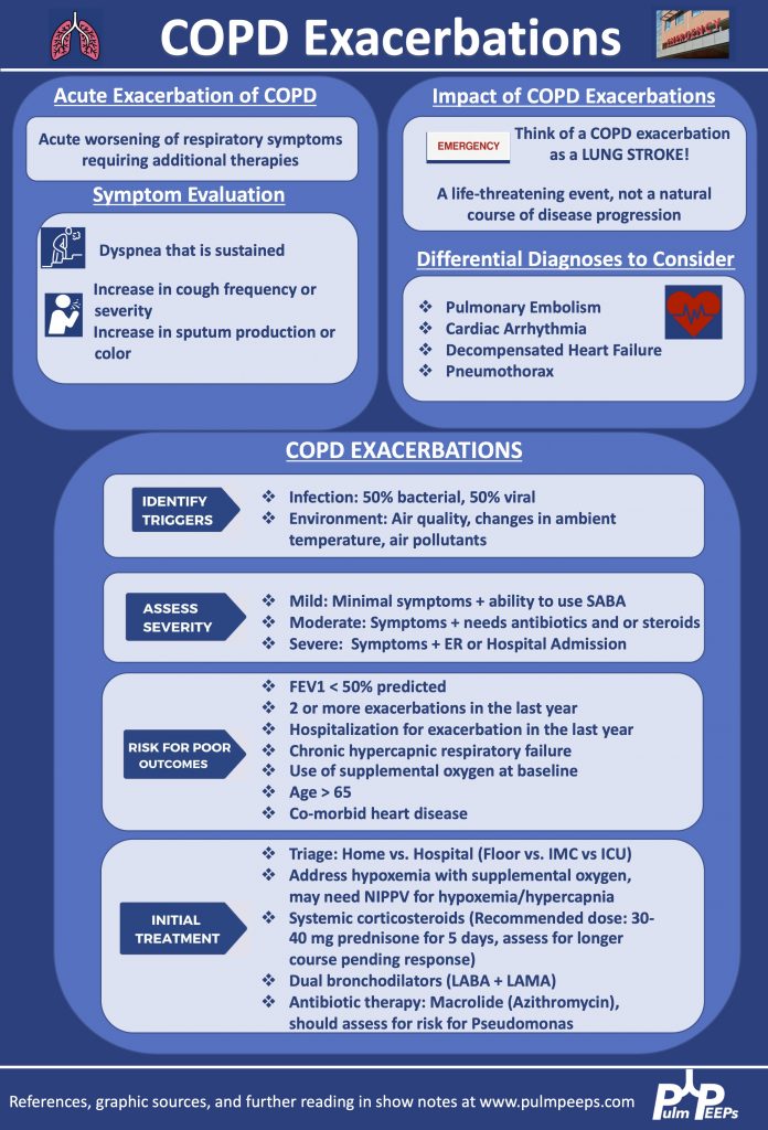
    Today we have episode two in our Pulm PEEPs and ATS Clinical Problems Assembly collaborative series on COPD. We are joined by Dr. Brad Drummond and Dr. Allison Lambert to discuss COPD exacerbations. Make sure to check out episode 1 in the series and stay tuned for more great content.

    Meet Our Guests

    Brad Drummond is an Associate Professor of Medicine at UNC School of Medicine. He is also the Associate Division Chief of Outpatient Services, the Co-Medical Director of the Pulmonary Specialty Clinics at UNC, and the Director of the Obstructive Lung Diseases Clinical and Translational Research Center. He is also the incoming Assembly Chair for the ATS CP Assembly.

    Allison Lambert is a Pulmonary and Critical Care physician at Providence Medical Group, where she is also the Director of the Adult Cystic Fibrosis Program and co-leads the Therapeutic Development Network. Her expertise spans CF, non-CF bronchiectasis as well as COPD. Allison is also a committee member in the ATS Clinical Problems Assembly

    Key Learning Points

    References

    1. Wedzicha JA, Miravitlles M, Hurst JR, et al. Management of COPD exacerbations: a European Respiratory Society/American Thoracic Society guideline. European Respiratory Journal. 2017;49(3). doi:10.1183/13993003.00791-2016
    2. Lindenauer PK, Dharmarajan K, Qin L, Lin Z, Gershon AS, Krumholz HM. Risk Trajectories of Readmission and Death in the First Year after Hospitalization for Chronic Obstructive Pulmonary Disease. Am J Respir Crit Care Med. 2018;197(8):1009-1017. doi:10.1164/rccm.201709-1852OC
    3. Leuppi JD, Schuetz P, Bingisser R, et al. Short-term vs Conventional Glucocorticoid Therapy in Acute Exacerbations of Chronic Obstructive Pulmonary Disease: The REDUCE Randomized Clinical Trial. JAMA. 2013;309(21):2223-2231. doi:10.1001/jama.2013.5023
    4. Johns Hopkins University. Roflumilast or Azithromycin to Prevent COPD Exacerbations (RELIANCE). clinicaltrials.gov; 2022. Accessed April 24, 2022. https://clinicaltrials.gov/ct2/show/NCT04069312
    5. Barnes PJ. Chronic Obstructive Pulmonary Disease. New England Journal of Medicine. 2000;343(4):269-280. doi:10.1056/NEJM200007273430407
    6. Celli BR, Wedzicha JA. Update on Clinical Aspects of Chronic Obstructive Pulmonary Disease. New England Journal of Medicine. 2019;381(13):1257-1266. doi:10.1056/NEJMra1900500
    7. Singh D, Agusti A, Anzueto A, et al. Global Strategy for the Diagnosis, Management, and Prevention of Chronic Obstructive Lung Disease: the GOLD science committee report 2019. Eur Respir J. 2019;53(5):1900164. doi:10.1183/13993003.00164-2019

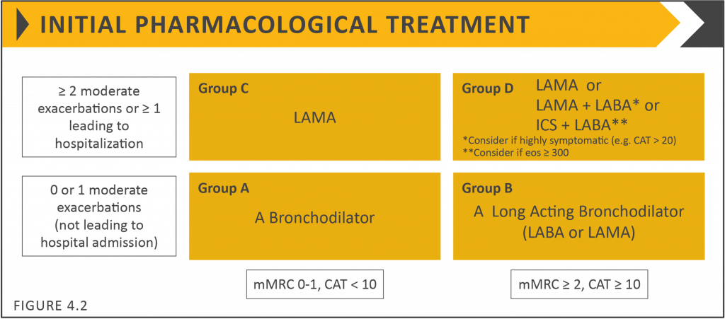
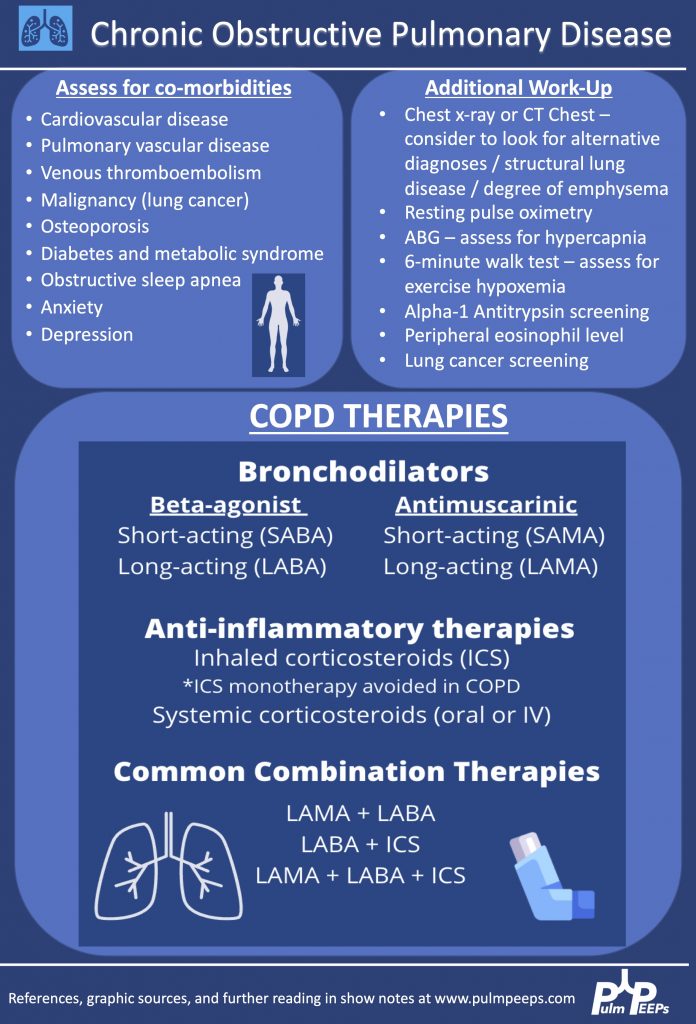
    13. COPD Classification and Practical Management Strategies

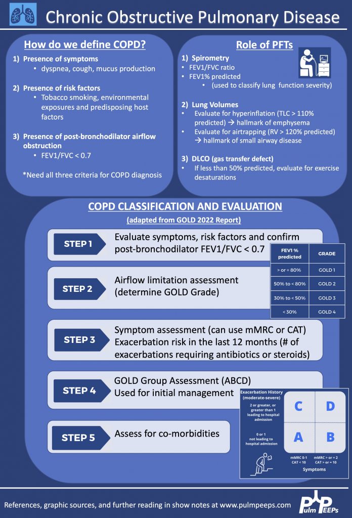
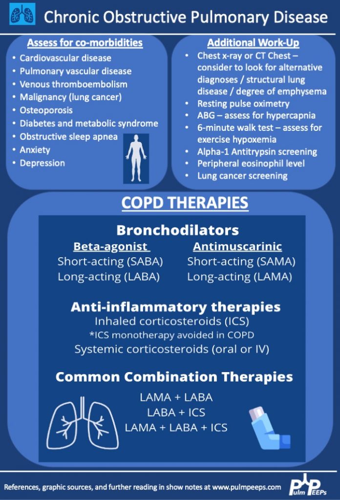
    The Pulm PEEPs are extremely excited today to be launching our series on Chronic Obstructive Pulmonary Disease in partnership with the ATS Clinical Problems Assembly. We are joined by Dr. Bob Wise and Dr. Wassim Labaki to discuss the classification and initial workup of COPD, and management strategies from inhalers to pulmonary rehabilitation. Make sure to listen today and in the coming weeks for the rest of our COPD discussion.

    Meet Our Guests

    Dr. Bob Wise is a Professor of Medicine at Johns Hopkins School of Medicine and has served as the Medical Director of the Pulmonary Function Lab at the Johns Hopkins Asthma and Allergy Center. Bob is a leader in the care of patients with obstructive lung disease and his research focus has been conducting multi-center clinical trials in airway disease and is also a master physiologist. Bob has been involved in various capacities with ATS throughout his tenure as well and received the ATS CP Assembly Sreedhar Nair Lifetime Achievement Award in COPD.

    Dr. Wassim Labaki is an Assistant Professor of Medicine in the Division of Pulmonary and Critical Care Medicine as well as the Medical Director of the Lung Volume Reduction Surgery Program at the University of Michigan. Wassim was the recipient of the Early Career Investigator Award in COPD from ATS in 2019 and currently is on the Program Committee of the ATS Clinical Problems Assembly.

    Key Learning Points

    Modified Medical Research Council (mMRC) Dyspnea Scale

    mMRC Grade 0 = Only breathless with strenuous exercise

    mMRC Grade 1 = Short of breath when hurrying on level ground, or walking up a slight hill

    mMRC Grade 2 = Walking slower than people of the same age due to dyspnea, or stopping due to dyspnea when walking at my own pace on level ground

    mMRC Grade 3 = Stopping for breath after walking 100 meters / a few minutes on level ground

    mMRC Grade 4 = Too breathless to leave the house or breathless with getting dressed / undressed

    Image source: Global Initiative for Chronic Obstructive Lung Disease https://goldcopd.org/

    References and links for further reading

    1. Clinicians. Global Initiative for Chronic Obstructive Lung Disease – GOLD. Accessed April 11, 2022. https://goldcopd.org/clinicians/
    2. Miami CF 3300 P de LB. COPD Foundation | Take Action Today. Breathe Better Tomorrow. Accessed April 11, 2022. https://www.copdfoundation.org
    3. Barnes PJ. Chronic Obstructive Pulmonary Disease. New England Journal of Medicine. 2000;343(4):269-280. doi:10.1056/NEJM200007273430407
    4. Celli BR, Wedzicha JA. Update on Clinical Aspects of Chronic Obstructive Pulmonary Disease. New England Journal of Medicine. 2019;381(13):1257-1266. doi:10.1056/NEJMra1900500
    5. Criner GJ, Martinez FJ, Aaron S, et al. Current Controversies in Chronic Obstructive Pulmonary Disease. A Report from the Global Initiative for Chronic Obstructive Lung Disease Scientific Committee. Ann Am Thorac Soc. 2019;16(1):29-39. doi:10.1513/AnnalsATS.201808-557PS
    6. Martinez FJ, Agusti A, Celli BR, et al. Treatment Trials in Young Patients with Chronic Obstructive Pulmonary Disease and Pre-Chronic Obstructive Pulmonary Disease Patients: Time to Move Forward. Am J Respir Crit Care Med. 2022;205(3):275-287. doi:10.1164/rccm.202107-1663SO
    7. Rodriguez-Roisin R, Rabe KF, Vestbo J, Vogelmeier C, Agustí A, all previous and current members of the Science Committee and the Board of Directors of GOLD (goldcopd.org/committees/). Global Initiative for Chronic Obstructive Lung Disease (GOLD) 20th Anniversary: a brief history of time. Eur Respir J. 2017;50(1):1700671. doi:10.1183/13993003.00671-2017
    8. Singh D, Agusti A, Anzueto A, et al. Global Strategy for the Diagnosis, Management, and Prevention of Chronic Obstructive Lung Disease: the GOLD science committee report 2019. Eur Respir J. 2019;53(5):1900164. doi:10.1183/13993003.00164-2019