We are back with another #RadiologyRounds! This week’s case comes from our Associate Editor @luke_hedrick
A portable film is obtained on a 65 yo woman with COPD presenting with progressive dyspnea and cough.
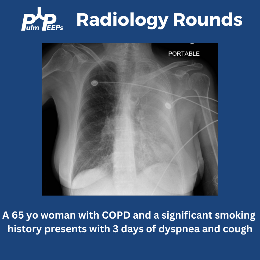
The image shows near complete opacification of the left lung. There is no ipsilateral or contralateral tracheal deviation which you would expect with atelectasis or a large pleural effusion, respectively. A CT chest is obtained to better visualize the parenchyma.
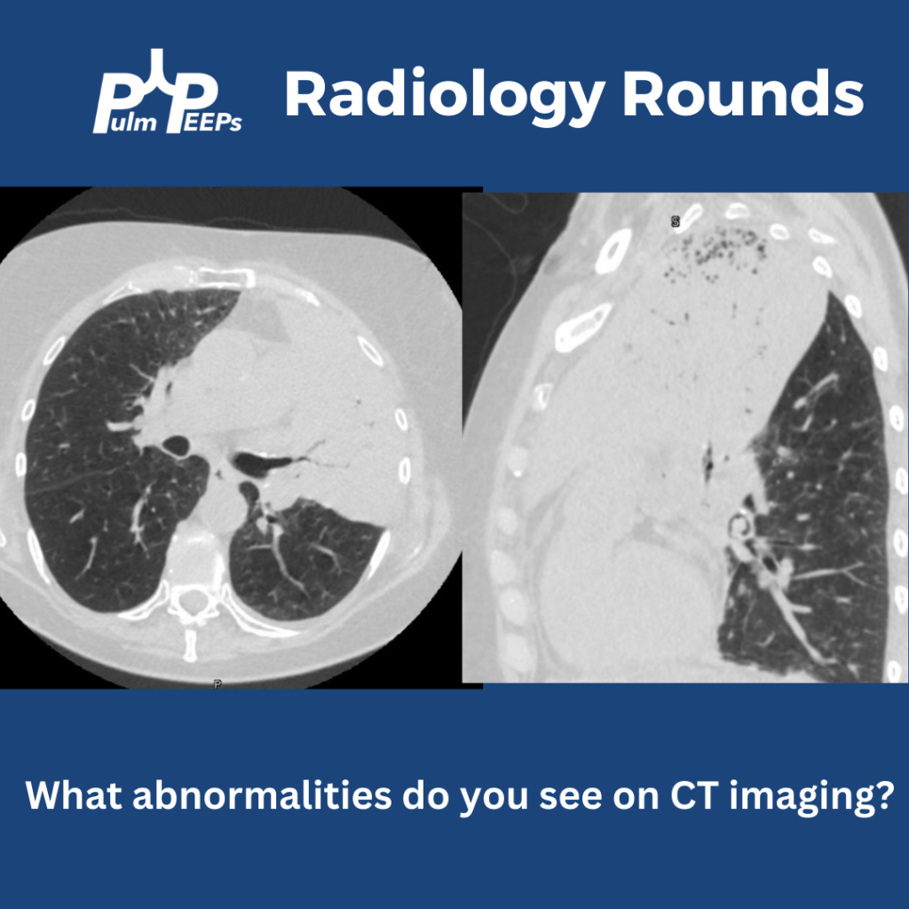
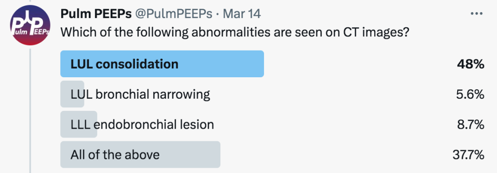
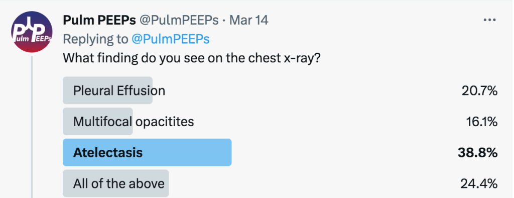
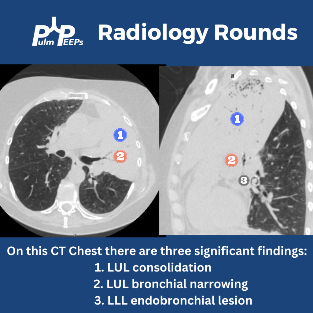
The CT chest shows all three findings as noted below.

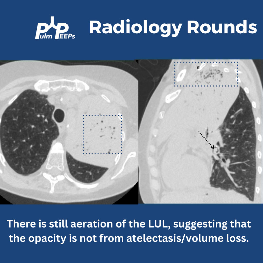
Yes! There is still aeration in some of the upper lobe, which would not be the case if this were caused by profound atelectasis. Also, atelectasis of such a large territory of the lung would usually cause traction on surrounding structures.
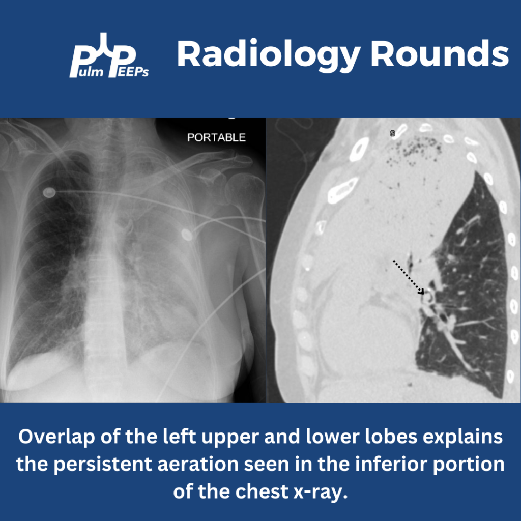
Having seen the CT, can you explain why the x-ray showed aeration in the inferior, left hemithorax? This is from the overlap of the left upper and lower lobes when viewed anteriorly. The sagittal view of the CT demonstrates this nicely.