103. Fellows’ Case Files: University of Virginia

Today, we’re virtually visiting the University of Virginia for another Fellows’ Case Files. This is a fantastic case that covers ARDS, the infectious work up of an immunosuppressed patient, and the evaluation of undifferentiated shock. Please let us know what you think of the episode and always feel free to reach out with interesting cases!
 

John Popovich completed his residency training and chief year at UVA and has stayed on there for his pulmonary and critical care fellowship.

Tim Scialla is an associate professor of medicine at UVA. He completed his residency and fellowship at Johns Hopkins Hospital where he was also an ACS. His clinical and research focuses are advanced airways disease. He is also the program director of the PCCM fellowship.

Matt Freedman completed his residency training at Virginia Commonwealth University and is currently a second year fellow at University of Virginia.


Patient: 52-year-old male with psoriasis, HIV/AIDS (CD4 count: 71), presenting with progressive shortness of breath, fever, non-productive cough, and weight loss.

Vital signs: Febrile (103°F), tachycardic (HR 110), hypoxemic on 6L O₂ (SpO₂ 90–92%).

Exam: Diffuse crackles, ill-appearing.

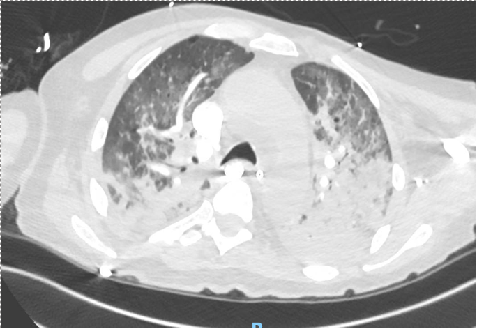
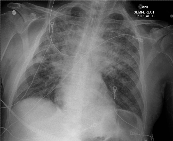
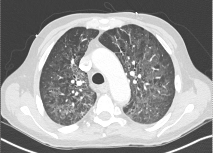
Imaging: CXR and CT showed bilateral upper lobe infiltrates, ground-glass opacities, septal thickening, and peripheral cystic changes.
 

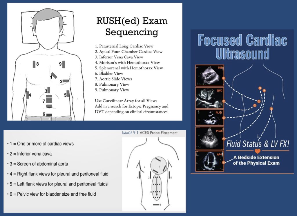
POCUS algorithms for investigating shock

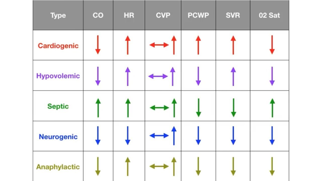
Shock physiology:


Diagnostic Reasoning in Immunocompromised Hosts

  • Framework: Anchor the differential based on type of immunosuppression.
    • HIV/AIDS → T-cell dysfunction, affecting susceptibility to PCP, TB, CMV, fungi (e.g. histo/blasto), and common CAP organisms.
  • PCP considerations:
    • PCP can occur despite prophylaxis (e.g. Bactrim), especially if adherence or resistance issues exist.
    • Classic symptoms in AIDS: acute, febrile, hypoxemic respiratory failure.

Use of Serum Markers and Imaging

  • LDH: Elevated in PCP, but non-specific. High negative predictive value when normal.
  • 1,3-β-D-glucan: Elevated in PCP and other fungal infections. Very sensitive for PCP (up to 95%).
  • Imaging: Ground-glass opacities with cystic changes support PCP diagnosis.

Role of Bronchoscopy and Diagnostic Yield

  • BAL studies to obtain:
    • DFA for PCP (rapid, high specificity, lower sensitivity)
    • PCR for PCP (higher sensitivity, slower turnaround)
    • Cultures: bacterial, fungal, mycobacterial
    • Cytology, galactomannan, histo/blasto urine antigens
  • Bronch Risk-Benefit:
    • Can change management in 40–60% of cases.
    • Complication rate: ~10–15%, most often hypoxemia.
    • Heuristic for pre-bronch ABG on non-rebreather:
      • PaO₂ >150 → likely safe
      • 100–150 → ~25% risk of intubation
      • <100 → high risk of decompensation

Steroids in PCP and Severe CAP

  • Steroids indicated in PCP with significant hypoxemia (PaO₂ <70 mmHg).
  • With new CAP guidelines (Cape Cod trial), steroids may also be considered in severe bacterial CAP.

Shock Evaluation in ICU

  • Framework: Simplify into likely causes — distributive most common, but rule out cardiogenic, obstructive, hypovolemic.
  • Physical exam + POCUS essential early.
    • POCUS: cardiac views, IVC, lung US, abdominal free fluid.
    • Low EF doesn’t exclude distributive shock.
  • PA catheter (Swan) utility:
    • Useful when physiology unclear or when tracking response to therapy is critical.
    • Swan data in this patient: low CVP and wedge, high SVR → distributive shock, not cardiogenic despite low EF.

Radiology Rounds – 1/16/24

We are back with another #RadiologyRounds for 2024 featuring high-yield imaging findings and teaching points for you to review

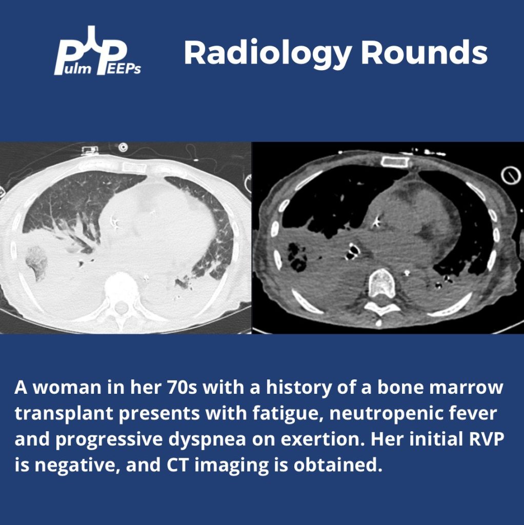
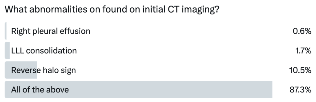
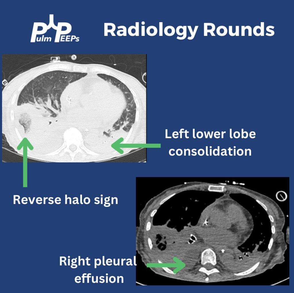
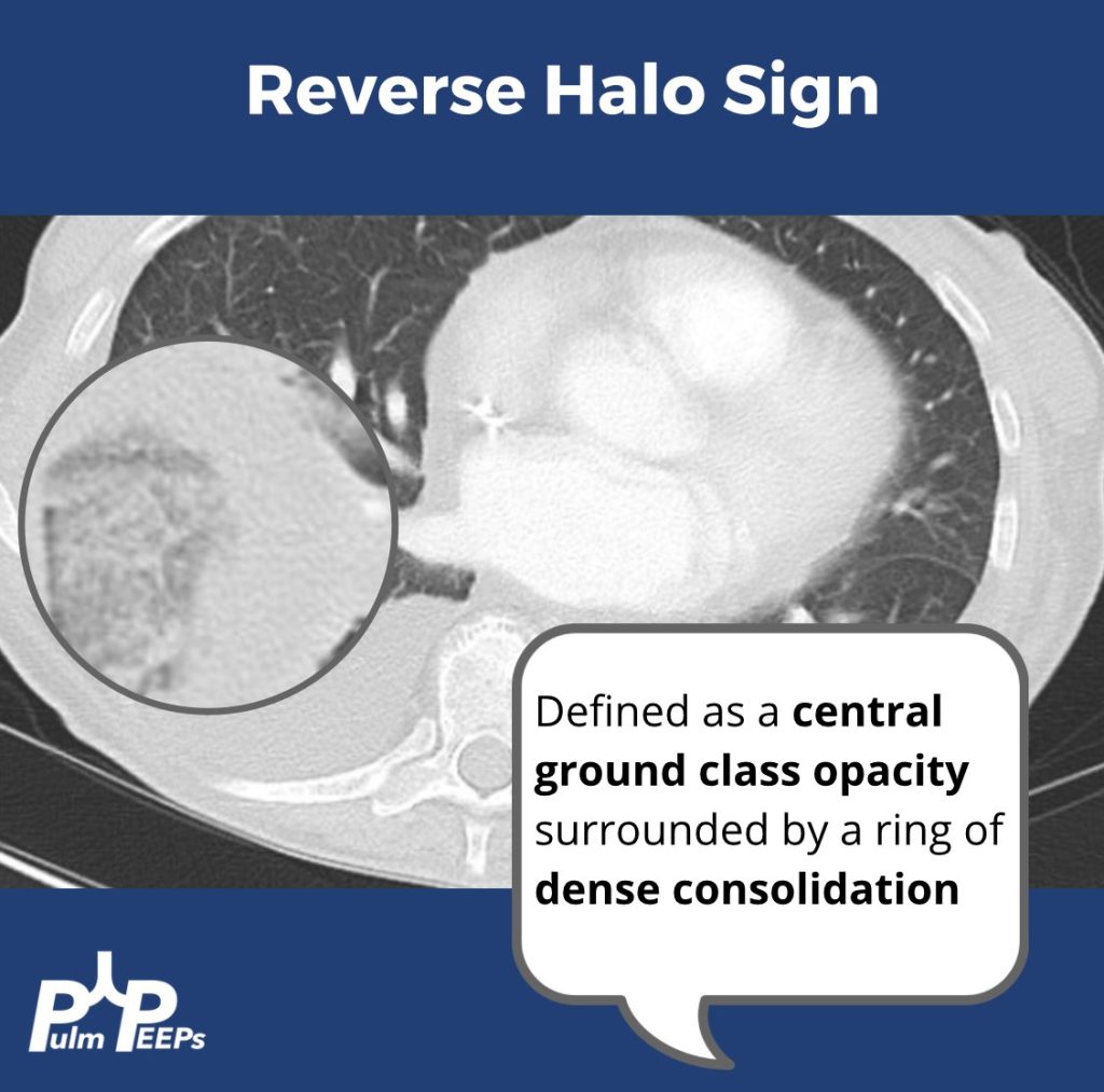
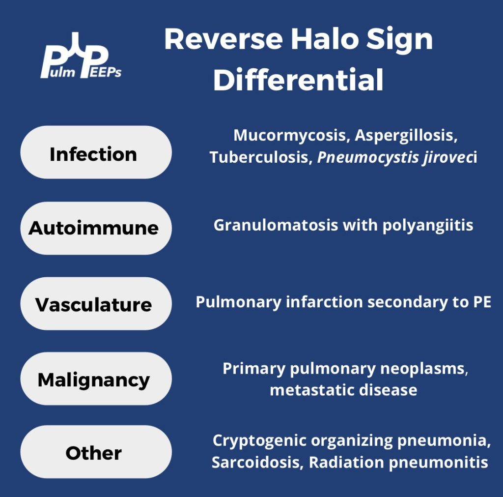
In this immunosuppressed patient, you find a reverse halo sign, right pleural effusion and left lower lobe consolidation.

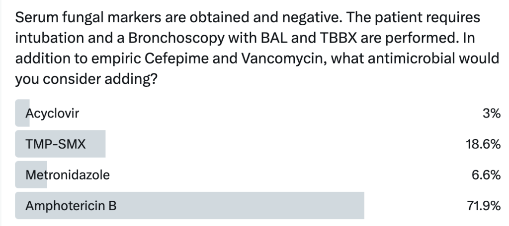
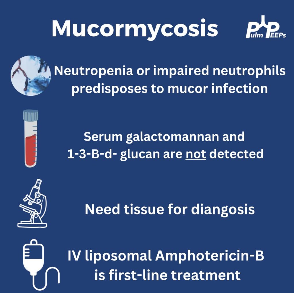
Serum fungal markers are negative but given the reverse halo sign, you empirically start Amphotericin B given concern for pulmonary mucormycosis.

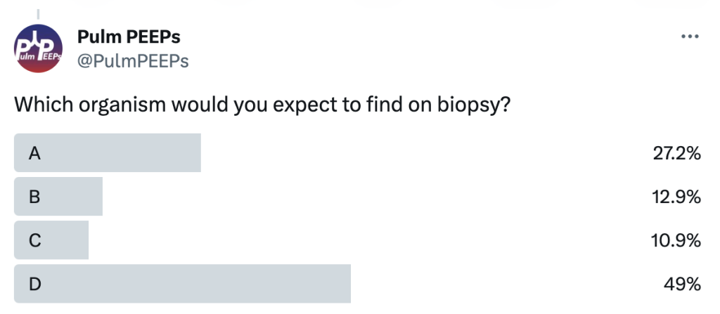
Given the profound neutropenia, the patient was predisposed and mucor was identified on lung tissue biopsy and IV Amphotericin-B was continued.

Radiology Rounds – 11/21/23

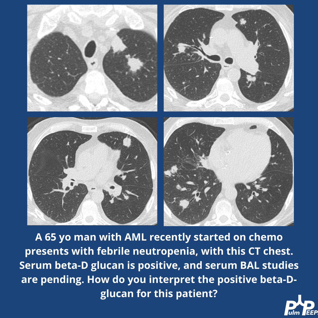
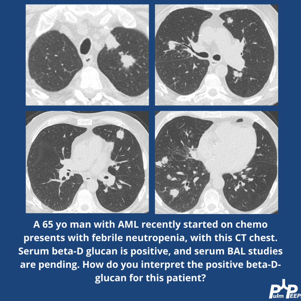
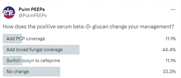
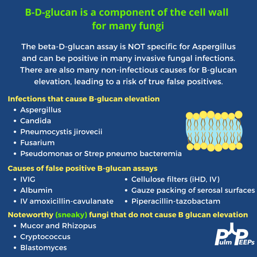
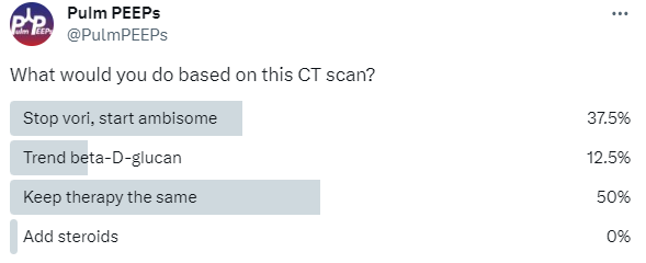
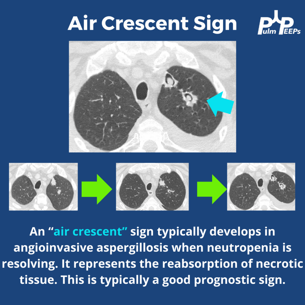
We’re back with another #RadiologyRounds by Pulm PEEPs Associate Editor Tess Litchman. An immunosuppressed 65-year-old man presents with neutropenic fever. He is started on empiric broad-spectrum antibiotics with vancomycin and zosyn. Serum beta-D-glucan is positive.

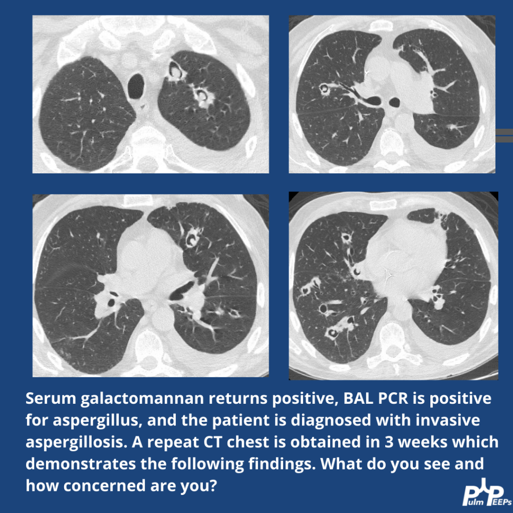
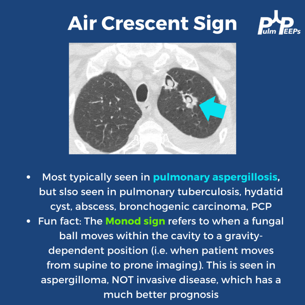
Further, workup reveals a positive serum galactomannan and a BAL PCR that is positive for Aspergillus. The patient is diagnosed with invasive aspergillosis and started on voriconazole. A repeat CT is obtained in 3 weeks. What do you think of the evolving findings?

Radiology Rounds – 11/14/23

We are excited to bring you another #RadiologyRounds

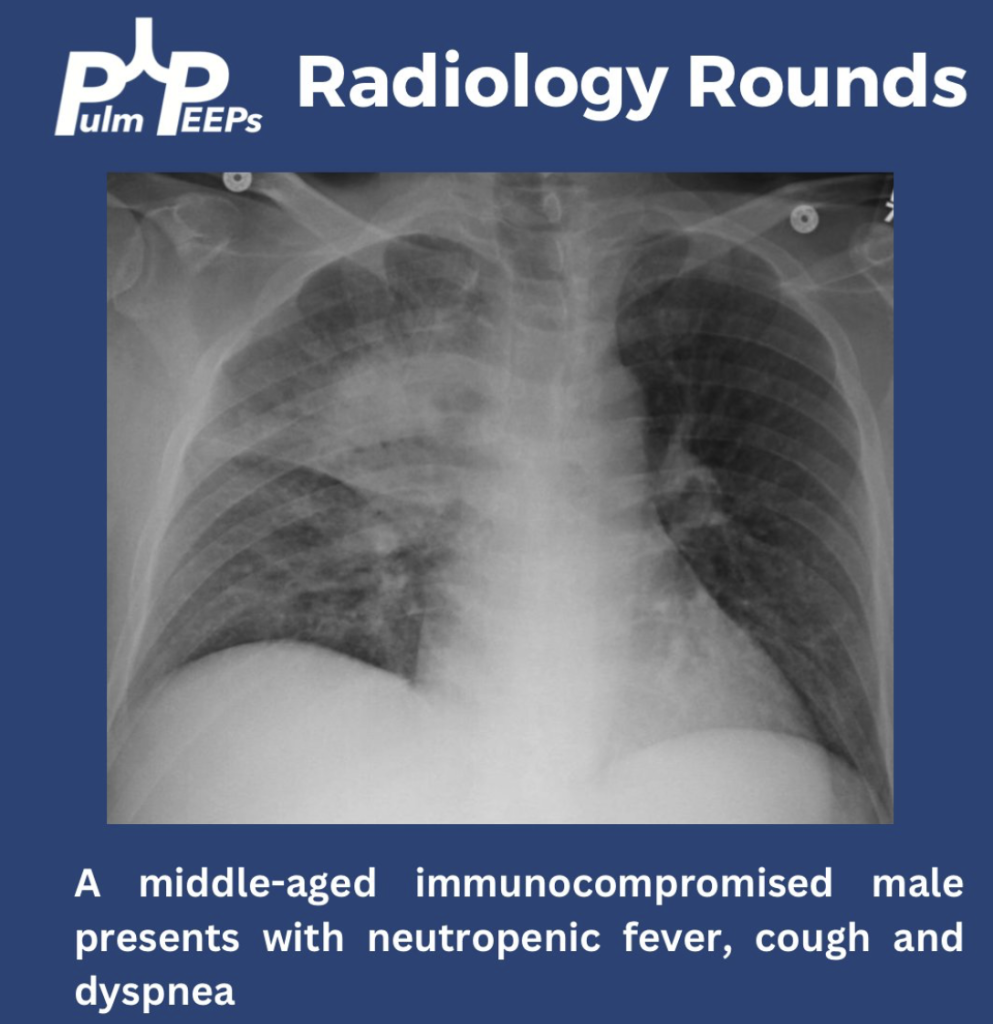
We have a middle-aged immunocompromised male presenting with neutropenic fevers, progressive cough and dyspnea. He has no sick contacts or recent travel.

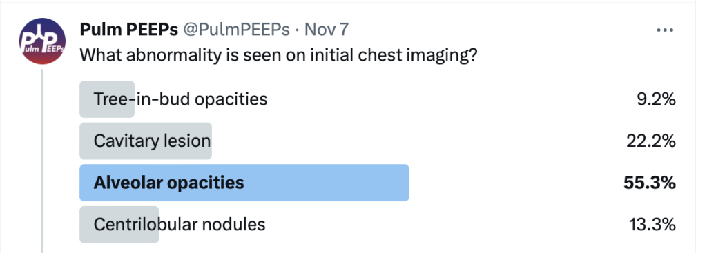
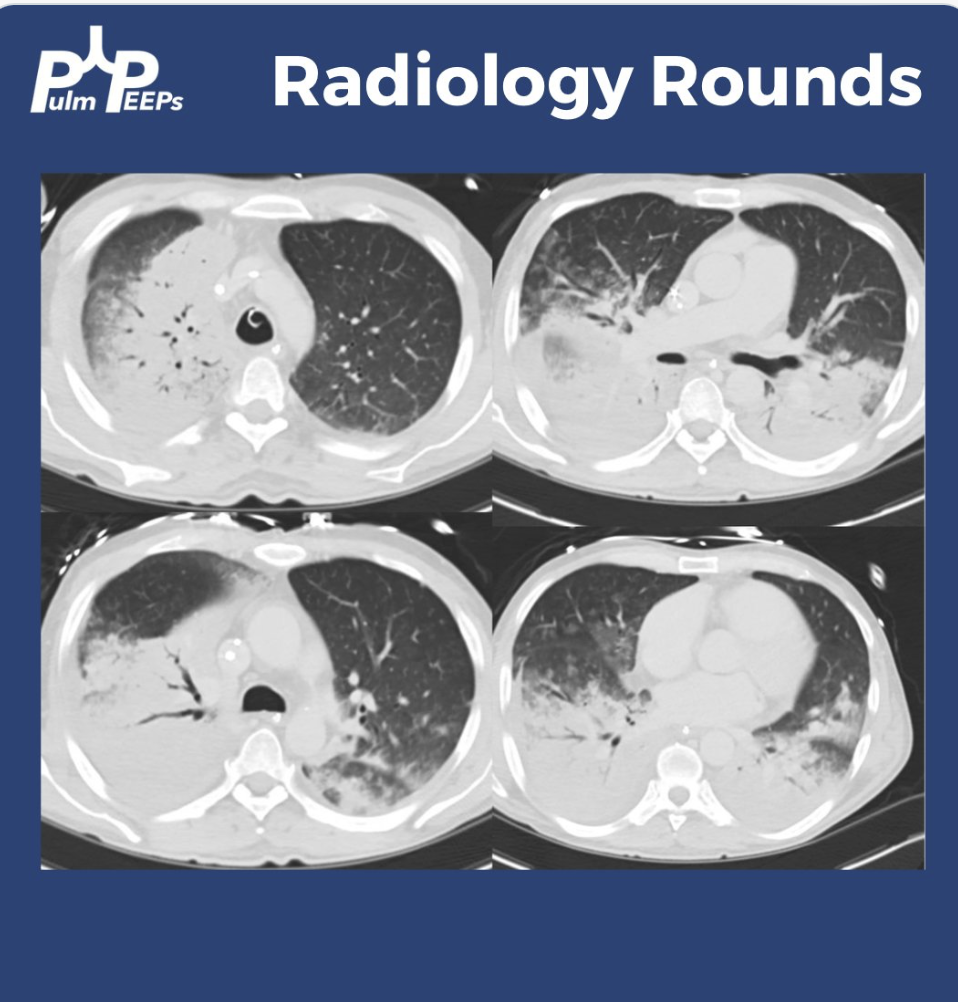
He was found to have primarily right upper alveolar opacities and blunting of the right costophrenic angle. He rapidly decompensated with acute hypoxemic respiratory failure requiring mechanical ventilation. A CT chest showed dense consolidations with air bronchograms.

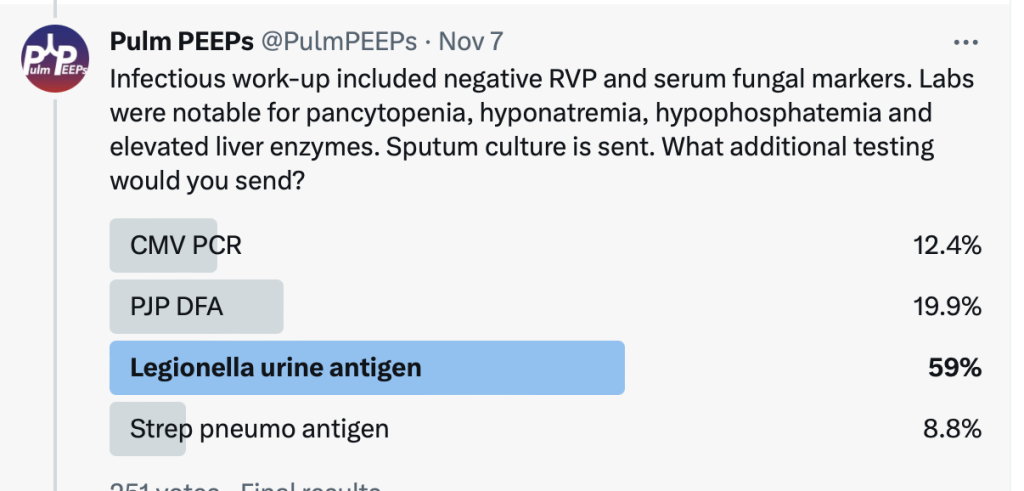
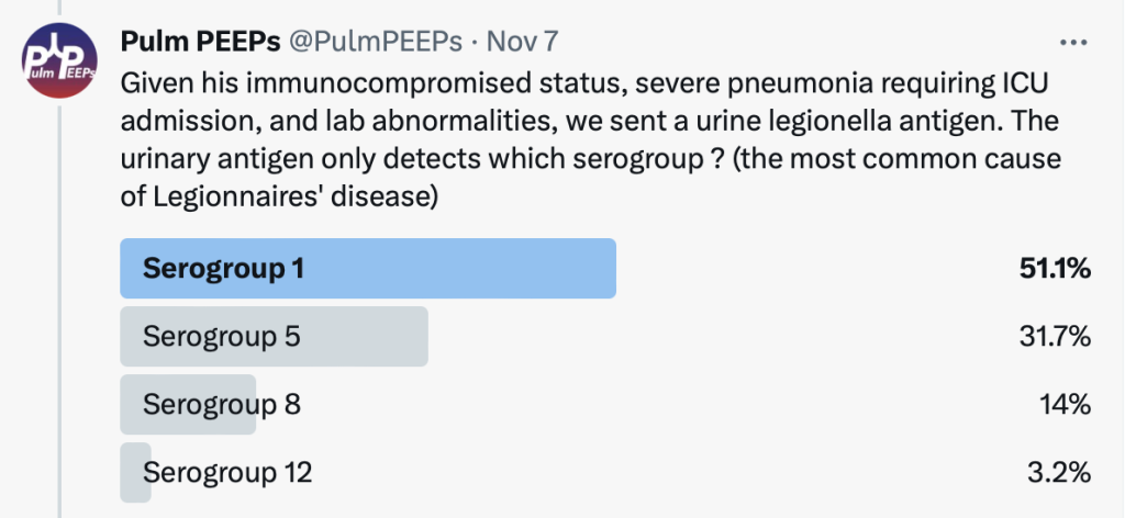
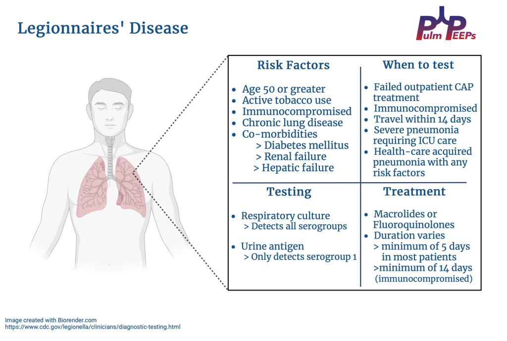
The urinary antigen and sputum culture were positive for Legionella and the patient was continued on Macrolide therapy. See our infographic for high-yield teaching points for Legionnaires’ Disease

52. Fellows’ Case Files: University of Pittsburgh

We’re excited to be back with another Fellows’ Case Files. Today, we’re visiting the University of Pittsburgh to meet a fantastic fellow and a dedicated educator, and to hear about a fascinating case. Let us know if you’ve ever had a similar case, and share your interesting cases with us!
Meet Our Guests

Rachel Wojcik obtained her B.S. in Biology from Mercyhurst University and a Master’s in Liberal Studies from the University of Denver in Global Affairs with a focus on Healthcare. She completed her MD at the University of Colorado before completing her residency and chief resident year at the University of Pittsburgh and has continued her training at Pitt for PCCM fellowship.

Dr. Stephanie Maximous is an Assistant Professor of  Medicine at the University of Pittsburgh School of Medicine and is the Clinical Education APD for the Pulmonary and Critical Care Fellowship program. She completed her fellowship at Pitt in addition to obtaining a Master’s Degree in Medical Education there. She teaches in and directs courses throughout the medical school, residency, and fellowship and was recently awarded the 2023 Outstanding Subspecialty Teaching Attending Award from the housestaff.

Case Presentation

Patient: A 70-year-old male with a history of idiopathic thrombocytopenia on chronic prednisone and a history of tobacco use disorder.

Presentation: Came to the hospital with 2-3 days of right-sided weakness and slurred speech.

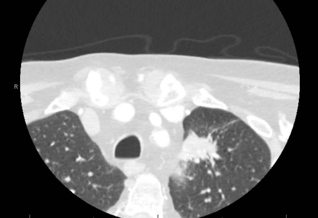
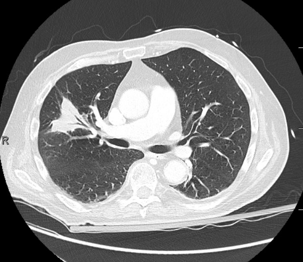
Findings: MRI showed a moderate-sized left pontine stroke. A CT angiogram of the neck showed no evidence of an occlusion, but a spiculated two-centimeter nodule at the apex of the left lung was found.

Additional Information: He requires a walker for mobility and needs help with activities like taking a shower and dressing. He had an unintentional 20-pound weight loss over six months, increased fatigue, and malaise.

Previous Investigations: A chest x-ray ordered two months prior by his hematologist was unremarkable, and a CT of the abdomen and pelvis showed no masses.

Key Learning Points

Bronchoscopy in Decision Making:

  • The decision to perform bronchoscopy in patients depends on a myriad of factors, including the location of any lesions, accessibility, potential risks, and the potential diagnostic yield.
  • Fiber optic bronchoscopy with BAL can rule out infections, and if no diagnosis is reached, more invasive methods like surgical biopsy might be necessary.
  • Consider the location of consolidated masses; navigational bronchoscopy might be needed for lesions without a clear airway leading into them.

Nocardia Insights:

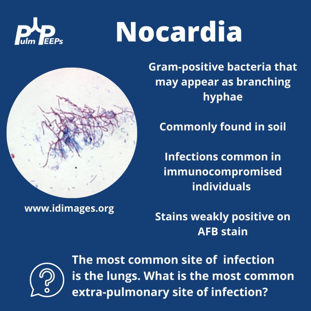
  • Nocardia is a gram-positive bacterium that stains weakly acid-fast.
  • It can be found in soil and certain water sources and can infect through the skin or by inhalation.
  • Two-thirds of patients with Nocardia are immunocompromised.
  • The dosage of Bactrim given for PJP prophylaxis doesn’t prevent Nocardia infections in immunocompromised individuals.
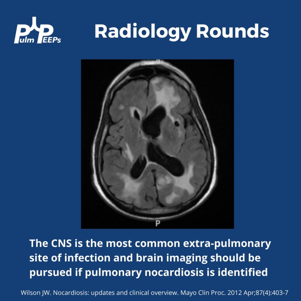
  • While the lungs are the most common infection site, Nocardia can manifest elsewhere, like the skin or CNS.

Treatment Approach:

  • Bactrim is the mainstay of treatment for Nocardia. If someone is allergic, desensitizing them can be crucial.
  • IV induction phases vary in length depending on the severity of the disease.
  • The overall treatment duration is protracted to prevent relapse.

Takeaway Points:

  • Bactrim for PJP prophylaxis doesn’t necessarily prevent Nocardia infections in immunocompromised individuals.
  • If someone is allergic to Bactrim, consider desensitizing them due to its importance in treating Nocardia.

References and Further Reading:

Menéndez R, Cordero PJ, Santos M, Gobernado M, Marco V. Pulmonary infection with Nocardia species: a report of 10 cases and review. Eur Respir J. 1997 Jul;10(7):1542-6. doi: 10.1183/09031936.97.10071542. PMID: 9230244.

Zia K, Nafees T, Faizan M, Salam O, Asad SI, Khan YA, Altaf A. Ten Year Review of Pulmonary Nocardiosis: A Series of 55 Cases. Cureus. 2019 May 26;11(5):e4759. doi: 10.7759/cureus.4759. PMID: 31363440; PMCID: PMC6663111.

Radiology Rounds – 7/18/23

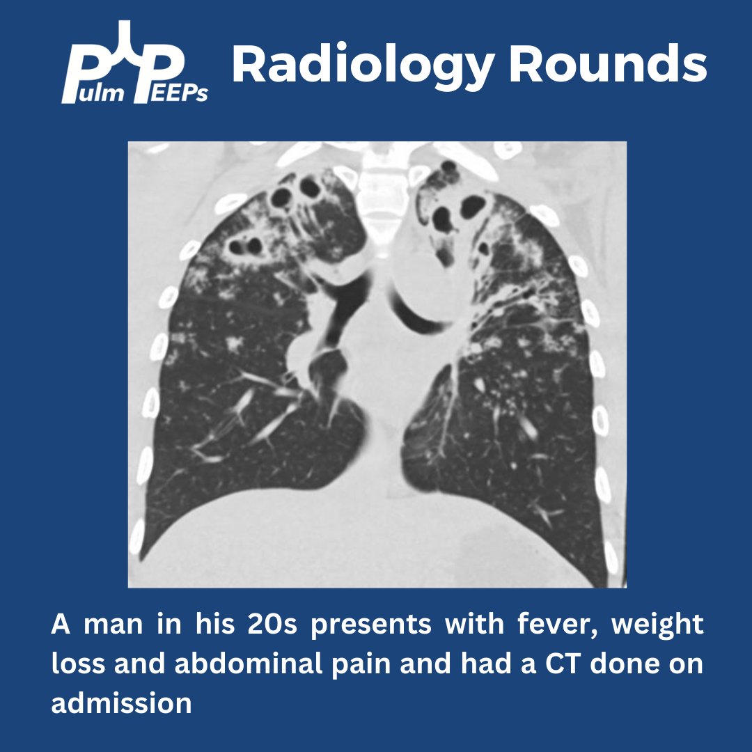
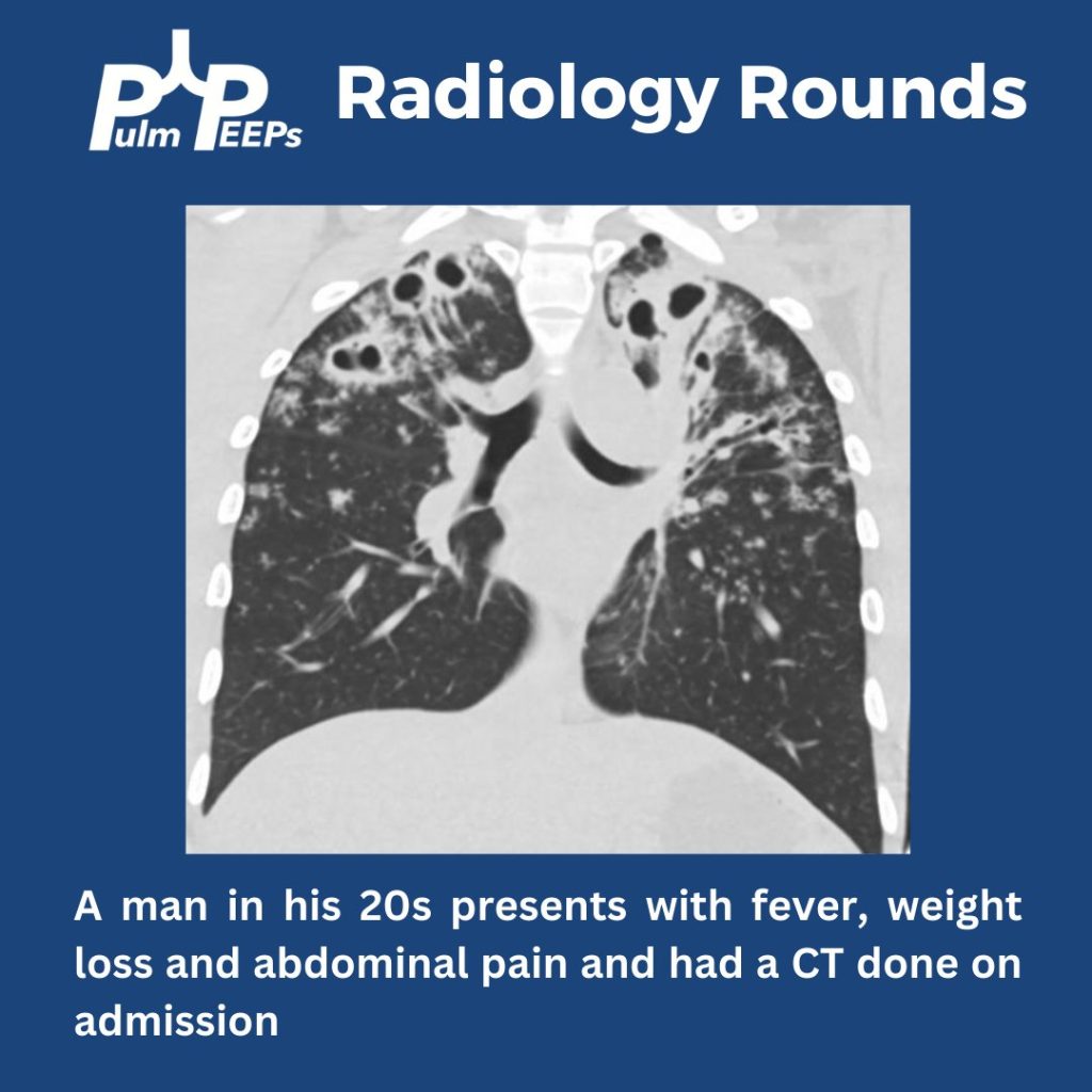
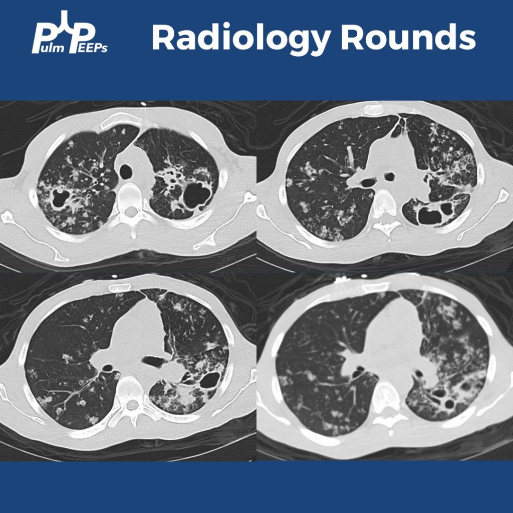
We are back with our first #RadiologyRounds of the new academic year. We have a young, immunocompetent man presenting with fever, weight loss, and abdominal pain.

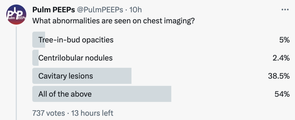
What abnormalities are seen on his chest imaging?

He was found to have bilateral apical cavitary disease, centrilobular nodules, and tree-in-bud opacities. He developed a productive cough with blood-tinged sputum as well as diarrhea.

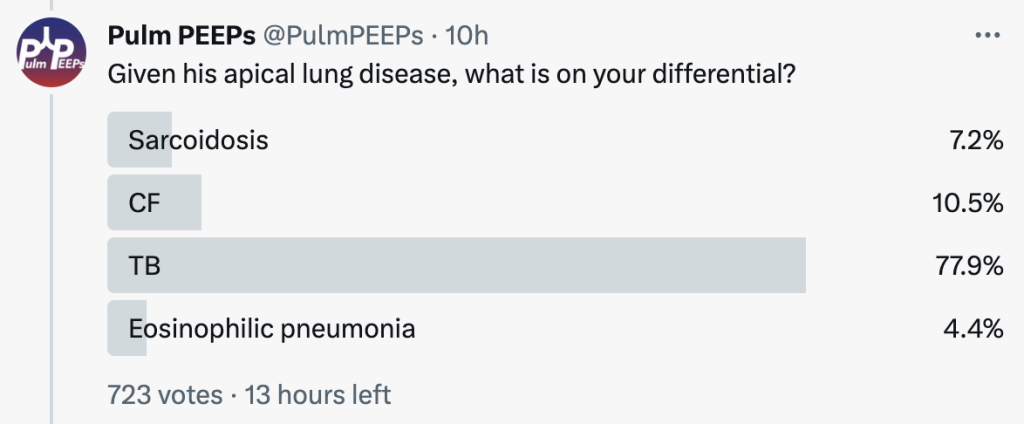
Given his apical lung disease, what is on your differential?

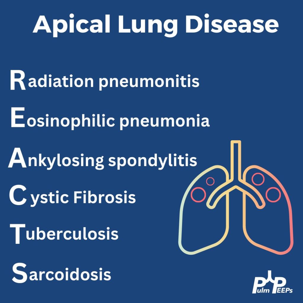
When thinking about apical lung disease, remember the mnemonic REACTS to help with your differential.

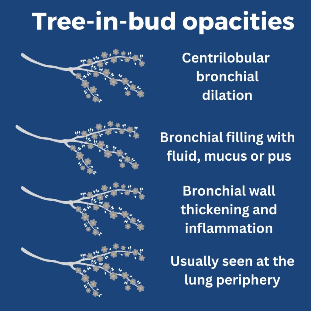
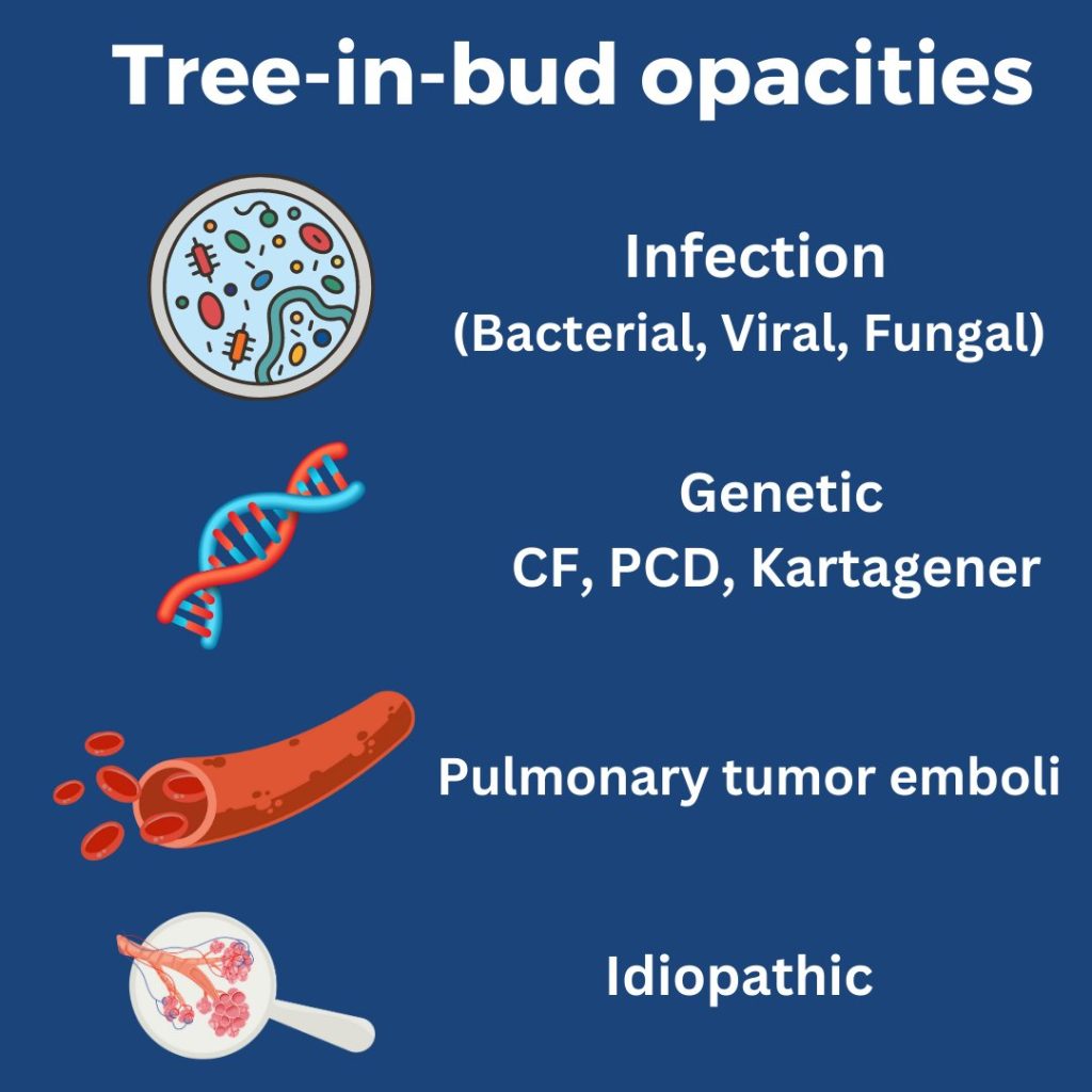
What are tree-in-bud opacities? They are findings seen on CT chest suggesting bronchial dilation, inflammation, and bronchial filling with fluid, mucus, or pus that can be caused by infections and non-infectious etiologies.

He had sputum and AFB cultures sent and his AFB smear was positive. He was ultimately diagnosed with disseminated TB and started on RIPE therapy.

39. Fellows’ Case Files: Indiana University

Join us as we head to Indiana University! Listen in as we discuss another great case and hear teaching points from our amazing guests.

Meet our Guests


Parth Savsani is currently an internal medicine resident at Indiana University School of Medicine. He received his undergraduate degree from the University of Wisconsin-Madison and his medical degree from the University of Illinois College of Medicine. He enjoys medical education and was selected to be the VA chief resident next year.

Maria Srour is a Pulmonary and Critical Care Fellow at Indiana University School of Medicine. She completed her internal medicine residency at Saint Louis University where she was also a chief resident, and received her medical degree from IU. She works in global health to improve care for sepsis patients in low resources settings, and is currently pursuing her MPH.

Laura Hinkle is a Indiana University die hard and has been there from her since medical school through residency and fellowship, and is now an Assistant Professor of Clinical  Medicine and the Associate Program Director for the Pulmonary and Critical Care Medicine Fellowship. She will be taking over as the Program Director July 1, 2023. She is a dedicated educator and is the Key Clinical Educator for Pulmonary and Critical Care, and the Director of the Clinical Transitions Curriculum.  Additionally, she is working on a Master’s Degree in Education through the University of Cincinnati. 

Case Presentation

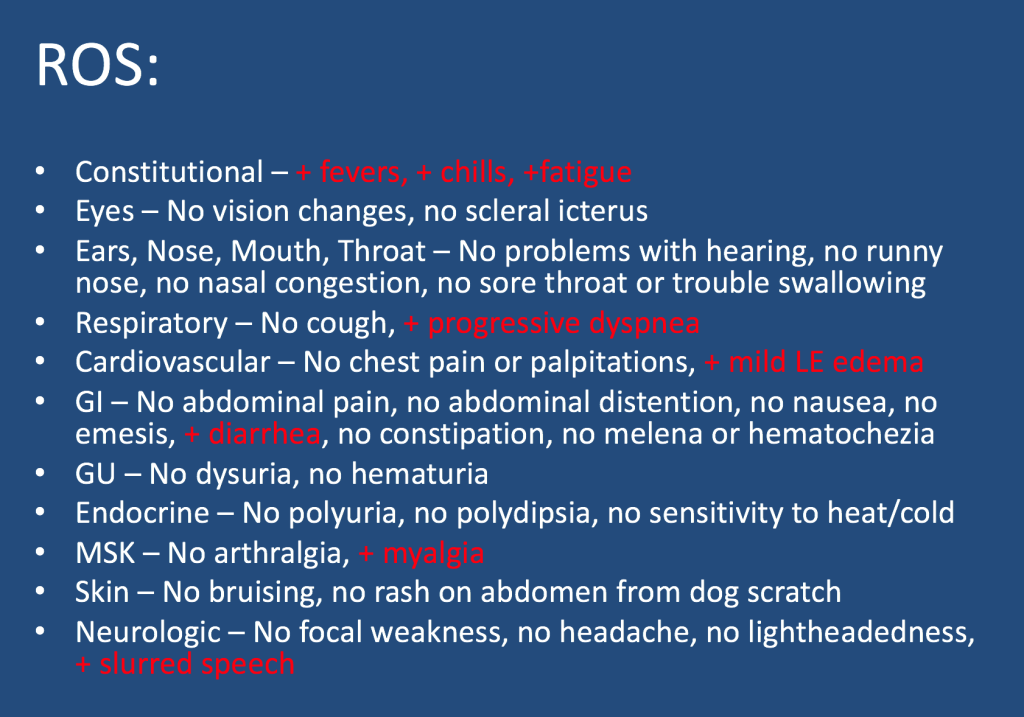
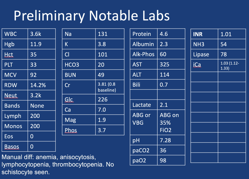
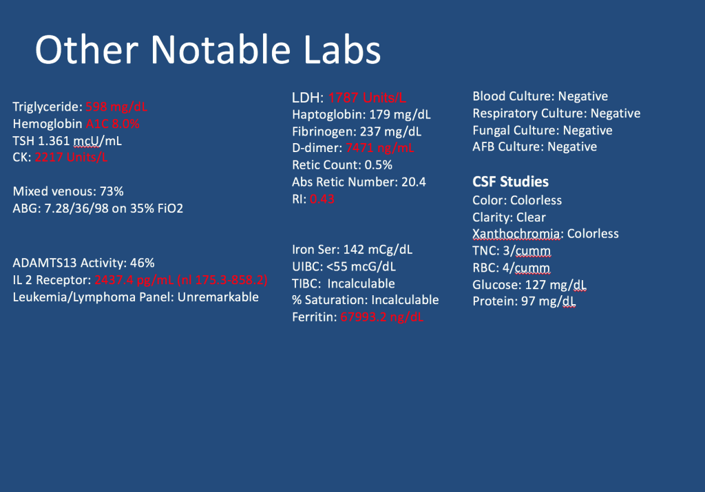
A male in her early 60s is transferred from a neighboring facility with a 1 week history of fatigue and lethargy. Three days prior to presentation he developed dyspnea and increased weakness with a near fall at home. HIs family also reported recent fevers, chills, dyspnea, and diarrhea. On his way to seek evaluation, he developed slurred speech without any other focal abnormalities.

Additional information is summarized as below:

Follow along our episode to hear the final diagnosis and key teaching points from the case!

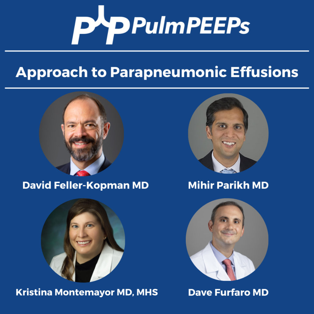
37. Top Consults: Approach to Parapneumonic Effusions

We continue our Top Consult Series on Pleural Disease and bring you a dedicated episode on Parapneumonic effusions. We are joined by two guest experts, Dr. David Feller-Kopman and Dr. Mihir Parikh. Listen in as we discuss the spectrum of parapneumonic effusions, including simple parapneumonic effusions, complicated parapneumonic effusions, and empyema. You will hear what to look for on imaging, what tests to send with pleural drainage as well as discuss the need for surgical consultation.

Meet our Guests

Dr. Mihir Parikh is currently an Assistant Professor of Medicine and academic interventional pulmonologist at Beth Israel Deaconess Medical Center. He is a highly esteemed educator and has worked to incorporate simulation training to improve procedural training for trainees and is a master of pleural disease.

Dr. David Feller-Kopman is a Professor of Medicine and the Section Chief of Pulmonary and Critical Care Medicine at Darmouth whose clinical and research expertise span the field of interventional pulmonology. Dr. Feller-Kopman is a true master of pleural disease, and has authored more than 225 peer-reviewed manuscripts and has been a leader for both ATS and CHEST committees.

Radiology Rounds – 3/21/2023

We are back with another #RadiologyRounds! This week’s case comes from our Associate Editor @luke_hedrick

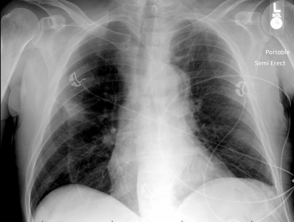
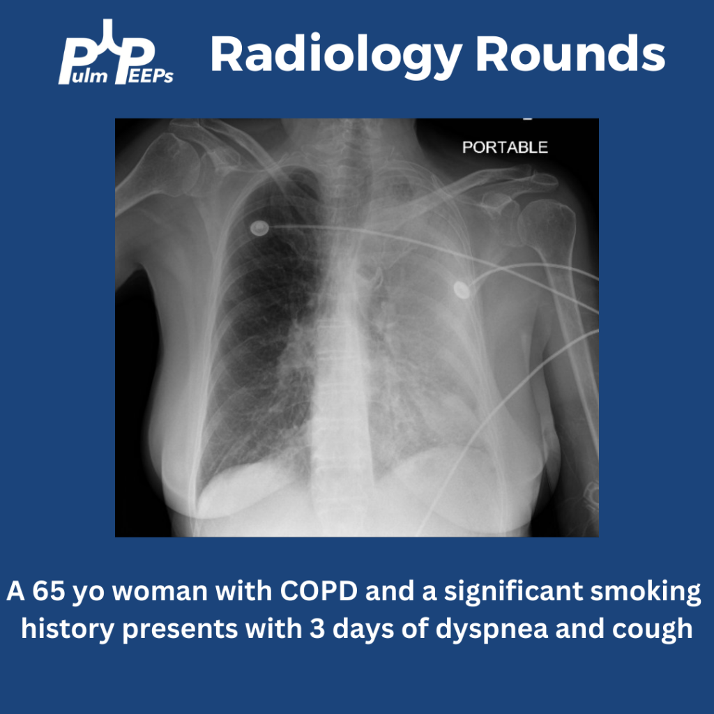
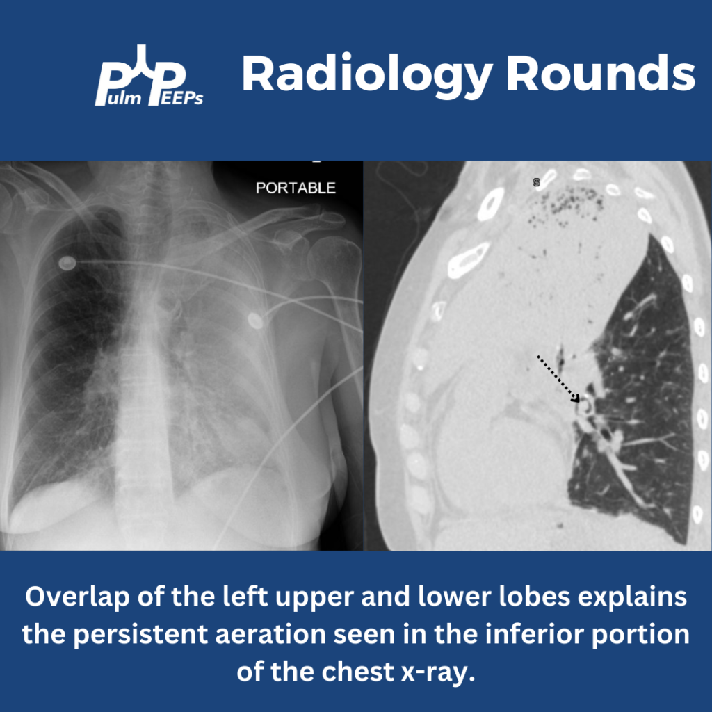
A portable film is obtained on a 65 yo woman with COPD presenting with progressive dyspnea and cough.

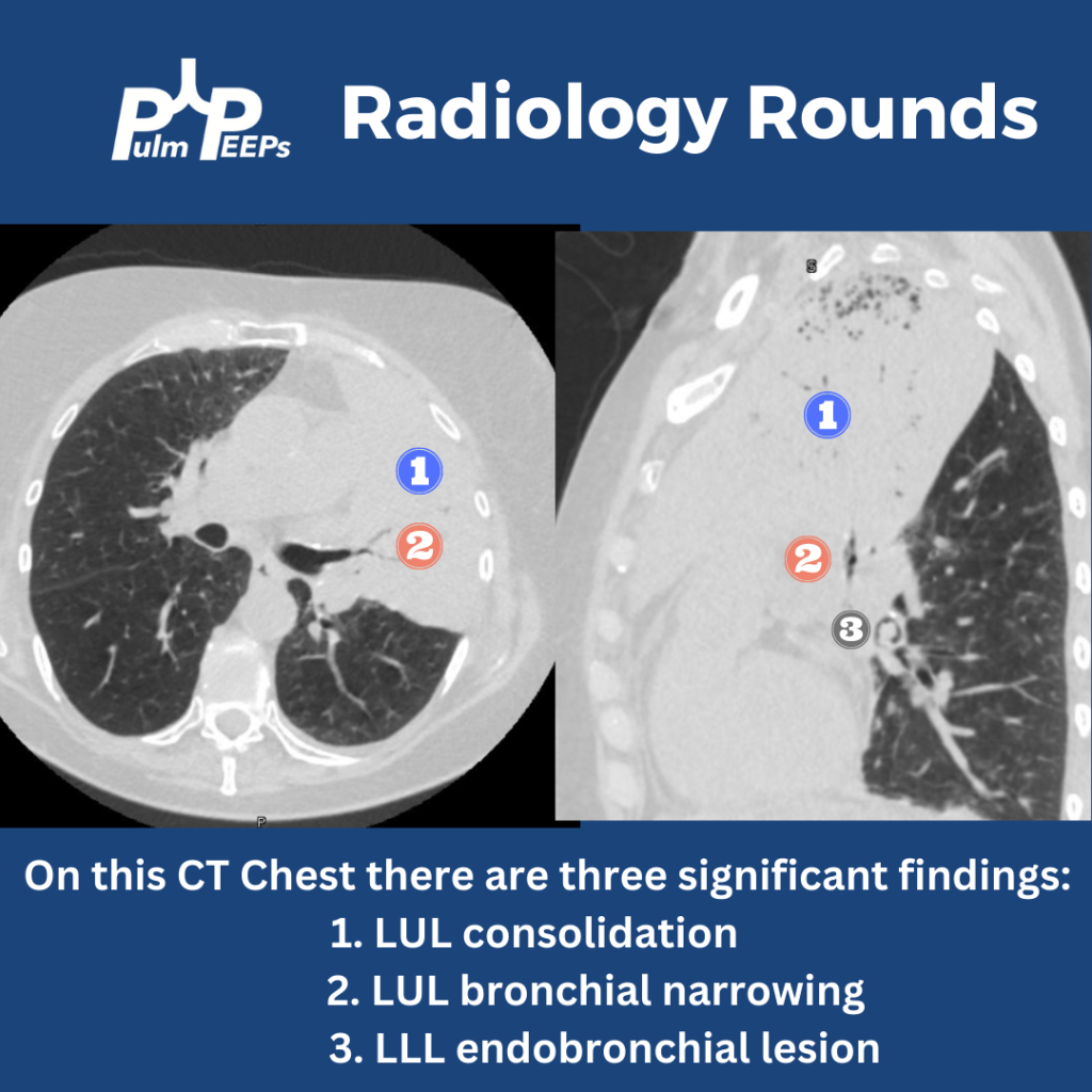
The image shows near complete opacification of the left lung. There is no ipsilateral or contralateral tracheal deviation which you would expect with atelectasis or a large pleural effusion, respectively. A CT chest is obtained to better visualize the parenchyma.

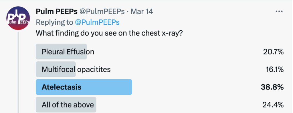
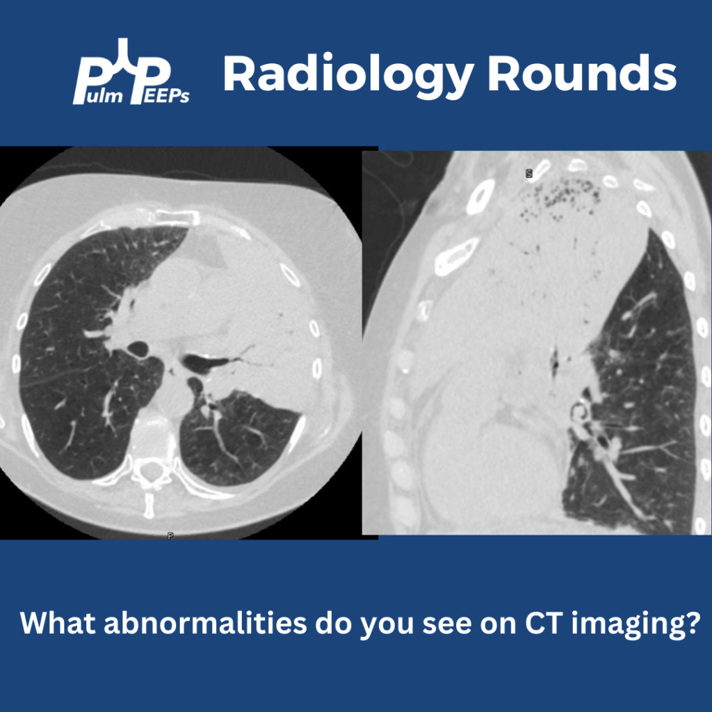
The CT chest shows all three findings as noted below.

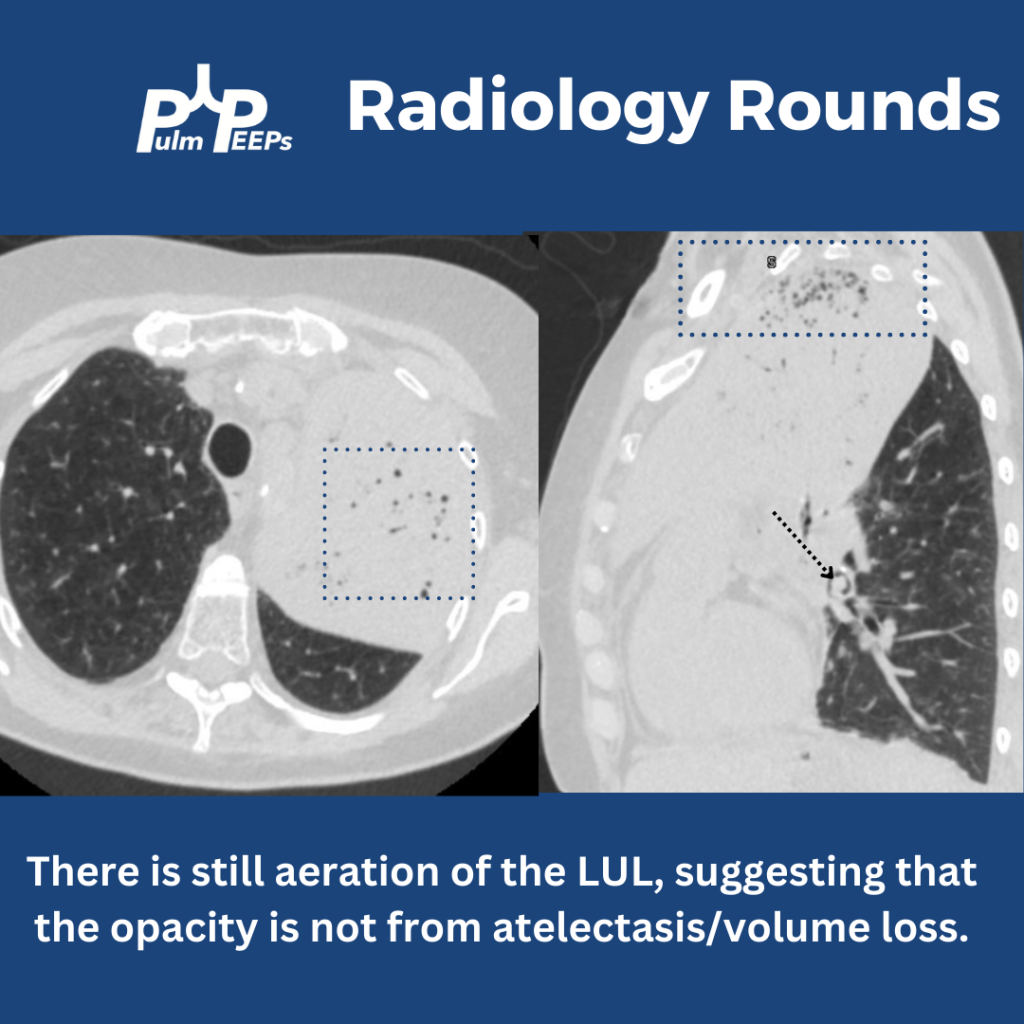
Yes! There is still aeration in some of the upper lobe, which would not be the case if this were caused by profound atelectasis. Also, atelectasis of such a large territory of the lung would usually cause traction on surrounding structures.

Having seen the CT, can you explain why the x-ray showed aeration in the inferior, left hemithorax? This is from the overlap of the left upper and lower lobes when viewed anteriorly. The sagittal view of the CT demonstrates this nicely.