107. Fellows’ Case Files: University of Kansas Medical Center KUMC

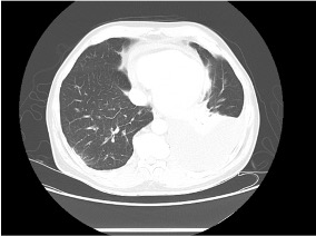
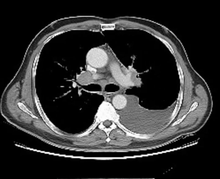
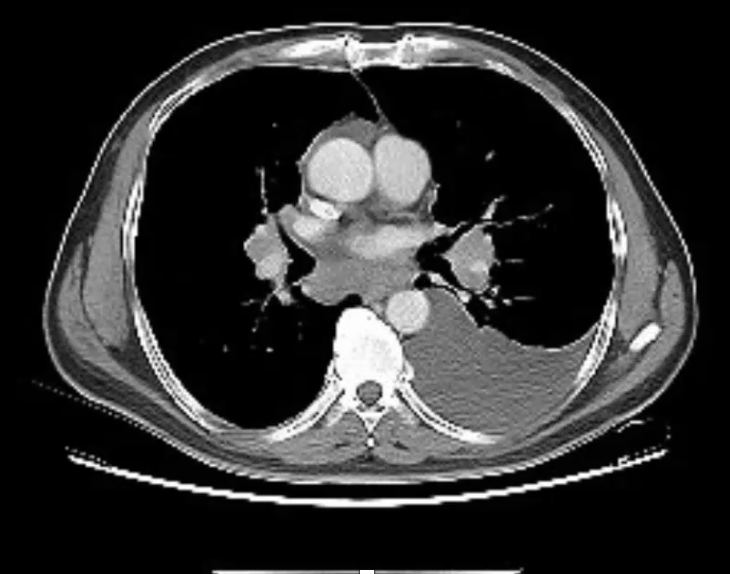
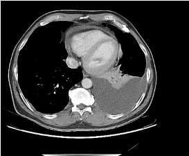
After a brief hiatus, we are excited to be back today with another Fellows’ Case Files! Today we’re virtually visiting the University of Kansas Medical Center (KUMC) to hear about a fascinating pulmonary presentation. There are some fantastic case images and key learning points. Take a listen and see if you can make the diagnosis along with us. As always, let us know your thoughts and definitely reach out if you have an interesting case you’d like to share.
Dr. Vishwajit Hegde completed his internal medicine residency at University of Kansas Medical Center where he stayed for fellowship and is currently a second year Pulmonary and Critical Care medicine fellow. 

Dr. Sahil Pandya is an Associate Professor of Medicine and Program Director of the PCCM Fellowship at KUMC.

1) Initial frame & diagnostic mindset

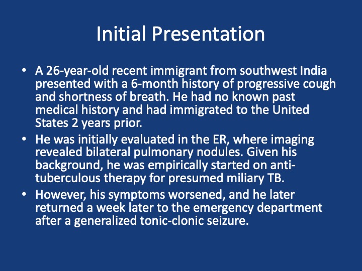
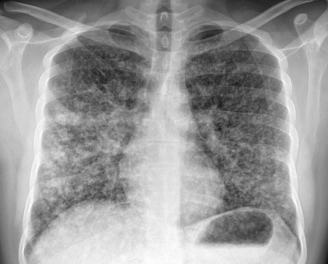
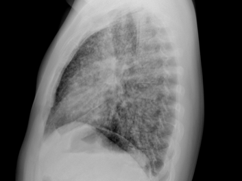
  • Young (26), subacute → chronic dyspnea/cough with diffuse pulmonary nodules; avoid premature closure on TB.
  • Use a Bayesian approach: combine pre-test probability (epidemiology, exposures, tempo) with targeted tests to decide next steps.
  • Always confirm TB when possible (micro/path + resistance testing); empiric RIPE may be reasonable but shouldn’t replace tissue when stakes are high.

2) Imaging pearls—nodular pattern recognition

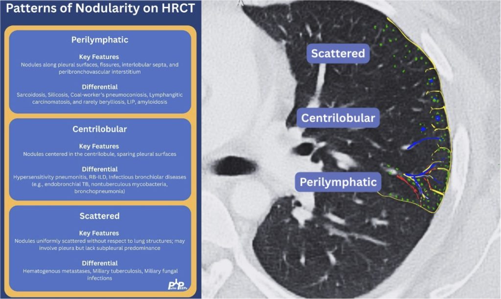
  • Ask three things: craniocaudal distribution, symmetry, central vs peripheral.
  • Centrilobular (spares pleura/fissures): airway-centered (e.g., NTM, bronchiolitis, tree-in-bud).
  • Perilymphatic (tracks fissures/pleura & septa): sarcoid, lymphangitic spread.
  • Random/diffuse (involves pleural surfaces): hematogenous spread → think miliary TB, disseminated fungal, septic emboli, metastatic disease.
  • Interval change matters: new cavitation and confluence can upweight infection or aggressive malignancy.

3) Neuro findings—ring-enhancing lesions

  • Differential: septic emboli/abscess, nocardia, fungal, TB, parasites, metastases, vasculitis, sarcoid.
  • Partner with neuroradiology for pattern nuances; treat seizures but keep searching for the unifying diagnosis.

4) Lab/serology strategy

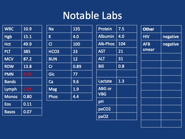
  • Broad infectious workup (AFB × multiple, fungal serologies), HIV and basic immune screen.
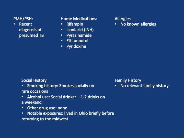
  • Negative/indeterminate tests don’t end the search—revisit history (e.g., Ohio travel → histo/blasto risk).

5) “Tissue is the issue”—choosing the procedure

  • For diffuse nodules with mediastinal adenopathy and stable patient: EBUS-TBNA + BAL, consider transbronchial or cryobiopsy.
  • Cryobiopsy pros: larger, less crush artifact, better for molecular testing; cons: ↑ bleeding/pneumothorax vs forceps.
  • VATS still best for certain ILD questions or if less invasive routes are non-diagnostic—but weigh patient preference and stage/likelihood of yield.
6) ROSE (rapid on-site evaluation) in bronchoscopy

  • Confirms adequacy in real time, steers you away from necrotic zones, helps decide when you’ve got enough for molecular studies, and when to pivot sites—reduces anesthesia time and repeat procedures.

7) Final diagnosis & management

  • Path: TTF-1+, CK7+, napsin A → pulmonary adenocarcinoma with a fusion driver.
  • Therapy: Targeted TKI (crizotinib) → dramatic radiographic response of miliary lung disease and CNS lesions.
  • Teaching point: even “miliary TB-like” lungs + CNS lesions in a 20-something can be driver-positive lung cancer—don’t let age or pattern blind you.

  1. Desai, S., Devaraj, A., Lynch, D., & Sverzellati, N. (2020). Webb, Müller and Naidich’s high-resolution CT of the lung (6th ed.). Lippincott Williams & Wilkins.
  2. Rajeswaran, G., Becker, J. L., Michailidis, C., Pozniak, A. L., & Padley, S. P. G. (2006). The radiology of IRIS (immune reconstitution inflammatory syndrome) in patients with mycobacterial tuberculosis and HIV co-infection: appearances in 11 patients. Clinical radiology, 61(10), 833-843
  3. Poletti, V., Ravaglia, C., & Tomassetti, S. (2016). Transbronchial  cryobiopsy in diffuse parenchymal lung diseases. Current opinion in pulmonary medicine, 22(3), 289-296.
  4. Norman, G. R., Monteiro, S. D., Sherbino, J., Ilgen, J. S., Schmidt, H. G., & Mamede, S. (2017). The causes of errors in clinical reasoning: cognitive biases, knowledge deficits, and dual process thinking. Academic Medicine, 92(1), 23-30.

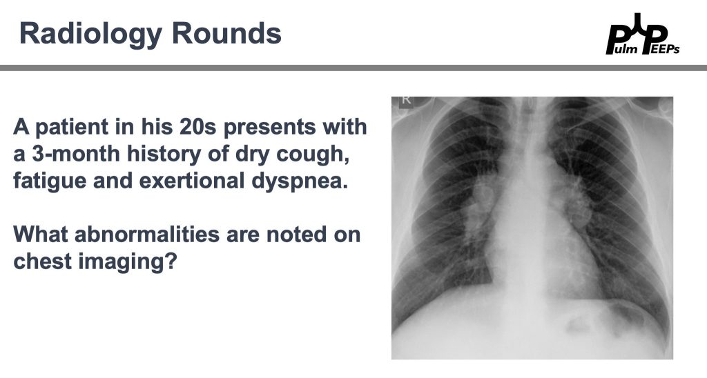
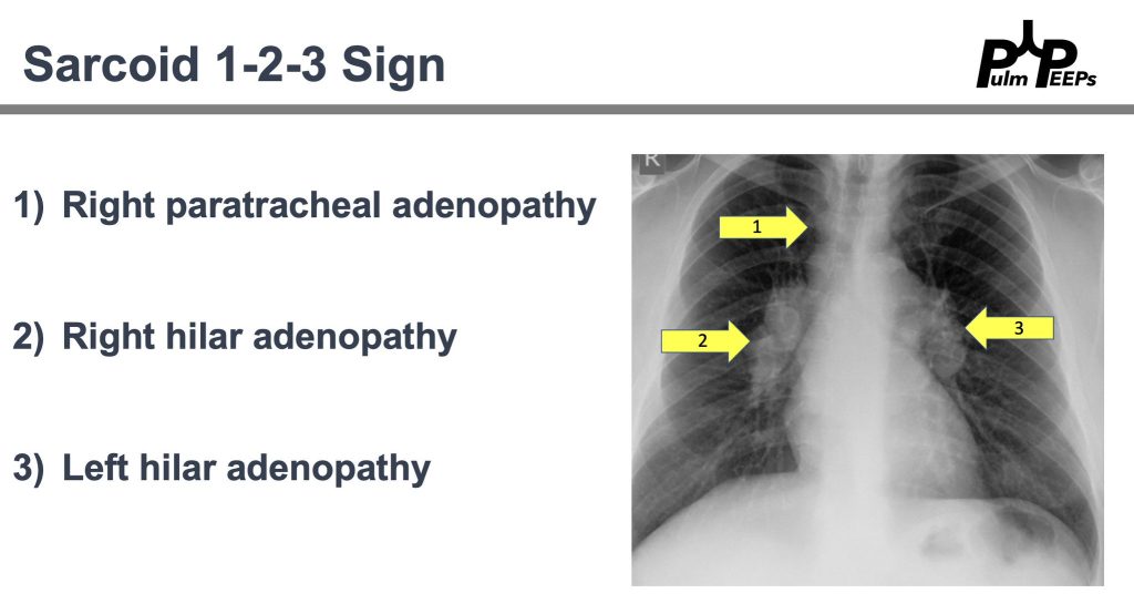
Radiology Rounds 52: Sarcoid 1-2-3 Sign

We’re back with another Pulm PEEPs Radiology Rounds!

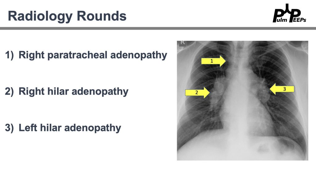
That’s right! All of the findings are present

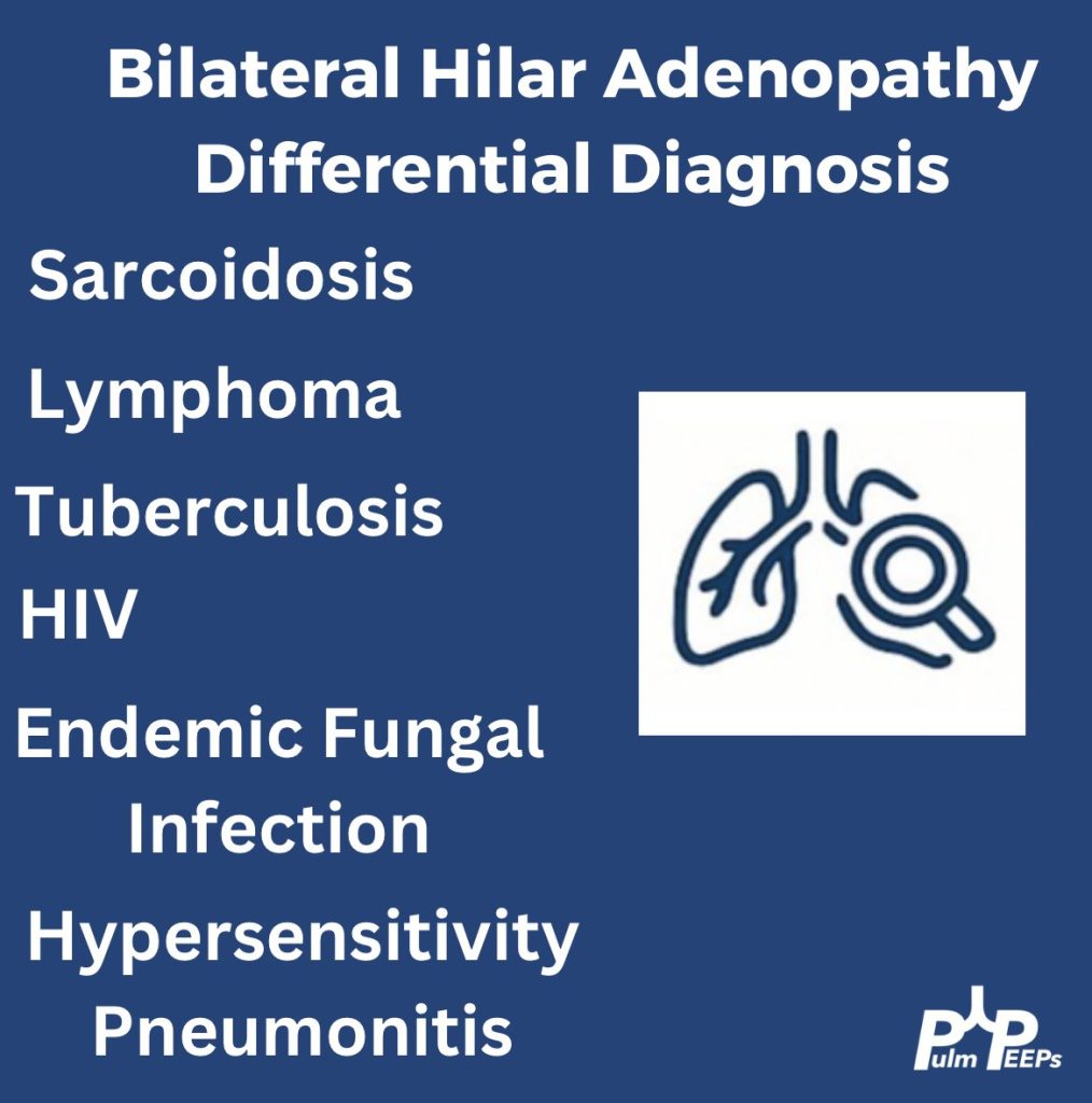
History and physical exam are essential. Here are a few can’t miss diagnoses when working up a young adult with bilateral hilar adenopathy.

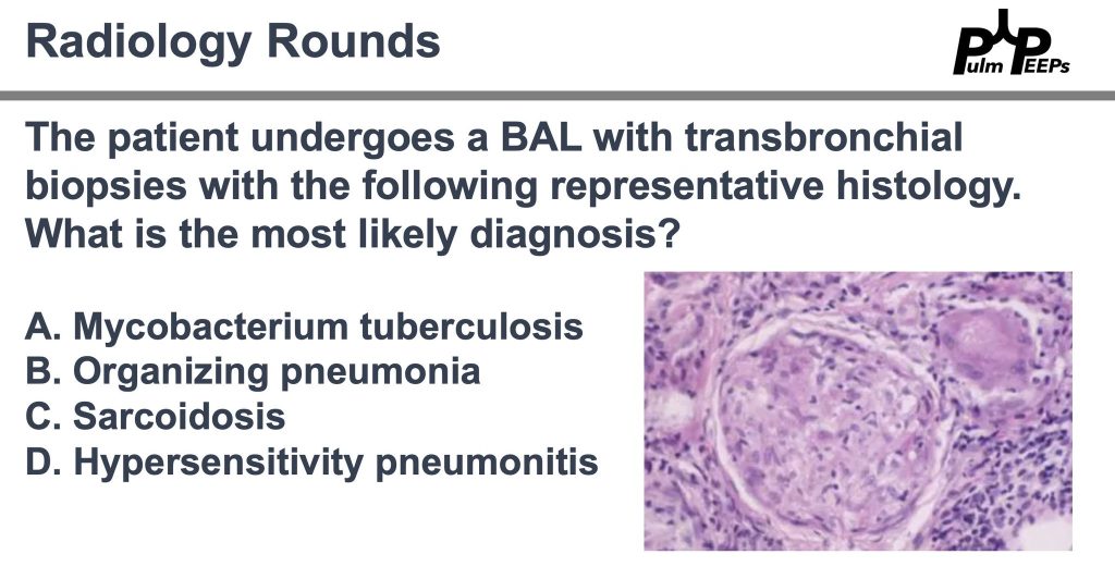
Pathology is consistent with a non-caseating granuloma. What is the most likely diagnosis?

Imaging and pathology were consistent with sarcoidosis and after negative work-up for alternative causes. The patient will follow-up with outpatient pulmonary for sarcoidosis management. Don’t forget about the Sarcoid 1-2-3 sign!

99. Fellows’ Case Files: Rutgers – Robert Wood Johnson Medical School

We’re back with another edition of Fellows’ Case Files! Today, we’re virtually visiting Rutgers University, Robert Wood Johnson Medical School to work through a fascinating pulmonary case. Enjoy, and let us know your thoughts.

Khalil El Gharib completed his residency training at Northwell at Staten Island University Hospital Program and is currently a first year fellow at Rutgers Robert Wood Johnson Medical School.

Sabiha Hussain completed her residency training at Robert Wood Johnson Medical School and her fellowship training at Columbia Presbyterian Medical Center in New York. She is currently a Professor of Medicine and the fellowship Program Director.

  • Patient: 28-year-old male with Asperger’s syndrome and IgA nephropathy.
  • Symptoms: 3-month history of progressive dry cough and dyspnea on exertion; later developed mild hemoptysis.
  • Notable exposure: Questionable black mold in the patient’s apartment.

Initial Workup and Diagnostic Reasoning

  • Vital signs: Hypoxemia (SpO₂ 91% on room air).
  • Exam: Inspiratory crackles.
  • ABG findings: Elevated A–a gradient (~50), indicating a gas exchange problem.
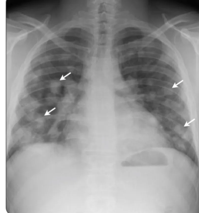
  • Chest X-ray: Bilateral, patchy infiltrates without specific lobar preference.
  • Initial management: Discharged with empiric antibiotics for presumed multifocal pneumonia.

Re-Presentation and Further Testing

  • Symptoms worsened; now with blood-tinged sputum.
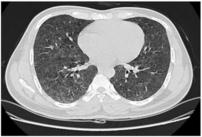
  • Chest CT: Showed diffuse ground-glass opacities (GGOs) without fibrosis, consolidation, or lymphadenopathy.

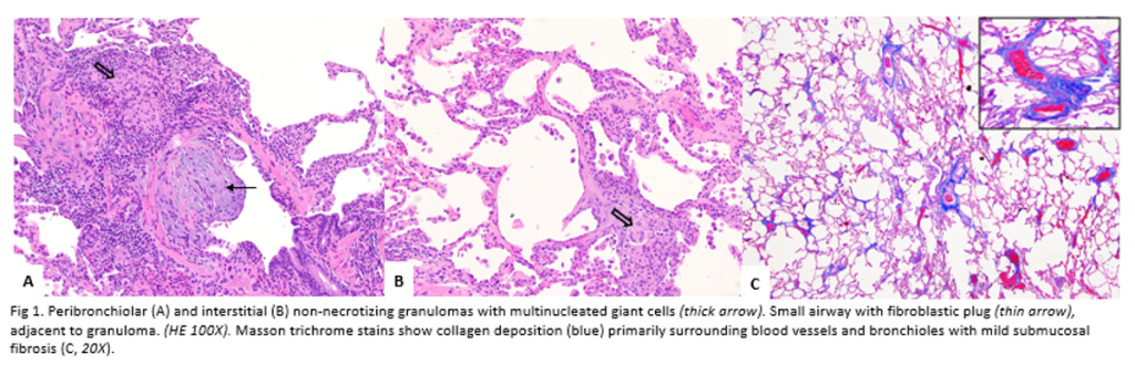
Pathology images a courtesy to Dr Isago Jerrett, pathology resident at RWJMS

Diagnostic Framework for Hypersensitivity Pneumonitis (HP)

  • New classification: Based on fibrotic vs. non-fibrotic phenotype (not acute/chronic).
  • CT features of HP:
    • GGOs with lobular air trapping.
    • “Three-density sign” (normal lung, low-density air-trapping, and ground-glass opacities).
  • BAL: Typically shows lymphocytic predominance in chronic HP, neutrophilic in early stages.
  • Serum IgG testing: Helps identify antigen exposure but doesn’t confirm disease alone.
  • Lung biopsy (VATS): Revealed poorly formed granulomas and airway-centered inflammation—consistent with HP.

Differential Diagnosis of Granulomatous Disease

  • Infectious: TB, fungal (must rule out with stains/cultures).
  • Non-infectious: Sarcoidosis, HP, granulomatosis with polyangiitis.
  • Key pathology clues for HP: Loosely formed granulomas, airway inflammation, giant cells.

Management and Outcome

  • Primary treatment: Antigen avoidance (patient moved out of mold-exposed apartment).
  • Adjunct therapy: Oral prednisone with a slow taper.
  • Outcome: Symptomatic and radiographic improvement over six months.

Teaching Pearls

  • Always take a detailed environmental and occupational exposure history.
  • Hypoxemia with an elevated A–a gradient in a young adult should trigger concern for interstitial/parenchymal lung disease.
  • CT and history are often enough to diagnose HP—biopsy is reserved for uncertain cases.
  • Remember evolving terminology: think fibrotic vs. non-fibrotic HP, not acute/chronic.

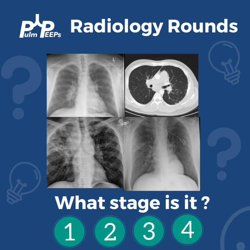
Radiology Rounds 19: Sarcoidosis Stages

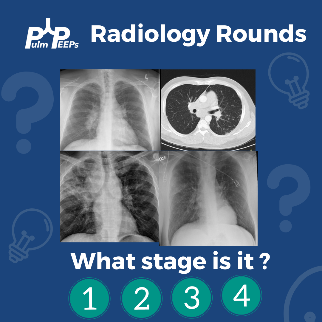
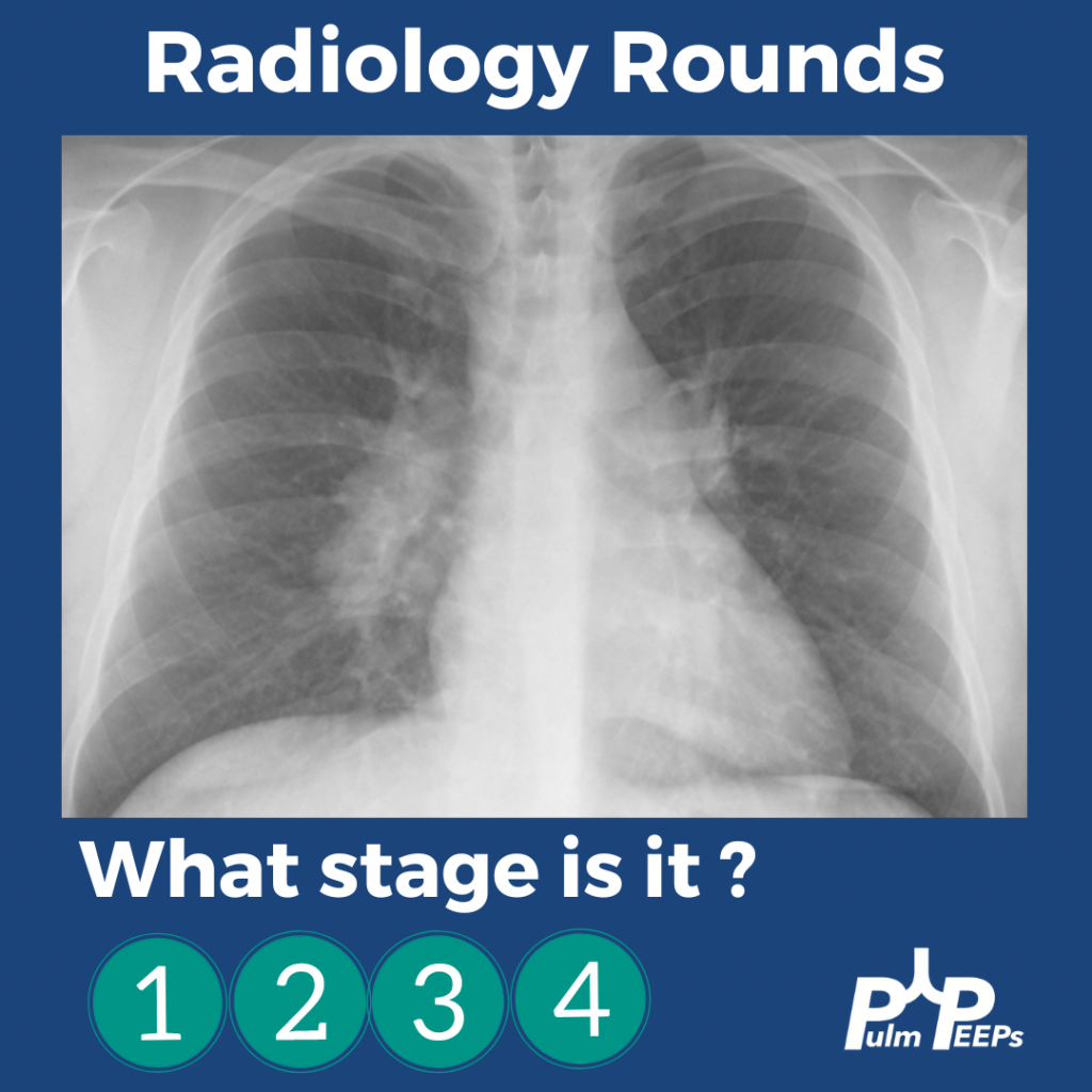
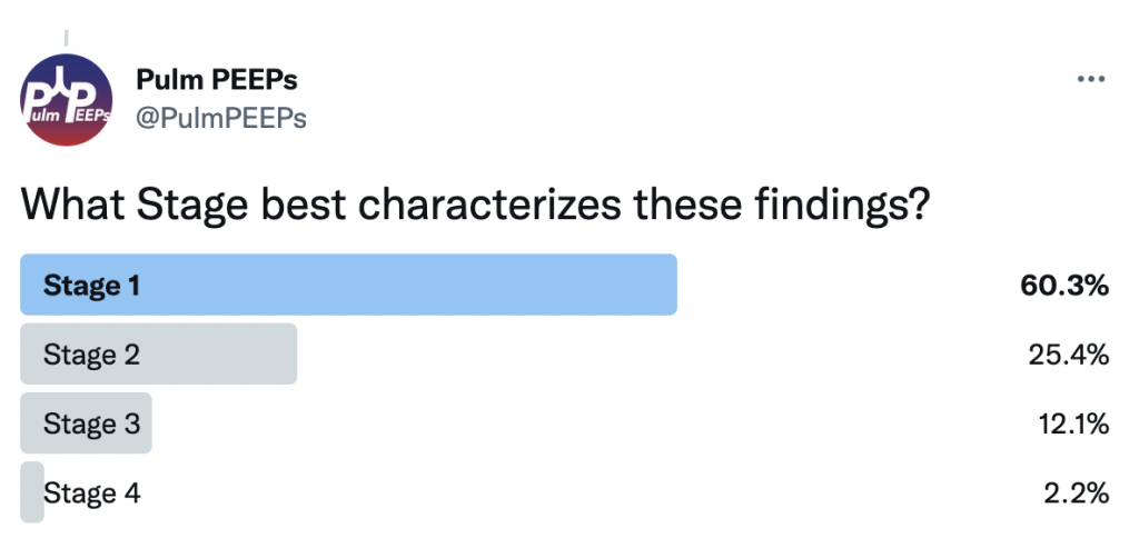
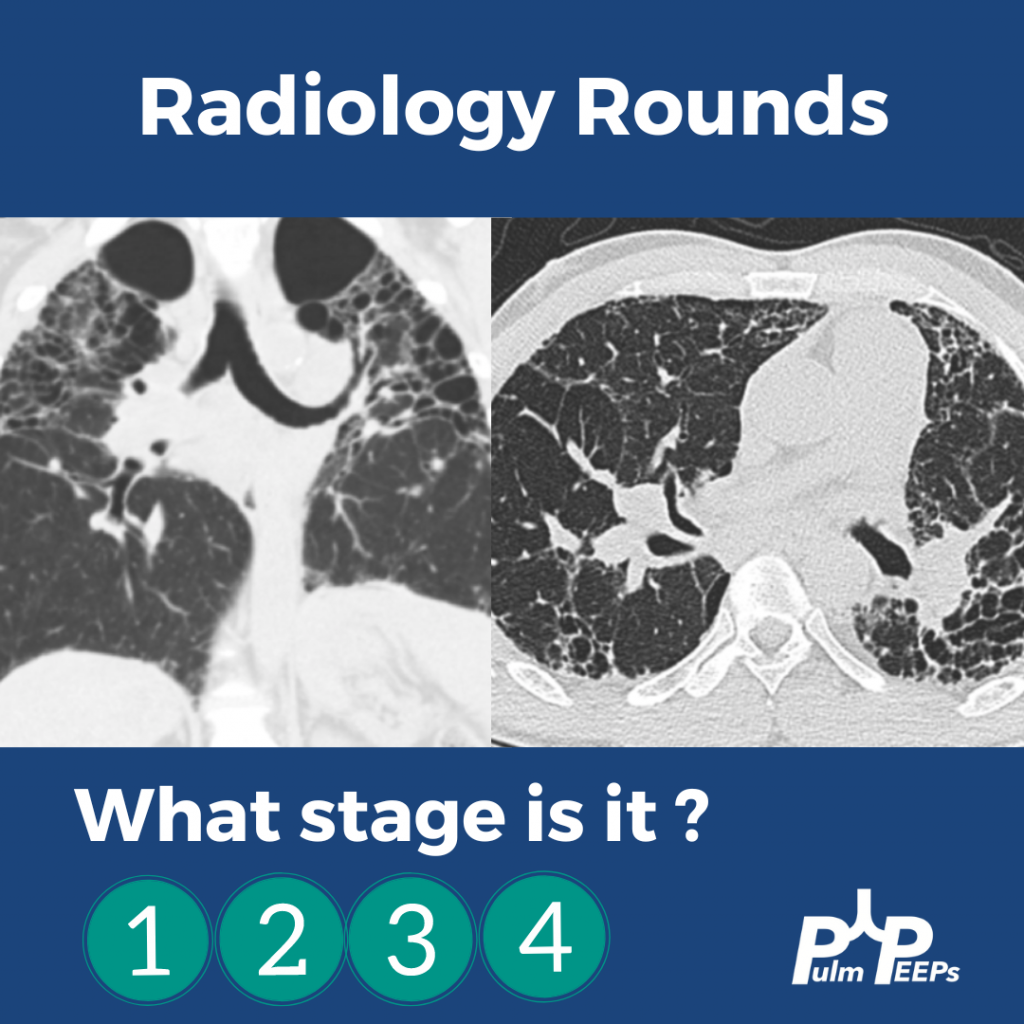
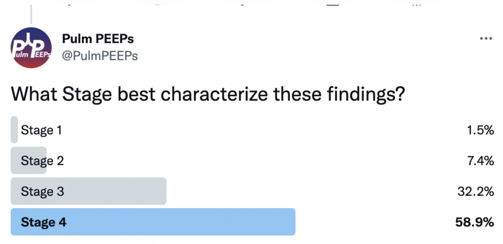
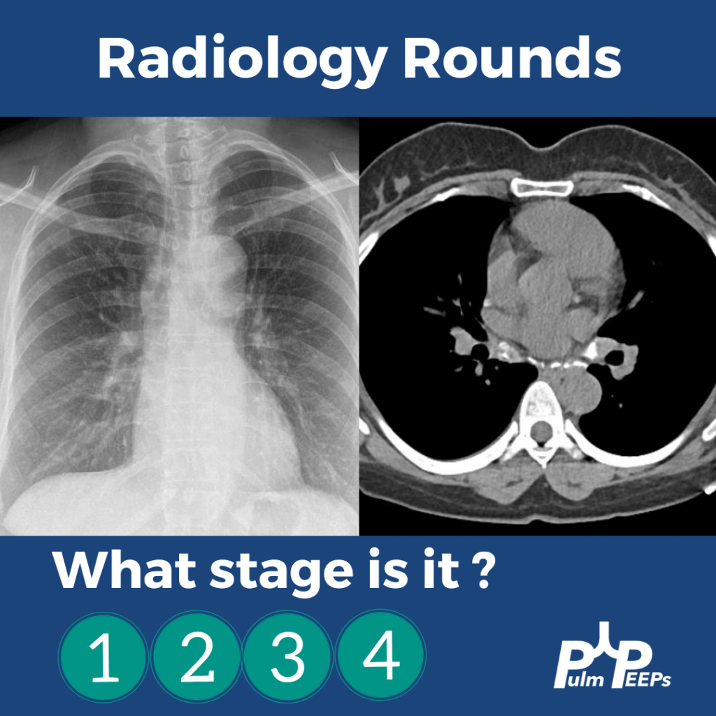
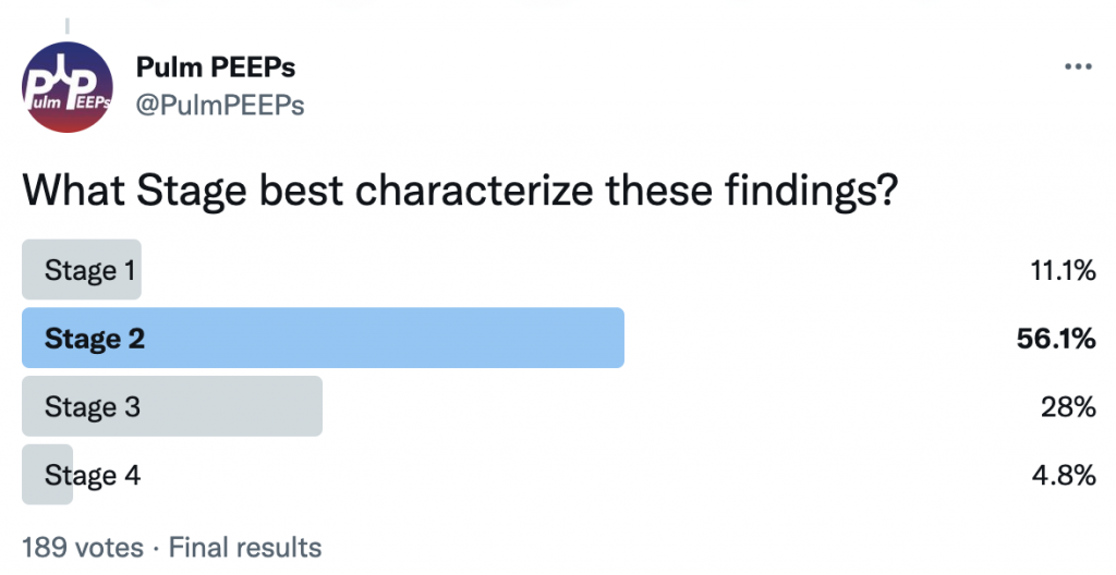
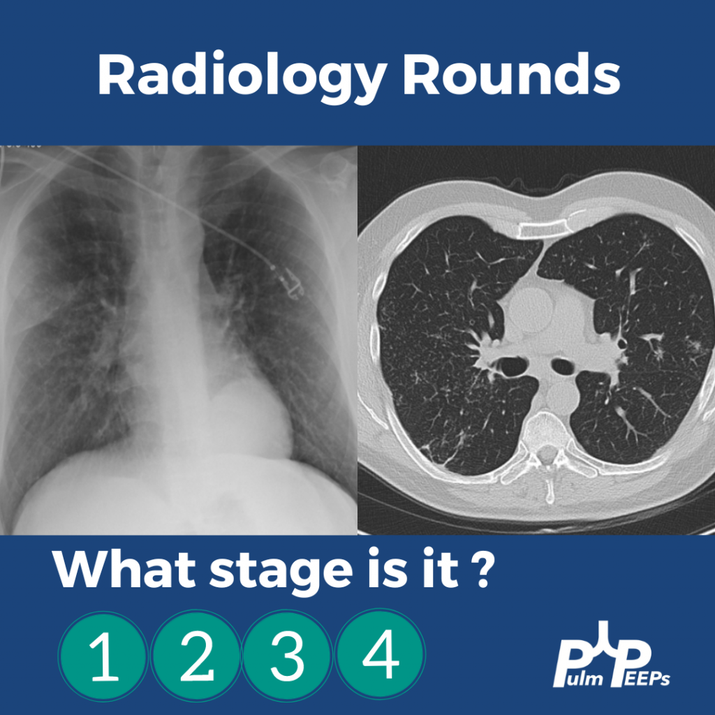
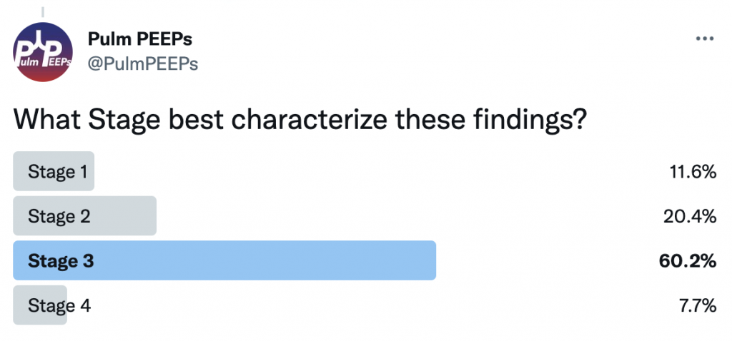
It is Tuesday and we have another Radiology Rounds we can’t wait to share with you. Follow along and see if you select the right answer as we go through different presentations of sarcoidosis and pick your answer! What stage is it?!

A middle-aged man presents to you after he was found to have hilar adenopathy on a routine chest x-ray.

A middle-age man presents with dyspnea on exertion, night sweats and weight loss. You see evidence of bilateral apical disease, and fibrosis with evidence of honeycombing on chest CT.

A young woman presents with dyspnea on exertion and was found to have hilar adenopathy with parenchymal disease.

An elderly man presents with dyspnea on exertion and was found to have nodular parenchymal disease without extensive lymphadenopathy.

Radiology Rounds 13: Mediastinal Widening – Aortic Dissection

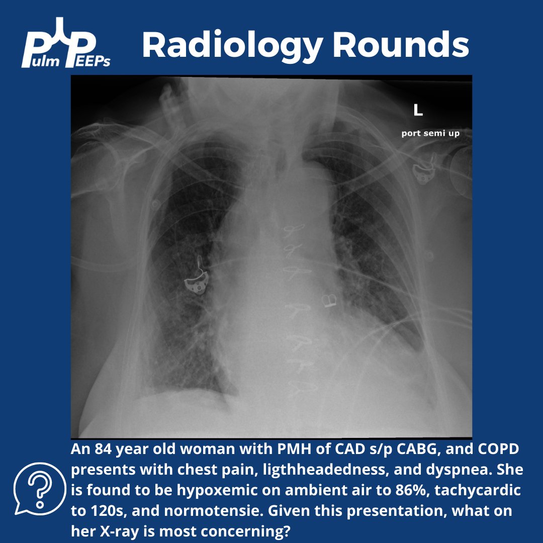
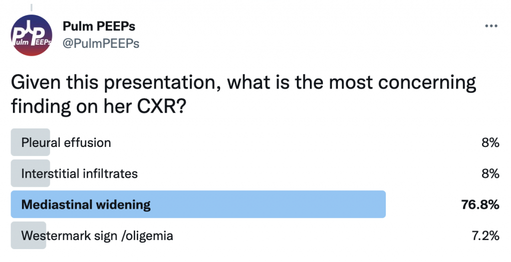
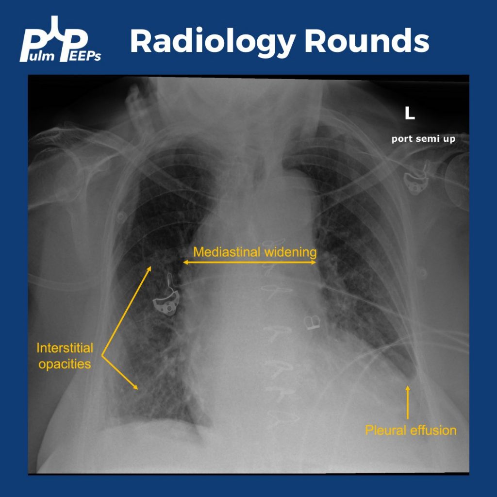
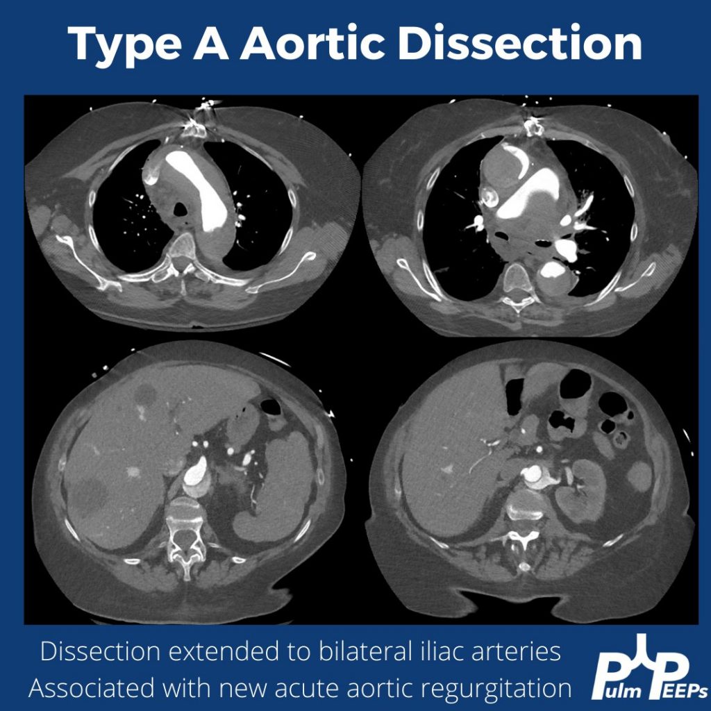
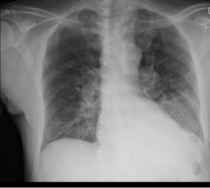
This week on #RadiologyRounds we have a patient with some key clues on the initial X-ray that help lead to the ultimate diagnosis.

While the patient has multiple findings on the X-ray, the mediastinal widening is the most concerning finding in the setting of acute chest pain, shortness of breath, and lightheadedness.

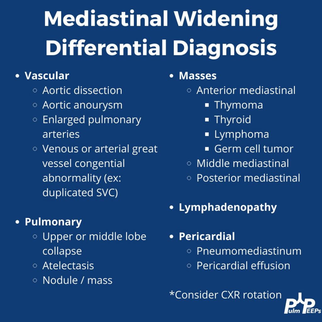
Mediastinal widening has a broad differential, which includes some can’t miss and life-threatening diagnoses

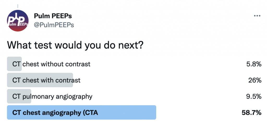
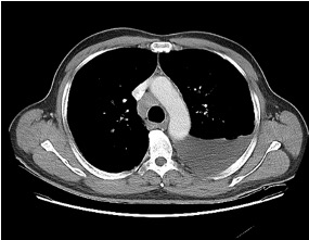
The key point is that aortic dissection, or ruptured thoracic aortic aneurysm, is high on the differential for this patient. Cross-sectional imaging is warranted and should be timed appropriately to evaluate the aorta. A triple rule-out CT would accomplish this as well. This patient had a CTA of the chest and was found to have a large type A aortic dissection with significant extension.

8. A Case of Dyspnea and Lymphadenopathy

Dave Furfaro, Kristina Montemayor, and Ansa Razzaq are back to tackle another pulmonary case! Listen in and solve the case yourself, and we’ll share some diagnostic pearls along the way. Let us know any additional thoughts on Twitter.

Patient Presentation

The is patient is 57-year-old man with hypertension and asthma who presents with dyspnea and left-sided pleuritic chest pain for 3 weeks. He was in his usual state of health until 3 weeks prior to admission, when he developed dyspnea and sharp left-sided chest pain that worsens with deep breathing. His symptoms are accompanied by unintentional 30-pound weight loss over the past several months as well as an intermittent cough that is nonproductive.

On physical exam, he is mildly tachypneic and saturating well on room air with otherwise normal vital signs. He has decreased breath sounds at the right lung base.

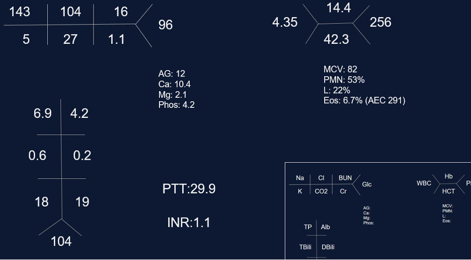
Initial labs

Key Learning Points

**Spoilers Ahead** If you want to think through the case on your own we advise listening to the episode first before looking at the infographics below

Physical Exam Pearls

Reasons for decreased breath sounds on physical exam

1. Increased thickness of chest wall

2. Reduced airflow to part of the lung

3. Overinflation to part of the lung

4. Something between the lung and chest wall — air or fluid

Determine bradypnea and tachypnea quickly by matching your breahting rate to the patient’s respiratory rate

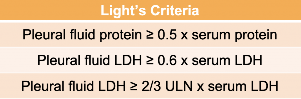
Pleural Effusions

Transudative effusion partial differential: heart failure, nephrotic syndrome, hepatohydrothorax, fluid overload, hypoalbuminemia, urinothorax, amyloidosis*, chlothorax*, hypothyrodism*, malignancy*, sarcoidosis*

*these effusions often present as exudates but can be transudates

Exudative effusion partial differential: Infection, hemothorax, malignancy, connective tissue disease, chylothorax, pancreatitis, esophageal perforation.

Sarcoidosis

Key Elements of Diagnosis:

1. Is there a compatible presentation (imaging, physical exam)

2. Detection of non-necrotizing granulomatous inflammation in one or more tissue samples

3. Exclusion of other disease that may present similarily

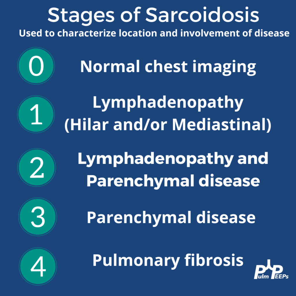
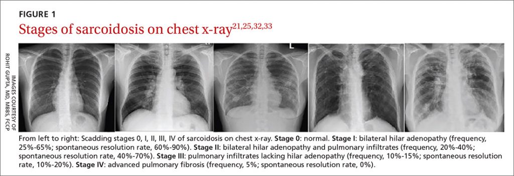
Pulmonary stages of sarcoidosis:

Key to remember that patients don’t always progress through these stages. The system is useful for prognosticating and determining treatment based on the risk for disease progression.

Image source: Gupta Et Al. J Fam Pract. 2021 April;70(3):E4-E15 | 10.12788/jfp.0177

Treatment for pulmonary sarcoidosis

Patients with stage 1, and even many with stage 2, often don’t require treatment

The first-line agent is oral glucocorticoids and the typical starting dose is prednisone 20 – 40 mg by mouth daily. The patient should be evaluated closely, and ideally, this dose can be tapered starting at about 4 – 6 weeks. Following this, the prednisone dose is tapered slowly over 6 months – 1 year while monitoring for symptom recurrence.

Second-line steroid-sparing agents are methotrexate, azathioprine, or mycophenolate. These are often used if the patient relapses, or is on more then 10mg daily for 3 months after the initial taper with intolerance of steroids

Third-line agents: tumor necrosis factor (TNF)- alpha antagonists (small molecule or monoclonal antibody therapy)

References and links for further reading

  1. Light RW. Pleural Effusion. New England Journal of Medicine. 2002;346(25):1971-1977. doi:10.1056/NEJMcp010731
  2. Feller-Kopman D, Light R. Pleural Disease. New England Journal of Medicine. 2018;378(8):740-751. doi:10.1056/NEJMra1403503
  3. Sarcoidosis: An FP’s primer on an enigmatic disease. MDedge Family Medicine. 2021;70(3). doi:10.12788/jfp.0177
  4. Iannuzzi MC, Rybicki BA, Teirstein AS. Sarcoidosis. New England Journal of Medicine. 2007;357(21):2153-2165. doi:10.1056/NEJMra071714
  5. Drent M, Crouser ED, Grunewald J. Challenges of Sarcoidosis and Its Management. New England Journal of Medicine. 2021;385(11):1018-1032. doi:10.1056/NEJMra2101555